Introduction
Overweight and obesity are the fifth leading risks for global deaths.Reference Franks, Hanson and Knowler1 The prevalence of obesity is highest among children of obese parents,Reference Whitaker, Wright, Pepe, Seidel and Dietz2 with the children of obese mothers being more at risk of overweight or obesity than those of obese fathers.Reference Catalano, Farrell and Thomas3, Reference Whitaker, Jarvis, Beeken, Boniface and Wardle4 Maternal pre-pregnancy overweight is an independent risk factor for infant overweight and abdominal obesity.Reference Pirkola, Pouta and Bloigu5 There are also associations between pregnant maternal body mass index (BMI) and offspring BMI, adiposity and insulin resistance,Reference Koupil and Toivanen6, Reference Mingrone, Manco, Mora, Guidone, Iaconelli, Gniuli, Leccesi, Chiellini and Ghirlanda7 and between maternal weight gain during pregnancy and offspring adiposity.Reference Reynolds, Osmond, Phillips and Godfrey8 One interpretation is that susceptibility to obesity and type 2 diabetes is partly programmed by the environment of the fetus during early pre- and postnatal life.Reference Lawlor, Smith and O'Callaghan9, Reference Nelson, Matthews and Poston10 In the United Kingdom, one in five pregnant women are now obese, which is a serious concern not only because of adverse pregnancy outcome, but also because of these associations with childhood risk of obesity and insulin resistance.Reference Dabelea11
Maternal hyperglycaemia during pregnancy was thought to be one of the most important predictors of infant obesity and metabolic disease, and possibly therefore the key vector linking the metabolism of the mother and her offspring.Reference Hillier, Pedula and Schmidt12 Now it is recognized that other maternal parameters associated with obesity and/or overnutrition during pregnancy may be involved, including hyperinsulinaemia, hypertriglyceridaemia, hyperleptinaemia and inflammation. Both maternal hyperglycaemia (which stimulates fetal insulin synthesis) and neonatal hyperinsulinaemia increase infant adiposity.13, Reference Silverman, Metzger, Cho and Loeb14 Maternal triglycerides, which are elevated in obese and insulin resistant women, will not cross the placenta but are hydrolysed by placental lipasesReference Herrera and Amusquivar15 and may affect fetal fuel supply and offspring adiposity.Reference Catalano, Farrell and Thomas3, Reference Hamilton, Odrobina and Yin16 Hyperleptinaemia has been reported in obese pregnancies and there is a positive association between cord blood leptin concentrations, birth weight and/or neonatal adiposity.Reference Stocker and Cawthorne17 In contrast to these reports there are fewer studies investigating a link between maternal overnutrition and/or obesity and inflammation during pregnancy and the effects on fetal development.Reference Briana and Malamitsi-Puchner18–Reference Poston20
Controlled intervention studies in animal models show strong parallels with human observational studies, supporting a causal relationship between maternal obesity and offspring adiposity and metabolic disease. Thus, offspring of high fat-fed dams had increased adiposity, insulin resistance and hypertension,Reference Bayol, Simbi, Bertrand and Stickland21–Reference Samuelsson, Matthews and Argenton23 while offspring of dams that have maternal diabetes had neonatal hyperinsulinaemia, hyperphagia and obesity.Reference Plagemann, Harder, Kohlhoff, Rohde and Dorner24 Changes in placental morphologyReference Farley, Tejero and Comuzzie25 and placental lipid accumulationReference Strakovsky and Pan26 have been observed in these models. These changes have been associated with increased placental fatty acid transporters and nutrient delivery to the fetus.Reference McCurdy, Bishop and Williams27, Reference Odaka, Nakano and Tanaka28
It is well reported that overweight and obesity are states of systemic and adipose inflammation, with increased levels of systemic adipokines and adipocytokines associated with impaired glucose tolerance and insulin resistance.Reference Bourlier and Bouloumie29, Reference de Luca and Olefsky30 Some reports have linked the inflammatory state associated with maternal obesity in both women and animal models with persistent influences on offspring metabolic function.Reference Catalano, Presley, Minium and Hauguel-de Mouzon31–Reference Urbanek, Hayes and Lee33 Increases in proinflammatory cytokines secreted by both maternal adipose and placental tissue can increase maternal circulating levels of these factors, resulting in increased exposure of the fetus and an adverse pregnancy outcome.Reference Dahlgren, Samuelsson, Jansson and Holmang34–Reference Zhu, Ma, Long, Du and Ford36 However, the role of inflammatory cytokines in fetal development is poorly understood and there are a limited number of studies defining the role of adipokines, such as adiponectin and adipocytokines, such as resistin, IL6, IL10, IL1β and IFNγ in obese pregnancies on offspring health.
The identification of the vectors in the maternal environment that mediate the effects of maternal obesity and her diet is of key importance to the development of clinical intervention strategies. In this study, we have investigated the effects of both maternal obesity and overnutrition in a diet-induced high fat-fed mouse model on her metabolic status during pregnancy. We have also investigated whether obesity and overnutrition during pregnancy influences the levels of maternal adipokines (adiponectin and RBP4) and adipocytokines (leptin, resistin, IL6, IL10, IL12, IL1β, IFNγ, CXCL1 and TNF-α) circulating in the plasma and resistin secreted from parametrial and subcutaneous adipose tissue.
Materials and methods
Materials
Reagents were obtained from Sigma-Aldrich, Poole, UK, unless otherwise stated.
Animals
Animal procedures were conducted in accordance with University of Buckingham project licence, under the UK Animals [Scientific Procedures Act (1986)] and as approved by the University's Ethical Review Board. Female C57/Bl6 mice (Charles River, UK) aged 6–7 weeks on arrival. Thirty-four females, housed in six cages of five or six animals, were fed a standard laboratory chow diet that contained 10% fat (from soya oil), 70% carbohydrate and 20% protein by energy (Beekay Feed, B&K Universal Ltd, UK) and forty-seven females, housed in eight cages of five or six animals, were fed a high fat diet 42% fat, 43% carbohydrate and 15% protein by energy (Western Rodent 829100, SDS diet, UK). They were housed at 21–23°C with lights on from 07:00 to 19:00 h in groups of five to eight mice per cage and fed the appropriate diet for 10 weeks before conception. Body weight was measured weekly before mating. Two separate groups of pregnant experimental animals were used. At day 18 of pregnancy, one group of pregnant dams (from matings of n = 18 chow, housed in three cages, and 24 high fat fed, housed in five cages) was fasted from 08:00 h and then dosed orally with glucose (3 g/kg body weight) at 13:00 h. A second group of dams (from matings of n = 16 chow, housed in three cages, and 23 high fat fed, housed in five cages) was sacrificed in the fed state for fat pad weight and samples were taken of both parametrial and inguinal subcutaneous adipose tissue. Whole blood was collected into EDTA-coated microvettes and the plasma was stored at −80°C before analysis.
Body composition
The body composition of the mice was measured under light gaseous anaesthetic (Isofluorane, Isoba, Shering-Plough Animal Health, UK) both at preconception and at day 18 of pregnancy using dual-energy X-ray absorptiometry (Lunar PIXImus 2 mouse densitometer and version 1.46 software, GE Medical Bedford, UK).
Preparation of adipose tissue-conditioned medium
Parametrial and inguinal subcutaneous adipose fat pad samples (∼0.2 g) were harvested into pre-warmed Krebs–Ringer–HEPES buffer pH 7.4 containing 1% endotoxin-free bovine serum albumin (BSA). Samples were immediately placed into DMEM/F-12 Ham's (1:1) (supplemented with l-glutamine, 15 mM HEPES and 1% BSA) and minced and the samples incubated at 37°C using a shaker for 90 min at 450 rpm (Heidolph Titramax, Germany). The culture medium was collected and frozen at −80°C before the analysis of resistin levels.
Plasma and adipose tissue-conditioned medium analysis
Blood glucose, insulin and leptin were measured as described previously. The concentrations of the adipokines adiponectin and RBP4 were measured by commercially available murine assays (ALPCO, USA and AdipoGen, Switzerland, respectively) using a spectrophotometer (SpectraMax250, Molecular Devices, USA) according to the manufacturer's recommendations. Levels of the cytokines IL6, IL10, IL12, IL1β, IFNγ, CXCL1 and TNF-α were quantified using a seven-plex assay (Meso Scale Discovery, USA) and resistin in a single-plex assay (Meso Scale Discovery, USA). Results were determined using the Sector Imager 2400 (Meso Scale Discovery, USA) according to the manufacturer's recommendations.
Statistical analysis
Data were analysed using GraphPad Prism software, version 5.0. Body weight, body composition, blood glucose, area under the glucose curve, plasma leptin and adiponectin, RBP4 and both plasma and secreted cytokine and resistin levels were analysed using an unpaired t-test. For IL6, IL10 and IL12 where the Barratt's test showed unequal variances apparently due to the variance being proportional to the mean, data were log transformed. Results are presented as means ± s.e.m. Statistical significance for effects in the figures is given as *P < 0.05; **P < 0.01; ***P < 0.001.
Results
Body weight and body composition at preconception
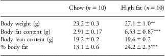
Ten weeks of high feeding before conception increased body weight, body fat and percentage body fat content significantly in the overnourished mice as compared with the chow-fed mice. Neither treatment affected lean body mass (Table 1).
Body weight and body composition of mice preconception after 10 weeks of chow or high fat feeding

Results are presented as means ± s.e.m.
Statistical significance for effects in the table is given as **P < 0.01; ***P < 0.001, using an unpaired t-test.
Body weight and body composition at day 18 of pregnancy
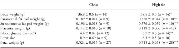
At day 18 of pregnancy, high fat-fed dams had increased body weight and body fat (Table 2). However, no significant differences were observed for placental weight or the number of viable fetuses per pregnancy. Fetal weight was, however, significantly increased in the pregnancies of high fat-fed dams as compared with chow-fed pregnancies (Table 2).
Maternal body weight and body composition, placental weight, 5 h fasted blood glucose, fetal number and weight on day 18 of pregnancy
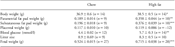
Results are presented as means ± s.e.m.
Statistical significance for effects in the table is given as *P < 0.05; **P < 0.01; ***P < 0.001 using an unpaired t-test.
Glucose tolerance at day 18 of pregnancy
At day 18 of pregnancy, fasting blood glucose was significantly higher in the high fat-fed dams as compared with those on a chow diet (Table 2). In a glucose tolerance test high fat-fed dams had worsened glucose tolerance over the 2-h test period (Fig. 1a). The plasma insulin concentrations measured 30 min before and 30 min after glucose administration were raised in dams fed a high fat diet as compared with those fed a chow diet (Fig. 1b).
Fasting blood glucose and oral glucose tolerance in high fat-fed and chow-fed dams at day 18 of pregnancy. Glucose tolerance after a 5 h fast (a) and plasma insulin levels in response to the glucose load (b) were measured at day 18 of pregnancy in chow (body weight 38.6 ± 1.4 g; open bars) and high fat (body weight 43.0 ± 0.7 g; black bars)-fed mice. n = 10 animals per group. *P < 0.05; **P < 0.01 for differences between chow and high fat-fed dams.

Plasma adipokines at day 18 of pregnancy
Neither fed plasma leptin nor RBP4 concentrations differed significantly between the maternal nutritional groups at day 18 of pregnancy (Fig. 2a and 2b), while fed levels of plasma adiponectin were significantly reduced in the high fat-fed dams as compared with the chow-fed dams (Fig. 2c).
Circulating plasma leptin, retinol binding protein 4 (RBP4) and adiponectin in high fat-fed and chow-fed dams at day 18 of pregnancy. Fed levels of plasma leptin (a), RBP4 (b) and adiponectin (c) were measured at day 18 of pregnancy in chow and high fat-fed mice. n = 6–9 animals per group. *P < 0.05 for differences between chow and high fat-fed dams.
Plasma cytokines at day 18 of pregnancy
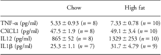
No differences in plasma levels of TNF-α, CXCL1, IL12 or IL1β were observed between maternal groups (Table 3). Although the mean value for IL6 was increased in the high fat-fed mice by 44.6% (Fig. 3a), the variance was high in the high fat-fed dams and even when the data were logged this difference did not reach statistical significance (P = 0.09). In contrast, circulating IL10 significantly increased by 52% (Fig. 3b) in high fat-fed dams as compared with control-fed dams (P = 0.04). IFNγ levels were significantly elevated in the high fat-fed dams (Fig. 3c).
Maternal circulating cytokine concentrations on day 18 of pregnancy
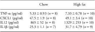
Results are presented as means ± s.e.m.
Circulating plasma cytokines in high fat-fed and chow-fed dams at day 18 of pregnancy. Fed levels of IL6 (a), IL10 (b) and IFNγ (c) were measured at day 18 of pregnancy in chow and high fat-fed mice. n = 9–10 animals per group. *P < 0.05 for differences between chow and high fat-fed dams.
Resistin secretion and plasma levels at day 18 of pregnancy
Circulating resistin levels were significantly reduced in the high fat-fed dams at day 18 of pregnancy as compared with the chow-fed dams (Fig. 4a). This was associated with a decrease in adipocyte secretion of resistin from both parametrial and inguinal subcutaneous adipose-conditioned media (Fig. 4b and 4c).
Circulating and adipocyte-secreted resistin levels in high fat-fed and chow-fed dams at day 18 of pregnancy. Fed levels of plasma resistin (a) and adipocyte-secreted resistin from parametrial adipose (b) and inguinal subcutaneous adipose (c) conditioned media were measured at day 18 of pregnancy in chow and high fat-fed mice. n = 9–10 per group. ***P < 0.001 for differences between chow and high fat-fed dams.
Discussion
Epidemiological studies in humans have shown that the prevalence of obesity, glucose intolerance and cardiovascular disease is influenced by the pre- and early postnatal phases of life.Reference Poston20 In rodents, maternal obesity and the overconsumption of diets high in fat and sugar are associated with increased adiposity, impaired glucose tolerance, insulin resistance and hypertension in her offspring.Reference Nivoit, Morens and Van Assche22, Reference Samuelsson, Matthews and Argenton23 In the present study, we have investigated how maternal overnutrition in an obese mouse model might affect the developing fetus. We have focussed in particular on adipokines and adipocytokines by investigating whether their secretion from adipose tissue and their plasma concentrations are altered in obese dams.
In agreement with previous reports,Reference Howie, Sloboda, Kamal and Vickers37 prolonged feeding of a high fat diet increased body weight and fat mass both before conception and at day 18 of pregnancy. Fasting blood glucose and plasma insulin were also elevated at day 18. All of these changes may contribute to increased fetal weight and predispose offspring to the development of metabolic disease in later life.Reference Silverman, Metzger, Cho and Loeb14, Reference Jones, Woollett and Barbour38, Reference Zambrano, Martinez-Samayoa, Rodriguez-Gonzalez and Nathanielsz39 It is notable that fetal weight was higher in the high fat fed than the chow-fed mice despite placental weight being unchanged, whereas others have reported a positive correlation between placental and fetal weight.Reference Strakovsky and Pan26
Overweight and obesity are associated with increased secretion of adipocytokines and other adipokines, some of which may affect insulin resistance through their pro- or, in the case of adiponectin, anti-inflammatory activity.Reference Hulsmans, Van Dooren, Mathieu and Holvoet40, Reference Mantzoros, Li, Manson, Meigs and Hu41 They may also affect fetal development as a consequence or independently of their effects on inflammation and insulin sensitivity. Resistin is a pro-inflammatory adipokine.Reference Bokarewa, Nagaev, Dahlberg, Smith and Tarkowski42 Obese high fat-fed dams at day 18 of pregnancy had reduced maternal circulating levels of resistin and this was reflected by reduced resistin secretion from both parametrial and inguinal subcutaneous fat depots. This is surprising because resistin is associated with insulin resistance,Reference Rajala, Qi and Patel43, Reference Steppan, Bailey and Bhat44 although there are a number of conflicting reports on its correlation with the level of adiposity.Reference Heilbronn, Rood and Janderova45–Reference Way, Gorgun and Tong47 Circulating resistin levels increase in both rodentsReference Caja, Martinez, Abelenda and Puerta48 and human pregnancies.Reference Megia, Vendrell and Gutierrez49, Reference Nanda, Poon, Muhaisen, Acosta and Nicolaides50 High concentrations of resistin in umbilical plasma samples suggest a potential role in fetal metabolism.Reference Briana and Malamitsi-Puchner18 Furthermore, umbilical resistin levels are associated with maternal circulating levels and are negatively correlated with birth weight and infant adiposity.Reference Cho, Yoo and Hong51 Thus, our results are consistent with the low concentration of plasma resistin playing a causal role in the increased fetal weight. They suggest that resistin could have different functions during early development as compared with adult life.
Maternal leptin levels support the development and maturation of the fetusReference Stocker and Cawthorne17 and others have associated hyperleptinaemia in obese pregnancies in rodent models with impaired health of the offspring.Reference Stocker and Cawthorne17, Reference Samuelsson, Matthews and Argenton23, Reference Savino, Liguori, Fissore and Oggero52 However, the plasma leptin concentration was not significantly elevated in our obese dams. Plasma RBP4 was also unaffected by diet in our study, despite others finding that cord blood levels in humans correlate positively with fetal growthReference Chan, Tsai and Wu53 and maternal adiposity,Reference Laudes, Oberhauser and Bilkovski54 and that rodent serum levels correlate with insulin resistance.Reference Yang, Graham and Mody55
The adipokine adiponectin is unusual in having anti-inflammatory properties and enhancing insulin sensitivity. The plasma concentration of adiponectin was lowered by the high fat diet and this may have contributed to impaired glucose tolerance coupled with elevated plasma insulin in the dams. Previous studies have reported that cord blood adiponectin levels correlate positively with birth weight,Reference Savino, Liguori, Fissore and Oggero52 whereas the low adiponectin levels in our study were associated with high fetal weight. Low adiponectin levels might promote high fetal weight by decreasing insulin sensitivity and thereby raising plasma insulin.
We have examined the circulating maternal levels of the pro-inflammatory cytokines IL6, IL12, IL1β, IFNγ, CXCL1 and TNF-α and the anti-inflammatory cytokine IL10 at day 18 of pregnancy. The level of IL-10 was increased, although both an increased and a decreased level have been reported in obese women.Reference Esposito, Pontillo and Giugliano56, Reference Juge-Aubry, Somm and Pernin57 In addition to its anti-inflammatory role, IL10 has insulin sensitizing properties and regulates differentiation and invasion of trophoblasts during the early stages of pregnancy.Reference Renaud, Macdonald-Goodfellow and Graham58 Therefore, any changes in the level of this cytokine may influence the growth and development of the fetus. Maternal IFNγ was also elevated. IFNγ is involved in the promotion of macrophage infiltration but has not been proposed to play a role in fetal development. The pro-inflammatory cytokines IL1β, IL6, IL12 and TNF-α have been reported to be increased in obese individuals and importantly increased levels of IL6 and TNF-α are reported in obese human pregnancies.Reference Bugatto, Fernandez-Deudero and Bailen59, Reference Ramsay, Ferrell and Crawford60 In mice, the in utero exposure to IL-6 in late pregnancy was associated with increased fetal growth through the stimulation of placental amino acid transport and enhanced lipid accumulation.Reference Lager, Asterholm and Schele35 In our study, we observed a tendency for an increase in maternal IL6 levels, but the levels varied markedly between individual obese dams. The levels of IL1β, TNF-α and CXCL1 were unchanged.
We acknowledge that we have not investigated the effects of a high fat diet and obesity separately on adipokine concentrations and secretion from adipose tissue. Early developmentally programmed events may occur as a result of the complex interactions between maternal overnutrition and maternal obesity or each may contribute independently.Reference White, Purpera and Morrison61 Our objective was to identify adipokines, including adipocytokines, that might be investigated further as possible links between maternal obesity and offspring weight and health.
In summary, we have investigated the effects of maternal obesity and overnutrition on maternal metabolic and inflammatory parameters at day 18 of pregnancy in mice, highlighting potential new programming mediators of metabolic disease in the offspring. The plasma concentrations of previously well-characterized maternal ‘vectors’, namely maternal hyperglycaemia and hyperinsulinaemia, were raised by overnutrition and obesity in the pregnant dam, but also the secretion and plasma concentration of the adipokine resistin was lowered, while plasma concentrations of the cytokines IL10 and IFNγ were raised. The roles of resistin, IL10 and IFNγ in metabolic phenotyping of the offspring merit further study. Other studies in pregnant women and non-human primates have implicated the inflammatory state associated with maternal obesity as causing persistent influences on offspring metabolic function.Reference Nelson, Matthews and Poston10, Reference Grayson, Levasseur and Williams32 In one recent study, circulating maternal cytokines were positively associated with maternal adiposity but not with fetal adiposity.Reference Farah, Hogan and O'Connor62 The translation of experimental findings such as ours to the clinical setting could inform future strategies for therapeutic interventions in obese pregnant women designed to improve the health of both mother and infant.
Acknowledgements
The authors thank Julie Cakebread, David Hislop and Anita Roberts for their excellent technical support.
Financial Support
This research did not receive any specific grant from any funding agency in the public, commercial or not-for-profit sector.
Conflicts of Interest
None.
Ethical Standards
Animal procedures were conducted in accordance with University of Buckingham project licence, under the UK Animals (Scientific Procedures Act, 1986) and as approved by the University's Ethical Review Board.