In 1945, Blalock and Taussig described their approach to maintain pulmonary circulation in duct-dependent congenital cardiac disease using an end-to-side anastomosis of the subclavian artery to the pulmonary artery.Reference Blalock and Taussig1 After 17 years, the use of a Teflon shunt was instead advocated, and subsequently polytetrafluoroethylene (Goretex) replaced the Teflon.Reference Gazzaniga, Lamberti and Siewers2, Reference Klinner, Pasini and Schaudig3 Since then, this is the most commonly used material, and usually this technique is referred to as a modified Blalock–Taussig shunt.
Patency of the shunt in shunt-dependent pulmonary circulation, either in right ventricular outflow tract obstruction or atresia, or in single-ventricle lesions after a Norwood stage I type operation, is pivotal. In case of a stenosis of the shunt, oxygen saturations decrease, and either a surgical or an interventional approach to replace, or reopen the shunt can be used.Reference Kaestner, Handke and Photiadis4, Reference Peuster, Fink and Bertram5
After an initial case that was published previously,Reference Krasemann and Qureshi6 we encountered seven more cases of children undergoing stent placement into the modified or classical Blalock–Taussig shunt. Here, we comment on the lessons learned from each case.
Methods
The institutional database of the Department of Paediatric Cardiology of the Evelina Children's Hospital was searched for children who underwent stenting of a Blalock–Taussig shunt. Of the eight children who were identified between February, 2006 and June, 2009, the case of one child was published previously, and is therefore not included in this series.Reference Krasemann and Qureshi6 Clinical, echocardiographic, and angiographic data of the remaining seven cases were reviewed.
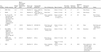
The characteristics of the children are depicted in Table 1. All children except one were male. A total of five modified and two classical Blalock–Taussig shunts were stented.
Children's characteristics.

AP = anteroposterior; AV = atrioventricular; AVSD = atrioventricular septal defect; BT = Blalock–Taussig; Cran = cranial; DILV = double inlet left ventricle; DORV = double outlet right ventricle; HLHS = hypoplastic left heart syndrome; LAO = left anterior oblique; Lat = lateral; LFA = left femoral artery; LFV = left femoral vein; LPA = left pulmonary artery; MRSA = methicillin-resistant Staphylococcus aureus; PA = pulmonary artery; RAO = right anterior oblique; RFA = right femoral artery; RFV = right femoral vein; TA = tricuspid atresia; TGA = transposition of the great arteries; TOF = tetralogy of Fallot; VA = ventriculoarterial
Tachypnoea was present in four children, whereas one remained ventilated after the shunt placement (case 3). Low oxygen saturations were present in all cases with levels between 35% and 60%. There was one child (case 7) with high inflammatory markers who was on antibiotic treatment.
Echocardiography confirmed the shunt stenosis or occlusion in all children. Modified suprasternal views showed stenosis at the distal end of the Blalock–Taussig shunt both on two-dimensional images and on colour Doppler.
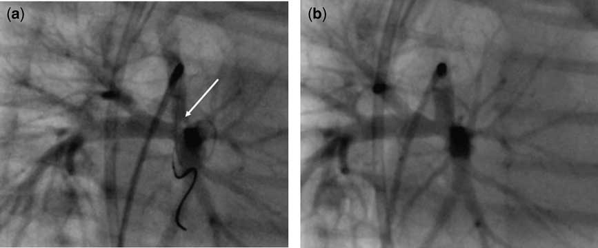
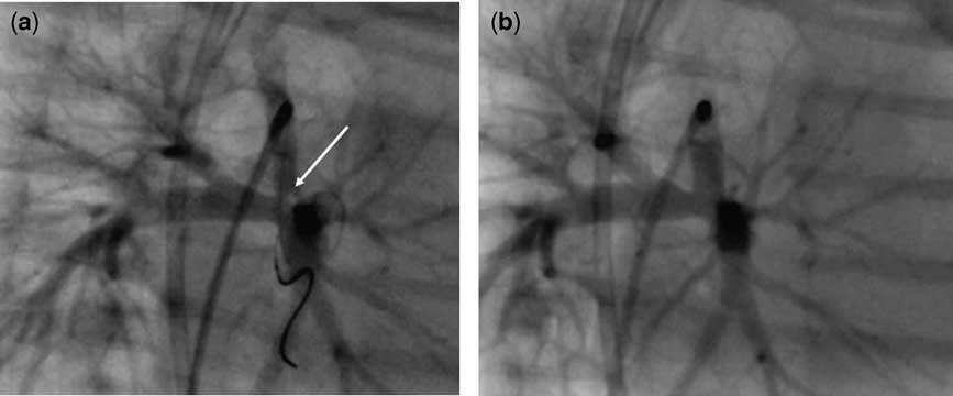
Femoral arterial and venous access was gained in all children. The shunt origin was entered via the arterial sheath with a 3F or 4F Cobra catheter (3F: Balt, Montmorency, France; 4F: Cordis Corporation, Piscataway, New Jersey, United States of America) in five of the cases. A right-sided modified shunt was reached with a cut-off 4F Pigtail catheter (Cordis Corporation). For a classical obstructed shunt, we used a 4F Judkins right coronary catheter (Cordis Corporation). The stenosis, which was identified using oblique projections (Figs 1 and 2), was located at the distal end in all four modified shunts, and at the anastomosis with the innominate artery in one of the classical shunts (case 5). The remaining classical shunt and one modified one were completely occluded (cases 3 and 7).
Refers to case 2. (a) Left anterior oblique projection, position of the 0.014 Luge-wire through the shunt into the left pulmonary artery. Stenosis of the Blalock–Taussig shunt at the pulmonary end (arrow). (b) Left anterior oblique projection, angiography after stent placement in the Blalock–Taussig shunt.
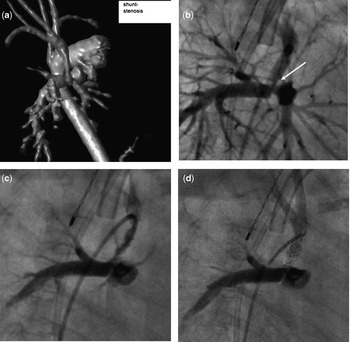
Refers to case 4. (a) Magnetic resonance imaging scan (volume rendered 3D) showing the distal narrowing of the modified Blalock–Taussig shunt and the proximal stenosis of the left pulmonary artery. (b) Right anterior oblique projection. Stenosis of the Blalock–Taussig shunt at the pulmonary end (arrow), and proximal left pulmonary artery stenosis. (c) Right anterior oblique projection. Dissection of the shunt neointima. (d) Right anterior oblique projection. Fluoroscopy after stent deployment as a rescue procedure after dissection.

In one case, a 0.018 inches Terumo wire (Terumo Medical Corporation, Somerset, New Jersey, United States of America) was used to engage the shunt, whereas in all other cases a 0.014 inches LugeTM Guide Wire (Boston Scientific, Natick, Massachusetts, United States of America) was advanced through the stenosed shunt into one of the branch pulmonary arteries (Fig 1). In one case, this was exchanged for a 0.014 inches FasDasher wire (Target Therapeutics, San Jose, California, United States of America), and in another for a 0.014 inches IronMan guidewire (Abbott Vascular Inc., Redwood City, California, United States of America) to allow better transmission of the stent around the tight angles.
Angiography during stent positioning was performed through a long 5F Mullins sheath (Cook, Bloomington, Indiana, United States of America) through which the stent was also passed in only one child, and with a second catheter passed antegradely from the femoral vein through the right and left atria and the left ventricle into the ascending aorta in two children. In the remaining four children, guidance was from the stored angiographic image before stent placement.
Liberte stents (Boston Scientific, Natick, Massachusetts, United States of America) with a length of 8 and 12 millimetres were used in all children. There were four children who received a single stent in the shunt, and in one of them the left pulmonary artery was stented through the shunt beforehand using a 5 × 8 millimetres Liberte stent. In one child, two stents were used to cover the entire length of the shunt, because neointimal dissection occurred over the whole length of the shunt after balloon dilation of the left pulmonary artery through the shunt (case 4, Fig 2). There was one child who received three overlappingly positioned stents (case 7). In the remaining children with a blocked classical shunt, a Driver coronary stent (Medtronic, Minneapolis, Minnesota, United States of America) measuring 4 × 9 millimetres was deployed in addition to two Liberte stents (case 3). There were four modified shunts with a diameter of 3.5 millimetres, and the remaining one measured 4 millimetres in diameter. The classical shunts were 3 millimetres or less in diameter. The stents were dilated to their full diameter of 4 or 5 millimetres, depending on the shunt size.
Postinterventionally, all children received 5 milligrams per kilogram of aspirin daily.
Results
There were no immediate complications related to the procedure.
Saturations improved immediately after the intervention to greater than 70% in all but one child, who underwent stenting of a classical shunt as a rescue procedure (case 3). This was a  -month-old boy with atrioventricular and ventriculoarterial discordance, left-sided atrioventricular valve atresia, and a large ventricular septal defect, who underwent a pulmonary artery banding at the age of 2 months, and developed a restrictive interatrial communication, and the ventricular septal defect showed echocardiographic signs of restriction. He underwent a Damus–Kaye–Stansel procedure, atrial septectomy, hemi-Fontan operation, and closure of a large venovenous collateral. An additional left-sided classical Blalock–Taussig shunt was created to improve persistently low oxygen saturations. After initial improvement from the surgery, the oxygen saturation decreased again to 30% and he became markedly acidotic. Angiography showed a completely occluded shunt. After stenting, the saturations improved to 60%, but he remained acidotic. Cardiac output remained poor, and he died on the day after the procedure. Another child showed good progress, but a week after discharge, he developed temperature, was admitted to his local hospital where shortly after feeding, his cardiac output dropped, and he suffered a cardiac arrest and died.
On follow-up, the stented shunts remained patent in the surviving children, of whom four underwent further surgery: There were two children who underwent a hemi-Fontan operation during which the stented modified shunts were removed (cases 1 and 2). During a Kawashima procedure, the stented origin of the right-sided classical Blalock–Taussig shunt was resected, and the subclavian artery reimplanted into the aortic arch (case 5). In the child who was on antibiotic treatment for high inflammatory markers (case 7), clots migrated from the shunt and proximal pulmonary arteries into the distal vessels during inflation of the balloon. After two days, the stented shunt reoccluded and was surgically removed. During surgery, the shunt was found to be covered by pus, and microbiological assessment revealed Klebsiella. He underwent full repair of the underlying cardiac defect (tetralogy of Fallot), but later developed right outflow tract dilation, which was again treated surgically, including patch plasty of both proximal branch pulmonary arteries. Furthermore, he underwent ballooning of the re-stenosed right pulmonary artery. There was one child who had not yet been operated upon.
Discussion
Occlusion of a Blalock–Taussig shunt leads to decreased pulmonary perfusion with subsequent clinical symptoms of respiratory distress and low oxygen saturations. The initial diagnostic tool in all our cases was echocardiography, which either showed the stenosis or revealed flow abnormalities on colour or conventional Doppler. Echocardiography was used to confirm the diagnosis, but neither two-dimensional nor colour Doppler absolute diameters predicted problems. In cases of complex anatomy, magnetic resonance imaging was also carried out.
Once the diagnosis was confirmed, cardiac catheterisation was carried out without delay.Reference Kaestner, Handke and Photiadis4 Interestingly, only one of the reported cases showed clinical signs of shunt obstruction shortly after its placement (case 4), whereas the majority of shunt obstructions occurred with a delay of up to 4 months after implantation (Table 1).
Access
In all our children, arterial access was gained with a 4F femoral sheath. This was upgraded to a 5F Mullins sheath in one child to allow angiograms during stent placement. Another option is to use a second catheter introduced from the femoral vein antegradely into the aorta through a patent interatrial communication, as shown in cases 4 and 6.
All shunts were accessed retrogradely from the arterial sheath. This approach was chosen to avoid ‘splintering’ of the heart by keeping both the atrioventricular and semilunatic valves open with the stiff guidewire-catheter ensemble in children with univentricular anatomy. Owing to its success, the same approach was chosen for the remaining cases, too. We did not perform a carotid cutdown in any of our cases, even though stents may track better through straighter vessels. However, pre-mounted coronary stents are quite flexible, and using short ones rather than longer stents provides further flexibility.
Angulations
The stenosis itself may not be well visualised on anteroposterior and lateral views, because the shunt stenosis can be oval shaped. Right and left anterior oblique views proved helpful to reveal the full degree and exact location of the narrowing in the majority of our cases. The initial angiogram was typically obtained anteroposteriorly, and further angulations were used to match the echocardiographic appearance.
Equipment
Owing to the acute angulations of the arch vessels and the shunt, intubating the origin of the shunt can be challenging. In our cases, we entered the shunt with pre-shaped catheters, such as Cobra or Judkins right coronary catheters. A cut-off pigtail may provide the right angulations, as shown in case 6. We used 0.014 inches guidewires with a short soft tip to cross the shunt and as a guide for the pre-mounted stent. The stiffer body allowed the pre-mounted stent to track around the sharp angles into the shunt. There was only one case in which a 0.018 inches Terumo wire was used for crossing, which was then exchanged to a 0.014 inches wire. Even when a long segment required stenting, we used short – 8 or 12 millimetres long – stents to negotiate the curves and overlapped them in the shunt – cases 3, 4.
Neointimal dissection
Despite the ballooning of a stenosed modified Blalock–Taussig shunt having been described, it carries the risk of neointimal dissection due to the development of an endothelial lining. We encountered this during case 4 (inadvertently) and in a previously published case.Reference Krasemann and Qureshi6 We assume that the neointima covering the shunt was torn during balloon inflation, because the balloons were slightly oversized compared with the shunt diameter. All our interventions were carried out using heparin, and therefore we interpreted the acute changes in the angiographic appearance (see Fig 2) as neointimal dissection, and not as hyperacute thrombus formation. It is suggested that stents may reduce the risk of thromboembolism by trapping thrombi permanently against the vessel wall.Reference Kaestner, Handke and Photiadis4 This also applies to the neointimal lining, which can otherwise dissect.Reference Krasemann and Qureshi6 Primary stenting may therefore be the intervention of choice. Balloon dilation of the pulmonary arteries through the shunt was described previously.Reference Petit, Gillespie and Kreutzer7 In our case 4, dissection of the neointima within the shunt occurred and required urgent non-planned placement of stents. Hence, in case 6, both the left pulmonary artery and the shunt were primarily stented. The risk of neointimal dissection, due to manipulation through the shunt, should always be considered, and stents should be available for rescue procedures. Prior balloon dilation was carried out in case 5, because the origin of the right-sided classical Blalock–Taussig shunt was very narrow, and the stent could not be positioned without this. In all other cases, stenting was the primary procedure.
In order to avoid neointimal dissection, primary stenting is now the practice of choice in our institution.
Oversizing of stents
In all our cases, we used a stent with a slightly larger diameter than the shunt diameter to avoid stent migration. We did not encounter any problems with the shunts or the surgical suture lines.
Infection
In one of our children (case 7), high inflammatory markers were present. We undertook the catheter intervention as a rescue procedure for acute occlusion of the modified Blalock–Taussig shunt with oxygen saturations of approximately 35% before the procedure. The procedure did not have a long-lasting effect. In cases with a presumed infected shunt, stenting can be carried out as a rescue procedure, but a primary surgical approach should be considered as well.
Classical Blalock–Taussig shunt
Stenting of classical Blalock–Taussig shunts is described in very few children many years after the initial operation.Reference Kouatli, Al Ata and Galal8, Reference Maree and Walsh9 Despite the stenting of modified Blalock–Taussig shunts being reported in several case seriesReference Petit, Gillespie and Kreutzer7, Reference Bader, Somerville and Redington10 and in emergency situations,Reference Peuster, Fink and Bertram5, Reference Zahn, Chang and Aldousany11 case 3 is the first report of a rescue procedure in acute blockage of a classical Blalock–Taussig shunt. Stenting was technically successful, but the child's poor haemodynamic condition obviated a better outcome.
Conclusion
Stenting of stenosed or occluded classical or modified Blalock–Taussig shunts is technically feasible, and is a potentially life-saving approach in neonates and infants in whom repeat shunt surgery is difficult. Compared with ballooning alone, stenting has the advantage of avoiding the potential deleterious consequences of neointimal dissection. To place the stent, multiple angiograms are necessary, which can be obtained either via a long sheath or an additional catheter placed anterogradely in the aorta. Left anterior oblique and right anterior oblique views are helpful to visualise the stenosis angiographically. Angled catheters allow entry into the shunts, and supportive guidewires with a flexible tip enable adequate stent positioning within the shunt.
Acknowledgements
We would like to thank Dr Gerald Greil for the magnetic resonance image.