Minimally invasive spine surgery (MIS) has gained increased popularity, and its indications have expanded in recent decades. Surgeons have attained proficiency with the method, resulting in its widespread adoption as the standard technique for spine decompression, with or without fusion. Reference Momin and Steinmetz1 Concomitantly, a variety of instruments have been developed to meet surgical demands and allow more efficient application of MIS in spinal operations. Reference Oppenheimer, DeCastro and McDonnell2 Despite these advancements, some steps of the procedures remain challenging, and movements to close the dura mater or repairing durotomies are still more technically demanding compared to traditional open surgery.
At the same time, the application of MIS has been shown to be a useful option even for pathologies such as intradural tumors (IDTs) with comparable outcomes to standard microsurgery. Reference Nzokou, Weil and Shedid3 Understandably, one of the steps that may deter surgeons from using it is the dural closure, which can be onerous under MIS tubes.
In this paper, we describe an efficient surgical technique using non-penetrating titanium clips (NPTC) to close the meninges after longitudinal incision for IDTs operated using the MIS technique. This technique can be a useful alternative to traditional sutures with threads. The application is easy, reduces operative time and minimizes the chances of unintended injury to structures underneath the dura. We reviewed the application of this technique in 12 consecutive cases with no postoperative CSF leak or complications related to durotomy closure.
In brief, IDTs were operated on using the MIS technique with a fixed tube with a diameter between 21 and 24 mm (Spotlight® access system). Docking was performed on the side most appropriate for lesion visualization, and progressive dilators were used to achieve the final working channel. Under the microscope, hemilaminectomy, drilling of the spinous process base, identification of the ligamentum flavum and exposure of the dura were performed sequentially. The incision in the middle of the operative field was made with a Beaver® ophthalmic blade, and the borders were gently retracted Nurolon® 4–0 (Ethicon, USA). Tumor dissection and debulking with microsurgical technique were subsequently performed. Once complete resection and hemostasis were achieved, steps for dural closure were performed as follows: identification of intact margins of the dura and release of adherences; approximation of borders with aid of sutures; placement of non-penetrating clip under microscopic view; progression of the closure with one NPTC every 1 or 2 mm and concomitant removal of anchoring sutures; and complete closure with last clips being placed up to 1 mm from the upper and lower incision limits (Figure 1A–C).
(A) Operator view after resection of intradural spine lesion by minimally invasive spine surgery (MIS) technique. Borders of the dura mater are anchored by sutures, which are helpful during the placement of the titanium clips. (B) Non-penetrating titanium clips are positioned every 1 or 2 mm. The clip applicator is easily handled in the tube used for the operation. (C) Titanium clips aligned in duroplasty after spine intradural tumor resection by MIS. Valsalva maneuver is performed at the end to verify watertight closure. (D) Some SurgicelR and a thin layer of TisselR are deposed by the end of the closure.

Figure 1 Long description
The first image shows the operator's view after resection of an intradural spine lesion using the minimally invasive spine surgery (MIS) technique. The borders of the dura mater are anchored by sutures, which assist in the placement of titanium clips. The second image depicts non-penetrating titanium clips being positioned every 1 or 2 millimeters. The clip applicator is easily maneuvered within the tube used for the operation. The third image displays titanium clips aligned in duroplasty following the resection of a spine intradural tumor by MIS. The Valsalva maneuver is performed at the end to verify a watertight closure. The fourth image shows some Surgicel and a thin layer of Tissel being applied at the end of the closure.
Horizon® clips (Teleflex Medical, USA) were ideally positioned in the most horizontal manner possible to prevent canal stenosis and increase dural coverage. The two most frequently used sizes were small and medium. The areas along the incision where the dura was eventually lacerated could be corrected using a larger clip. Attention was given to avoid stenosis of the canal with the amount of membrane included in the clips. After the entire incision was closed, Valsalva was requested from the anesthetic team to confirm that there was no CSF leakage. Finally, a layer of Surgicel (Ethicon, USA) absorbable hemostat was placed on top of the incision line, and a small amount of Tissel® fibrin sealant (Baxter, USA) was applied (Figure 1D). The tube was carefully withdrawn while monitoring for inadvertent bleeding from the muscles or subcutaneous tissue. Final coagulation was performed under the microscope if necessary.
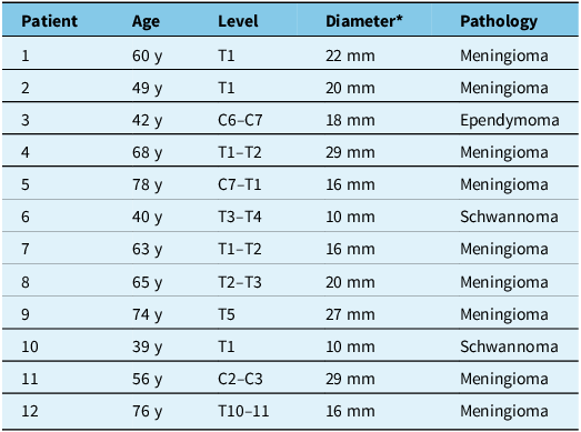
Most of the lesions were extramedullary, with diameters ranging from 10 to 29 mm (Table 1). No postoperative CSF leak or pseudomeningocele was observed. There were no incision infections or complications related to closure during the follow-up. During the first cases in which the technique was adopted, some reinforcement by sutures was used. With repetition of the method, we observed that this was no longer necessary. No change in the postoperative protocol was made, and the patients were mobilized on postoperative day 1. Regular follow-up imaging for tumor resection, including MRI, was obtained (Figures 2 and 3).
(A) Preoperative MRI scan of a meningioma at C7–T1 level operated on by minimally invasive spine surgery. (B,C) Postoperative scan with gross total resection and absence of pseudomeningocele at early follow-up.

Figure 2 Long description
An MRI scan of the cervical and thoracic spine showing a meningioma at the C7T1 level. The preoperative scan (A) displays the meningioma. The postoperative scans (B and C) show the results after a minimally invasive spine surgery, indicating a gross total resection and the absence of a pseudomeningocele at early follow-up.
Postoperative MRI scan of a meningioma operated on by minimally invasive spine surgery. It was approached from the left side. Arrows point to the trajectory of tubes used during the surgery. Dissection and reconstruction of the skin and superficial tissue are notable. Arrowheads show the area where the dura was incised and closed. Non-penetrating titanium clips do not produce artifacts, and follow-up MRI examinations are carried out normally.

Figure 3 Long description
A postoperative MRI scan of a meningioma operated on by minimally invasive spine surgery. The scan shows the left side approach. Arrows indicate the trajectory of tubes used during the surgery, highlighting the dissection and reconstruction of the skin and superficial tissue. Arrowheads point to the area where the dura was incised and closed. Non-penetrating titanium clips are visible, which do not produce artifacts, allowing for normal follow-up MRI examinations.
Titanium clips to close the dura mater were used consecutively in twelve intradural spinal lesions operated on using the MIS technique

Table 1 Long description
The table presents data for twelve patients who underwent surgery for intradural spinal lesions using the MIS technique. It includes columns for patient number, age, lesion level, diameter in millimeters, and pathology type. The ages range from 39 to 78 years. Lesion levels vary from T1 to T10-11 and C2-C3. Diameters range from 10 to 29 millimeters. Pathologies include meningioma, ependymoma, and schwannoma. The table provides a comprehensive overview of the patient demographics and lesion characteristics.
* Craniocaudal diameter.
The use of the MIS technique has become standard in the practice of many spine surgeons, and its indications have expanded to several spine pathologies. Reference Oppenheimer, DeCastro and McDonnell2,Reference Nzokou, Weil and Shedid3 Advantages include diminished blood loss, decreased hospital stay and better postoperative pain profile. Reference Momin and Steinmetz1,Reference Oppenheimer, DeCastro and McDonnell2 At our institution, MIS has become the standard of care for intradural spinal lesions with diameters of up to 30 mm. Following lesion resection, dural closure is a crucial step in the operative time. In our experience, the use of titanium clips (TC) offers a waterproof reconstruction of durotomy and is technically easier than closing with regular sutures.
IDTs have traditionally been operated on using the open technique and under the microscope when muscle dissection and laminectomies are more extensive compared with MIS. CSF leak has a low incidence, but still, it is present in up to 2%–5% of cases. Reference Setzer, Vatter, Marquardt, Seifert and Vrionis4 Meanwhile, the minimally invasive technique has proven safe and effective in the treatment of a growing number of spinal conditions. When used for intradural lesions, recent literature has demonstrated that MIS has similar results in terms of tumor resection compared to open surgery. Reference Helal, Yolcu, Kamath, Wahood and Bydon5 In our series, gross total resection was achieved in all 12 extramedullary tumors, and we observed that the use of TC reduced operative time compared with standard suturing under a microscope. This may be attributed to the fact that the clip applier can be easily positioned in the surgical access, and its use is technically not demanding for the durotomy necessary in MIS surgery for IDTs (Figure 4).
Pack of non-penetrating metal clips and applier used for dural closure in spine surgery for intradural tumors operated by minimally invasive technique.

Figure 4 Long description
The image shows a pack of non-penetrating metal clips and an applier used for dural closure in spine surgery. The clips are neatly arranged in a red and white plastic holder, while the applier, with its metallic tips and red handles, is placed beside the pack. These tools are specifically designed for minimally invasive techniques used in operating on intradural tumors.
Challenges for dural closure include restriction of the corridor for instrument positioning and remaining perforations from the needle during closure. The mismatch in diameter between the needle and suture, which usually does not lead to relevant effects in cranial surgeries, can be significant after durotomies in spine operations. Reference Kaufman, Matthews, Zwienenberg-Lee and Lew6 In contrast to the cranial dura, which develops from both mesoderm and neural crest and forms a robust, dual-layered structure, the spinal dura originates solely from mesodermal mesenchyme and remains a thinner, single-layered membrane. As a result of these developmental differences, the spinal dura is generally more delicate and susceptible to mechanical stress, a factor that can influence surgical handling. Reference Dasgupta and Jeong7
We found that the positioning of the applicator was easily adaptable to the operative tube, and the deployment of NPTC was less demanding than the movements necessary with a regular needle and suture. The clips present the advantage of not penetrating the tissue and do not carry the risk of damaging the underlying structures, as is theoretically possible during the movement of a needle in the deep operative field.
Previous studies, including lab models, have demonstrated the sealing potential of metal clips in different circumstances. Faulkner et al. reported better watertight profile in calf spine models under the application of pressure by saline through a dural reconstruction when compared to prolene and nylon sutures. Reference Faulkner, Finn and Anderson8 Another study showed that NPTC were able to resist hydrostatic pressure almost twice as intense as regular suture. Reference Ito, Aoyama, Horiuchi and Hongo9 These studies also pointed out the advantage of preventing subdural adhesions of clips to neural structures after cicatrization using the technique. Notably, this was particularly important importance in the correction of meningoneural defects in the pediatric population.
The use of NPTC for dural closure is not new in clinical practice. Studies have presented results in a significant series of adult and pediatric populations with no symptomatic CSF leaks. Lew et al. reported that, based on their experience with over a hundred cases, the dura must be tension-free to allow proper approximation with clips. Reference Lew, Matthews, Kaufman and Zwienenberg10 In one patient who required a second operation several months later for deterioration unrelated to the wound, the use of clips did not lead to additional difficulties in the reopening stage. They also believed small corridors usually seen in spinal surgery are welcoming indications for clips with applicators. These findings were consistent with our observations.
Finally, we did not observe significant artifacts in postoperative imaging (Figure 3). It is crucial that a method to close the durotomies do not restrict follow-up evaluations in patients with resected tumors. TC were not associated with a decrease in the quality of CT and MRI scans in follow-up exams. This is especially important in the observation of lesions that are at risk of recurrence. Despite our limited series, complications related to wound healing or radiographic pseudomeningocele were not diagnosed during imaging follow-up.
In conclusion, the safety and efficacy of TC for dura closure in the resection of tumors by MIS were demonstrated in 12 consecutive cases in which there was no CSF leak, wound infection or pseudomeningocele on follow-up imaging. Intraoperative complications due to closure were also not reported, and the use of clip appliers was not hindered by tube channels. Despite the limitations of a small sample analyzed in this study, we believe that NPTC are an efficient alternative for duroplasty and should be part of the spine surgeon armamentarium.
Author contributions
LO contributed to the study conceptualization and writing. PK and TBC contributed to data collection and figures. AG and DS contributed to the study revision and feedback. SJY contributed to the study conceptualization and revision and coordinated the project.
Funding statement
The authors did not receive any funds or financial support for this study.
Competing interests
The authors have no conflict of interest for this article.