Introduction
The expansion of endoscopic endonasal approaches for resecting benign or malignant skull base tumours has led to improvements in the techniques available for reconstructing skull base defects. Developments in the approach, resection and reconstructive phases of endoscopic endonasal skull base surgery are based on a better understanding of the endoscopic surgical anatomy and improved endoscopic surgery techniques. These developments have allowed the surgical management of more substantial and complex tumours, which in turn create larger skull base defects requiring the use of more sophisticated reconstructive techniques.
Currently, reconstruction with a vascularised pedicle flap provides the most reliable outcomes, with post-operative cerebrospinal fluid leak rates of less than 5 per cent.Reference Kassam, Carrau, Snyderman, Gardner and Mintz1 Commonly used skull base vascularised flaps include the Hadad–Bassagaisteguy nasoseptal flap,Reference Hadad, Bassagaisteguy, Carrau, Mataza, Kassam and Synderman2, Reference Kassam, Thomas, Carrau, Snyderman, Vescan and Prevedello3 nasoseptal rescue and modified rescue flaps,Reference Rivero-Serrano, Snyderman, Gardner, Prevedello, Wheless and Kassam4 reverse rotation flap (Caicedo flap),Reference Caicedo-Granados, Carrau, Snyderman, Prevedello, Fernandez-Miranda and Gardner5, Reference Kasemsiri, Carrau, Otto, Tang, Prevedello and Muto6 anterior pedicle lateral nasal wall flap (Hadad–Bassagaisteguy 2 flap),Reference Hadad, Rivero-Serrano, Bassagaisteguy, Carrau, Fernandez-Miranda and Prevedello7 posterior pedicle lateral nasal wall flap (Carrau–Hadad flap),Reference Rivera-Serrano, Bassagisteguy, Hadad, Carrau, Kelly and Prevedello8 middle turbinate flap,Reference Prevedello, Barges-Coll, Fernandez-Miranda, Morera, Jacobson and Madhok9 transpterygoid temporoparietal fascia flap,Reference David and Cheney10, Reference Fortes, Carrau, Snyderman, Kassam, Prevedello and Vescan11 and transfrontal pericranial flap.Reference Yoshioka and Rhoton12–Reference Smith and Ducic14 Temporoparietal fascia and pericranial flaps are regional or extranasal vascularised flaps, whereas all others are intranasal. It should be noted that multiple flaps or a combination of vascularised flaps and free tissue grafts may be needed to complete skull base reconstruction.
Surgical techniques
The eight most commonly used skull base vascularised flaps are described and their indications and surgical techniques are discussed.
Pedicle nasoseptal flap
The pedicle nasoseptal flap (or Hadad–Bassagaisteguy flap) was developed at the University of Rosario, Argentina, by Hadad, Bassagaisteguy and colleagues.Reference Hadad, Bassagaisteguy, Carrau, Mataza, Kassam and Synderman2, Reference Kassam, Thomas, Carrau, Snyderman, Vescan and Prevedello3 The flap consists of the mucoperiosteum and mucoperichondrium of the nasal septum; it is pedicled on the posterior nasoseptal artery, a branch of the posterior nasal artery. This versatile flap is ideal for reconstructing extensive defects of the anterior, middle, clival and parasellar skull base.
Several factors influence the laterality of the flap being harvested. Firstly, tumours that invade the rostrum of the sphenoid, nasal septum, lateral pterygoid recess or pterygopalatine fossa preclude ipsilateral flap harvesting; in such cases, the flap should be elevated on the contralateral side. Secondly, when both sides of the nasal septum are feasible for flap harvesting, the right side is preferable because it is usually easier for a right-handed surgeon to dissect, especially if right middle turbinectomy forms part of the endoscopic endonasal approach. Lastly, the presence of nasal septal spurs or severe deviation increases the technical difficulty and perforation risk during flap harvesting; in such cases, the contralateral side is preferred.
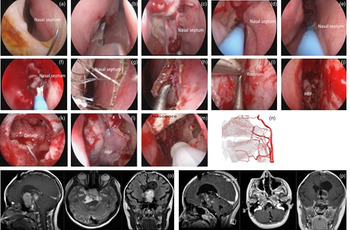
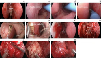
The surgical steps involved in harvesting the Hadad–Bassagaisteguy nasoseptal flap are shown in Figures 1 and 2. The design of a Hadad–Bassagaisteguy nasoseptal flap may be based on the anticipated size and shape of the defect. However, we usually harvest its maximum surface area, which spans from the vomer posteriorly to the mucocutaneous junction of the columella anteriorly, and from the floor of the nasal cavity to 1–2 cm below the olfactory cleft (to preserve the olfactory epithelium). To facilitate visualisation of the nasal septum and surgical field (and therefore flap harvesting), the inferior (bilateral) and the left middle turbinates are out-fractured and the right middle turbinate is removed.
A series of photographs and magnetic resonance images showing treatment of a pituitary adenoma with acromegaly by endoscopic endonasal trans-sphenoidal removal of the tumour and reconstruction of the skull base defect with a right nasoseptal flap. (a) The nasal septum is infiltrated with a 0.5–1 per cent lidocaine solution containing 1/100,000–1/200,000 adrenaline (3–5 ml dental premix). (b) The right middle turbinate is removed as the initial surgical step to facilitate visualisation of the surgical field during the bimanual technique and flap harvesting. (c) Improved visualisation of the entire nasal septum after removal of right middle turbinate. (d) Monopolar electrocautery with an extended curved insulated needle tip facilitates the incisions. The inferior flap incision begins at the lateral posterior choana just anterior to the torus tubaris. (e) The inferior incision is continued anteriorly along the maxillary crest towards the columella. (f) The superior incision follows a line 1–2 cm below the most superior aspect of the septum and continues anteriorly. (g) A vertical incision is usually made parallel to the mucocutaneous junction of the columella to join the superior and inferior incisions. (h) A Cottle dissector is used to elevate the mucoperichondrium and mucoperiosteum from anterior to posterior. (i) The nasoseptal flap is raised posteriorly to meet the rostrum of the sphenoid sinus. (j) The harvested flap is stored in the nasopharynx to avoid interference with the extirpative surgery. (k) Endoscopic view after tumour removal and before reconstruction of the skull base defect. (l) The final position of the nasoseptal flap after removal of a tumour resulting in a significant skull base defect. (m) Nasal packing with Nasopore® followed by the balloon of a 12 Fr Foley catheter to bolster the flap against the defect. (n) Three-dimensional (3D) anatomy of the branches of maxillary artery from the external carotid artery and anterior and posterior ethmoidal arteries from the internal carotid artery that supplies most of the intranasal vascularised pedicle flaps. (o, p) Pre- (o) and post-operative (p) 3D magnetic resonance images showing the tumour and post-operative nasoseptal flap. Left image, sagittal views; middle images, axial view; right images, coronal views. HBF = Hadad–Bassagaisteguy flap

A series of photographs showing treatment of a right sinonasal adenocarcinoma by endoscopic endonasal resection of the tumour and reconstruction of the skull base defect with a left nasoseptal flap. (a) Pre-operative endoscopic view of the tumour. (b) A posterior choana incision from below the floor of the sphenoid sinus anterior to just reaching the torus tubarius. (c) The inferior incision continues anteriorly along the maxillary crest towards the columella. (d) The superior incision is made by following an imaginary line 1–2 cm below the most superior aspect of the septum. (e) Vertical incision to join the superior and inferior incisions. (f) A Cottle dissector is used to elevate the mucoperichondrium and mucoperiosteum. (g) The harvested flap is stored in the nasopharynx. (h) Endoscopic view after removal of the tumour showing the skull base defect. (i) Inlay subdural graft of collagen matrix. (j) Abdominal fat is used to obliterate the sphenoid sinus before applying the nasoseptal flap to the dura or defect. (k) Nasoseptal flap in its final position. HBF = Hadad–Bassagaisteguy flap

To harvest the Hadad–Bassagaisteguy nasoseptal flap, the mucosa is incised with a monopolar electrocautery equipped with an extended insulated needle tip bent at a 90° angle (Arthroscopic Hook Electrode; Valleylab, Boulder, Colorado, USA). The inferior incision of the pedicle is performed at the level of the posterior choana, below the floor of the sphenoid sinus and starting just anterior to the torus tubarius and extending towards the nasal septum. This incision is continued infero-anteriorly following the free edge of the posterior nasal septum towards the junction of the nasal septum and maxillary crest. At this point, it joins a sagittally oriented incision that follows the maxillary crest towards the columella. The superior incision crosses the rostrum of the sphenoid sinus extending from the lateral nasal wall towards the septum at the level of the natural ostium of the sphenoid sinus. This incision joins another sagittally oriented incision that follows an imaginary line drawn 1–2 cm below the most superior aspect of the septum. This incision is continued anteriorly towards the mucocutaneous junction of the columella. These two sagittally oriented incisions, traversing the entire length of the septum, are joined at their most anterior aspect by a vertical incision that follows the mucocutaneous junction of the columella.
A Cottle dissector is used to elevate the flap paddle, following the subperiosteal plane from anterior to posterior. Once harvested, the nasoseptal flap is stored in the nasopharynx or inside the maxillary sinus during extirpative surgery. A multilayer technique is preferred for reconstructing skull base defects. Cerebrospinal fluid (CSF) flow is stopped by inlaying a collagen matrix (Duragen; Intergra Life Sciences, Plainsboro, New Jersey, USA) graft into the subdural or epidural space. Subsequently, the nasoseptal flap is applied over the defect. Abdominal fat may be used to obliterate the dead space (the bony tunnel after a transclival or transodontoid approach) or the sphenoid sinus when appropriate. Biological glue can be applied to further fix the flap, but this is optional. Subsequently, nasal packing, expandable sponges or the balloon of a size 12 Fr Foley catheter is inserted under direct endoscopic visualisation to bolster the flap against the defect.
Nasoseptal rescue technique
A nasoseptal rescue flap is a modified Hadad–Bassagaisteguy nasoseptal flap that involves elevating the pedicle (but not the paddle) of the Hadad–Bassagaisteguy nasoseptal flap.Reference Hadad, Bassagaisteguy, Carrau, Mataza, Kassam and Synderman2, Reference Rivero-Serrano, Snyderman, Gardner, Prevedello, Wheless and Kassam4 As a significant CSF leak is rare in pituitary surgery, the nasoseptal flap is not routinely raised, thus avoiding potential sinonasal morbidity. A nasoseptal rescue flap preserves the possibility of raising a Hadad–Bassagaisteguy nasoseptal flap if needed; therefore, it is indicated when a CSF leak is possible but not probable.
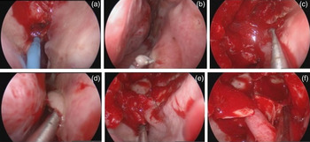
The surgical steps involved in harvesting a nasoseptal rescue flap are shown in Figure 3. Two incisions are made encompassing a strip of mucoperiosteum that comprises the pedicle. One incision is made at the level of the natural ostium of the sphenoid sinus and the other follows the posterior choana, as described for the Hadad–Bassagaisteguy nasoseptal flap. A Cottle dissector and ball probe (ostium seeker) are then used to raise the mucoperiosteal flap, thus exposing the rostrum of the sphenoid sinus and the entire height of the ipsilateral posterior septum. The technique is repeated bilaterally. Wide sphenoidotomy and posterior septectomy are subsequently performed. The unencumbered mobility of the pedicle greatly facilitates the latter, allowing its dynamic transposition to work above or below the flap. Once wide bilateral sphenoidotomies are performed, the flaps are displaced inferiorly towards the floor of the nasal cavity, where they remain for the duration of the dissection.
A series of photographs showing treatment of a pituitary adenoma by endoscopic endonasal trans-sphenoidal excision of tumour and raising of a nasoseptal rescue flap. (a) The inferior incision of the flap begins from the posterior choana (which is anterior to the torus tubarius) using a monopolar electrocautery with an extended curved insulated needle tip. (b) The inferior incision is continued infero-anteriorly following the free edge of the posterior nasal septum towards the junction of nasal septum and maxillary crest. (c) The superior incision starts at the level of the natural ostium of the sphenoid sinus and joins another sagittally oriented incision following an imaginary line 1–2 cm below the most superior aspect of the septum. (d) A Cottle dissector is used to raise the mucoperiosteum flap to free the flap exposing the rostrum of the sphenoid sinus and the entire height of the posterior septum. (e) Wide sphenoidotomy and posterior septectomy transpose the bipedicled flap to provide access above or below the flap. (f) Once bilateral sphenoidotomies are performed, the flap is displaced downward into its final position.

Thus, intrasphenoidal dissection and sellar tumour removal are performed above the pedicle (rescue flap) using a bimanual technique. If there is no intra-operative CSF leak, at the end of pituitary surgery the nasoseptal rescue flap is translocated posteriorly to cover any denuded bone at the floor of the sphenoid sinus. Conversely, if a significant CSF leak is encountered, a standard Hadad–Bassagaisteguy nasoseptal flap can be harvested by extending the incisions.
Reverse rotation flap
Use of the Hadad–Bassagaisteguy nasoseptal flap has become popular because it reliably prevents post-operative CSF leakage. However, the Hadad–Bassagaisteguy nasoseptal flap leaves denuded septal bone and cartilage at its donor site. Re-mucosalisation of the donor site as a secondary intention is prolonged, often taking three to six months. The reverse rotation flap (also named the Caicedo reverse flap) was designed to reline the bare donor site of the nasoseptal flap.Reference Caicedo-Granados, Carrau, Snyderman, Prevedello, Fernandez-Miranda and Gardner5, Reference Kasemsiri, Carrau, Otto, Tang, Prevedello and Muto6 It incorporates the nasoseptal mucoperiosteum opposite to the Hadad–Bassagaisteguy nasoseptal flap to emerge as an anteriorly based septal flap vascularised by branches of the anterior ethmoidal and facial arteries. It hastens post-operative re-mucosalisation of the donor, shortening the interval to one to two weeks.
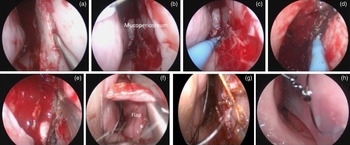
The surgical steps involved in harvesting the Caicedo flap are shown in Figure 4. Once the Hadad–Bassagaisteguy nasoseptal flap has been elevated, posterior bony septectomy is performed to remove the vomer and ethmoid perpendicular plate. This exposes the contralateral nasoseptal mucoperiosteum, which can then be harvested as an anteriorly based vascularised random flap. Superior and inferior incisions are performed as mirror images of the Hadad–Bassagaisteguy nasoseptal flap incisions but extending only to the free edge of the remaining nasal septal cartilage. A third posterior incision is then performed to connect the superior and inferior incisions at the level of the rostrum of the sphenoid sinus. This frees the reverse flap, which can then be transposed 180° to the opposite side (the donor site of the Hadad–Bassagaisteguy nasoseptal flap) to cover the denuded septum. Its free edge (originally at the rostrum of the sphenoid sinus) is sutured to the columella using 4–0 gauge absorbable sutures. Trans-septal quilting stitches with 3–0 gauge absorbable sutures are used to fix the flap against the nasal cartilage septum to prevent accumulation of a haematoma. Silicone nasal splints are used to protect the flap and help keep it in position against the nasal septum. These splints also prevent the flap from interfering with subsequent phases of the surgery.
A series of photographs showing treatment of a pituitary adenoma with acromegaly by endoscopic endonasal trans-sphenoidal removal of the tumour and reconstruction of the skull base defect with a right nasoseptal flap. A Caicedo reverse rotation flap was harvested to reline the denuded donor site of the Hadad–Bassagaisteguy nasoseptal flap. (a) Posterior bony septectomy is performed to remove the vomer and ethmoidal plate. Septectomy can be extended anteriorly to obtain the desired flap transposition. (b) The contralateral mucoperiosteum of the nasal septum is then exposed. (c) An inferior incision of the flap is made to follow the free edge of the remaining nasal septal cartilage. (d) A superior incision is made following the mirror image of the Hadad–Bassagaisteguy nasoseptal flap superior incision until it reaches the free edge of the remaining nasal septal cartilage. (e) A vertical incision is made at the rostrum of the sphenoid sinus to connect both the superior and inferior incisions. (f) The flap is rotated 180° to the opposite side to cover the denuded nasal septal cartilage. (g) It is attached to the columella using 4-0 absorbable chromic catgut sutures and trans-septal quilting with 3-0 absorbable chromic catgut sutures. (h) Silicone nasal splints are sutured to the septum in both nostrils to protect the flap and prevent it interfering with subsequent surgery.

Anterior pedicle lateral nasal wall flap
The anterior pedicle nasal lateral wall (or Hadad–Bassagaisteguy 2) flap was developed by Hadad, Bassagaisteguy and colleagues.Reference Hadad, Rivero-Serrano, Bassagaisteguy, Carrau, Fernandez-Miranda and Prevedello7 Its anterior pedicle is based on the branches of the facial and anterior ethmoidal arteries. This flap can be used as an alternative reconstructive technique; it can be harvested when the Hadad–Bassagaisteguy nasoseptal flap is not feasible or when the dimensions of the defect require multiple flaps. It provides a large surface area that can be used to reconstruct a defect extending from the crista galli to the tuberculum sella anteroposteriorly and from orbit to orbit laterolaterally.
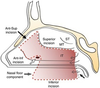
The surgical steps involved in harvesting the Hadad–Bassagaisteguy 2 flap are shown in Figure 5. The flap pedicle is aligned vertically in the sagittal plane and extends from the roof of the nasal cavity, just anterior to the middle turbinate, to the head of the inferior turbinate. The anterior incision follows the anterior border of the ascending maxillary process; the posterior incision follows the uncinate process and extends over the superior aspect of the inferior turbinate. A perpendicular incision starting at the most posterior aspect of the incision over the lateral nasal wall travels medially to cross the floor of the nose and reach the inferior border of the septum. Another perpendicular incision is made anteriorly from the head of the inferior turbinate to cross the floor of the nose and reach the inferior border of the septum. Two horizontal incisions at the floor of the nose are joined by another incision that follows the maxillary crest at the junction of the floor of the nose with nasal septum. This incision can be extended to the nasal septum superiorly, depending on the size of the defect. A separate vertical incision is made over the head of the inferior turbinate and extended laterally to intersect the anterior incision of the pedicle. This latter incision is critical to allow elevation of the mucoperiosteal lining of the nasal and meatal sides of the inferior turbinate. The dissection continues along the medial aspect of the inferior turbinate.
Schematic diagram showing the margins of right anterior pedicled lateral nasal wall flap.Reference Hadad, Rivero-Serrano, Bassagaisteguy, Carrau, Fernandez-Miranda and Prevedello7 Arrows indicate the extent of the flap. IT = inferior turbinate; MT = middle turbinate; ST = superior turbinate
The inferior turbinate is then fractured medially and the mucoperiosteum (together with the inferior turbinate bone) is elevated from the lateral nasal wall. The turbinate bone is later removed with rongeurs or through-cutting instruments. The nasal floor mucosa is subsequently elevated and the flap is freed posteriorly. Placement of the Hadad–Bassagaisteguy 2 flap over the defect requires rotation of the pedicle from the sagittal to the axial plane; thus, it needs to be bolstered in place with packing.
Posterior pedicle lateral nasal wall flap
The posterior pedicle lateral nasal wall flap (or Carrau–Hadad flap) was designed to encompass the mucoperiosteum of the inferolateral wall and floor of the nasal cavity.Reference Rivera-Serrano, Bassagisteguy, Hadad, Carrau, Kelly and Prevedello8 The pedicle is based posteriorly on branches of the sphenopalatine artery, a terminal branch of the internal maxillary artery. It provides a large surface area adequate for reconstructing skull base defects extending from the planum sphenoidale to the nasopharynx anteroposteriorly and laterolaterally from orbital apex to orbital apex or the internal carotid artery canal to the internal carotid artery canal.
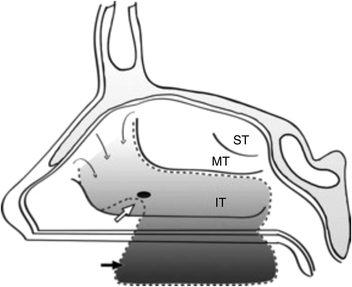
The surgical steps involved in harvesting the Carrau–Hadad flap are shown in Figure 6. An incision is made along the anterior border of the ascending maxillary process, starting at a level corresponding to the most caudal aspect of the nasal bone and extending inferiorly to the level of the head of the inferior turbinate. It is extended further along the lateral nasal wall towards the floor of the nose to reach the inferior border of the septum. At its most inferomedial point, it joins a perpendicular sagittal incision along the maxillary crest. The latter incision extends to a point corresponding to the level of the sphenopalatine foramen posteriorly. Another vertical incision (anterior to the uncinate process and following its anterior border) is made extending over the superior aspect of the inferior turbinate to reach the sphenopalatine foramen posteriorly. The incision can be extended superiorly to incorporate the fontanel of the maxillary sinus to increase the width of the flap, although this is technically difficult. Subsequently, a third incision is made to connect the most superior aspects of the first two incisions. A separate vertical incision is made over the head of the inferior turbinate and extending laterally to intersect the pedicle's anterior incision. This latter incision is critical to allow elevation of the mucoperiosteal lining of both the nasal and meatal sides of the inferior turbinate. The dissection continues along the medial aspect of the inferior turbinate.
Schematic diagram showing the margins of the right posterior pedicled lateral nasal wall flap.Reference Rivera-Serrano, Bassagisteguy, Hadad, Carrau, Kelly and Prevedello8 Arrows indicate the extent of the flap. IT = inferior turbinate; MT = middle turbinate; ST = superior turbinate

The inferior turbinate is then fractured medially and the mucoperiosteum is elevated from the lateral nasal wall and turbinate bone. The turbinate bone is removed with rongeurs or through-cutting instruments. Lastly, the mucoperiosteum of the nasal floor is elevated and the flap is freed anteriorly.
Middle turbinate flap
The middle turbinate flap is another intranasal alternative for reconstructing anterior skull base defects. It comprises the mucoperiosteum of the middle turbinate and a pedicle based on the anterior branch of the sphenopalatine artery that courses through the posterior attachment. This flap can be used to reconstruct small defects of the fovea ethmoidalis, planum sphenoidale and sella, especially when the other intranasal flaps are not feasible.Reference Prevedello, Barges-Coll, Fernandez-Miranda, Morera, Jacobson and Madhok9
The surgical steps involved in harvesting the middle turbinate flap are described next. Firstly, a vertical incision is made along the anterior face of the middle turbinate head. Then, a horizontal incision is made in an anterior to posterior direction along the vertical attachment of the middle turbinate below the skull base. A Cottle periosteal elevator is used to raise the mucoperiosteum of the middle turbinate in a superior to inferior direction. The thin bone of the middle turbinate is then removed from the inner aspect of the flap and the vertical attachment of the turbinate is transected from the skull base. Further elevation of the flap posteriorly and transection of the basal lamella forms a posterior pedicle flap. The pedicle can be dissected further to provide a better arc of rotation.
Temporoparietal fascia flap
The temporoparietal fascia flap is a regional vascularised pedicle flap comprising a strong fascial layer connecting the overlying fibrous septae of the subcutaneous tissue at the temporoparietal region of the scalp. Its pedicle is based on the anterior branch of superficial temporal artery and one or two veins accompanying the artery. This flap is 2–3 mm thick over the parietal region and extends in a fan-like manner from the preauricular region to cover a surface area as large as 17 × 14 cm.Reference David and Cheney10, Reference Fortes, Carrau, Snyderman, Kassam, Prevedello and Vescan11
Multiple reports have addressed the use of the temporoparietal fascia flap in a variety of other reconstructive settings.Reference David and Cheney10, Reference Fortes, Carrau, Snyderman, Kassam, Prevedello and Vescan11 Its use in skull base reconstruction after endonasal endoscopic resection, however, requires a transpterygoid tunnel.Reference David and Cheney10 This flap type is most useful for reconstructing large defects of the posterior and middle cranial base when intranasal vascularised flaps are not feasible or when the extent of the defect necessitates the use of multiple flaps.
The surgical steps involved in harvesting a temporoparietal fascia flap are shown in Figure 7. An ipsilateral approach is preferable to a transpterygoid endonasal approach because it facilitates flap transposition into the nasal cavity (where it contributes to the corridor). Anterior and posterior ethmoidectomies and large maxillary antrostomy facilitate transpterygoid transposition. The ipsilateral sphenopalatine artery is identified and ligated at the level of the sphenopalatine foramen. The posterior wall of the maxillary sinus is removed to expose the pterygopalatine fossa. The most anterior aspect of the lateral pterygoid plate may be reduced by using a high-speed drill to further enlarge the space for transposition of the flap.
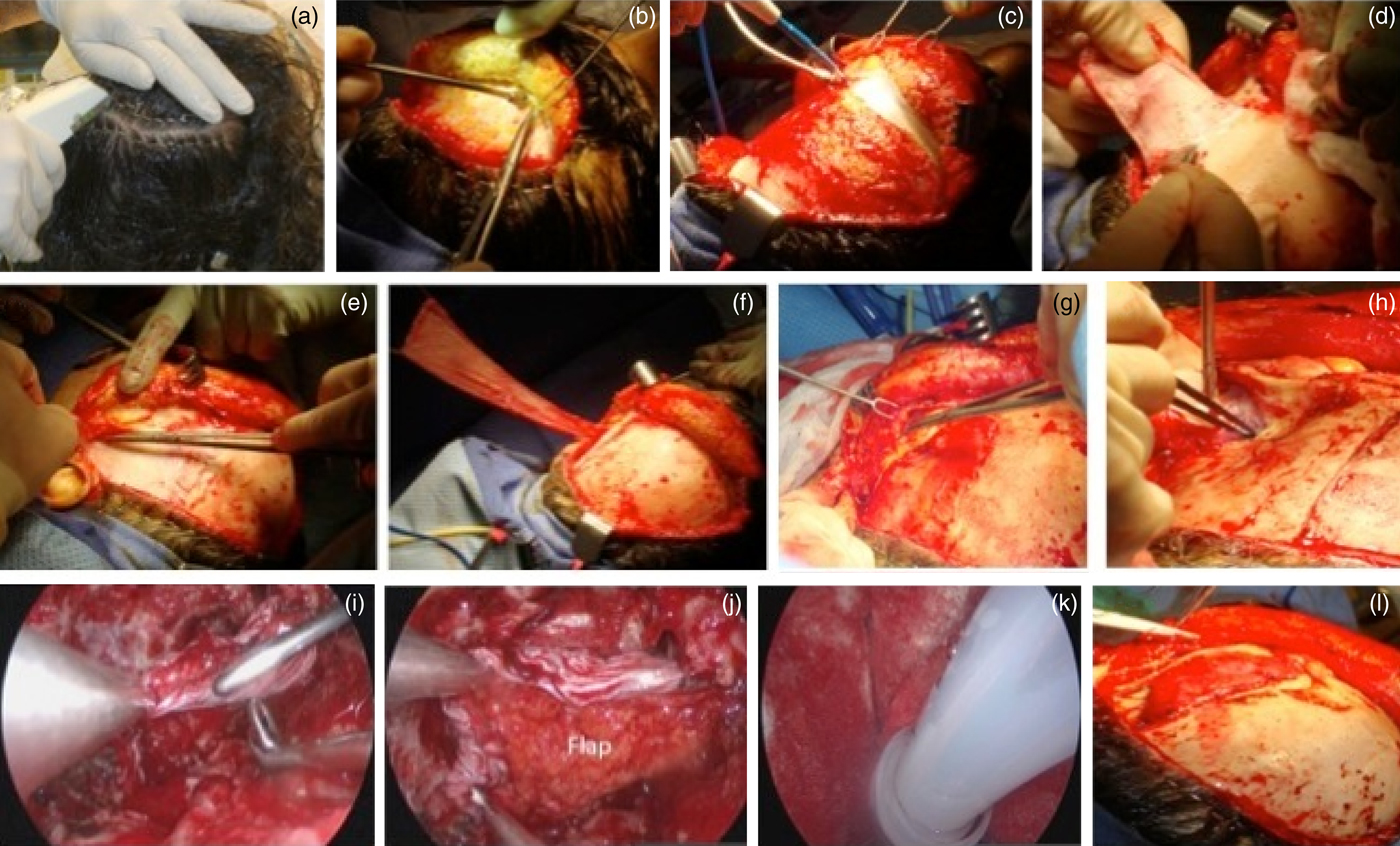
A series of photographs showing treatment of an extensive clival chordoma by endoscopic endonasal removal of the tumour and reconstruction of the skull base defect using a temporoparietal fascia flap (the Hadad–Bassagaisteguy nasoseptal flap was not feasible because of tumour involvement). (a) The scalp is prepared and marked. (b) A hemicoronal incision is made and the temporoparietal fascia flap is dissected from the overlying subcutaneous tissue cautiously. (c) The temporoparietal fascia flap is incised at its lateral margins after sufficient surface area is exposed. (d) The temporoparietal fascia flap is elevated from the cranium and deep temporal fascia down to its pedicle. (e) The superficial layer of the deep temporal fascia is incised vertically and separated from the underlying muscle. This dissection plane is then followed inferiorly to elevate the periosteum from the surface of the zygomatic arch. (f) The temporoparietal fascia flap is harvested with a long vascular pedicle based on the anterior branch of superficial temporal artery; it has a good arc of rotation. (g) The temporalis muscle is separated from the lateral orbital wall to create a tunnel, allowing communication between the temporal, infratemporal and pterygopalatine fossae. (h) The soft tissue tunnel is usually dilated by passing a guide wire and dilator before transposing the flap through the tunnel to the nasal cavity. (i) The temporoparietal fascia flap is transposed from the temporal to the infratemporal fossa and then advanced into the nasal cavity through the pterygopalatine fossa via a transpterygoid tunnel. (j) The flap is reconstructed and adjusted into its final position at the clival region. (k) Nasal packing with Nasopore® followed by the balloon of a 12 Fr Foley catheter bolsters the flap against the defect under direct endoscopic visualisation. (l) The temporalis muscle is sutured back to the bony plate that is secured at the temporal region.
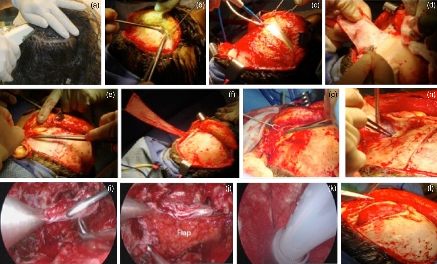
A hemicoronal incision is then made down to the level of the hair follicles. The temporoparietal fascia is dissected from the subcutaneous tissue, thus elevating the temporoparietal fascia flap. Once enough surface area is exposed, the fascia is incised at its lateral margins and elevated from the cranium and the superficial layer of the deep temporal fascia down to its pedicle. To preserve the frontal branch of the facial nerve, the dissection must not cross anterior to an imaginary line between the tragus and the lateral eyebrow or proceed anterior to the hairline. A lateral canthotomy incision is performed to expose and separate the temporalis muscle from the lateral orbital wall and pterygomaxillary fissure. This also creates a tunnel that allows communication between the temporal and infratemporal fossae and the pterygopalatine fossae. By passing a guide wire and dilator under direct endoscopic visualisation, further dilation of the soft tissue tunnel is achieved. After an adequate tunnel size is obtained, the flap is tied to the external end of the guide wire. As the nasal end of the guide wire is pulled out through the nostril, it draws the flap through the tunnel into the nasal cavity. Finally, the flap is placed and bolstered over the defect. This flap requires preservation of the venous outflow; therefore, avoiding excessive pressure over the pedicle is critical. External incisions are closed with a single layer of 4-0 sutures after insertion of a suction drain.
Transfrontal pericranial flap
The transfrontal pericranial flap is a regional vascularised pedicle flap comprising the anterior pericranial fascia at the frontal region of the scalp. Its pedicle comprises the supraorbital and supratrochlear neurovascular bundles. There have been many reports of the excellent outcome associated with using a pericranial flap for reconstructing skull base defects resulting from anterior craniofacial resection.Reference Yoshioka and Rhoton12–Reference Smith and Ducic14 This type of flap is extremely useful for large anterior skull base defects when intranasal pedicle flaps are not available or when the extent of the defect may require multiple flaps. It provides a large surface area that is adequate for reconstructing anterior skull base defects extending anteroposteriorly from the crista galli to the tuberculum sella and laterolaterally from orbit to orbit.
The surgical steps involved in harvesting the transfrontal pericranial flap are shown in Figure 8. First, a standard coronal incision is made down to the calvarium. Next, the scalp flap is elevated to the superior orbital rims in a subpericranial fashion. If additional length necessitates dissection posterior to the coronal incision, care must be taken during the coronal incision so that the pericranium is not divided. The additional extension of pericranium is dissected before elevating the coronal flap anteriorly. To extend the inferior exposure of the coronal flap without placing undue traction on the supraorbital and supratrochlear vessels and nerves, the latter must be freed from their foramina. This is more viable when the foramen is less than 10 mm above the supraorbital rim.Reference Yoshioka and Rhoton12 Subsequently, the pericranial flap is raised from the overlying scalp tissue and a 2-cm osteotomy is opened at the nasion (which communicates with the frontonasal area) using a high-speed drill. After the pericranial flap is inserted and positioned, it is bolstered with packing for support.
A series of photographs showing treatment of an extensive sinonasal small cell neuroendocrine carcinoma involving the nasal septum, lateral nasal wall and intradural extension by craniofacial tumour resection and reconstruction of skull base defect with a transfrontal pericranial flap. (a) Pre-operative endoscopic view of right sinonasal small cell neuroendocrine carcinoma. (b) The scalp is prepared and marked. (c) A standard bicoronal incision is made sub-pericranially to the calvarium. (d) The flap is elevated to the superior orbital rims in a subpericranial fashion with a blunt elevator. To prevent desiccation, the flap remains attached to the anterior scalp until required. (e) To extend the inferior side of the bicoronal flap without placing traction onto the supraorbital and supratrochlear vessels and nerves, the latter can be freed from their foramen. (f) The defect size is assessed before raising the pericranial flap. (g) Dura that is involved in the tumour is excised until the negative margin is reached. (h) The pericranial flap is raised from the overlying scalp tissue. (i) The pericranium is incised and elevated from the galea. (j) Final view of the anterior-based pericranial flap after harvesting. (k) An inlay subdural graft of collagen matrix is used to cover the defect because a multilayer reconstructive technique is preferred. (l) The pericranial flap is inserted to reconstruct the anterior cranial floor with several tacking sutures placed through the bone in the posterior bony defect to support the flap.

Discussion
Over the past 20 years, the surgical techniques used for skull base defect reconstruction have developed from conventional external approaches to expanded endoscopic approaches; the materials used vary from autologous free tissues (with or without synthetic substitutesReference Cappabianca, Cavallo, Esposito, Valente and de Divitiis15, Reference Seiler and Mariani16) to various vascularised flaps.Reference Hadad, Bassagaisteguy, Carrau, Mataza, Kassam and Synderman2–Reference Smith and Ducic14 Several factors influence the choice of reconstruction technique and its outcome. These include the location and extent of the skull base tumours and defects, the availability of the different reconstructive techniques, surgeon bias and skill level, the possibility of increased post-operative CSF pressure, the general health status of the patient and the need for prompt administration of adjunctive therapies.Reference Kassam, Thomas, Carrau, Snyderman, Vescan and Prevedello3
Reconstructing large skull base defects with free tissue grafts (autologous or synthetic) is associated with high post-operative CSF leak rates.Reference Hadad, Bassagaisteguy, Carrau, Mataza, Kassam and Synderman2 Persistent CSF leaks are commonly associated with failure to integrate the graft or with migration of part of the graft. Vascularised flaps were developed to overcome the initial unacceptable high rates of post-operative CSF leaks associated with free tissue graft reconstruction after an endoscopic endonasal approach. Adoption of the Hadad–Bassagaisteguy nasoseptal flap led to a dramatic drop in post-operative CSF leak rates to less than 5 per cent.Reference Hadad, Bassagaisteguy, Carrau, Mataza, Kassam and Synderman2, Reference Kassam, Thomas, Carrau, Snyderman, Vescan and Prevedello3 Therefore, we favour vascularised flaps for reconstructing large skull base defects because they promote adequate and rapid healing, especially in post-chemoradiotherapy patients.
There are clearly several vascularised flap options for reconstructing skull base defects. The Hadad–Bassagaisteguy nasoseptal flap is our preferred choice if flap harvesting is not compromised. This flap is useful for reconstructing defects located anywhere between the posterior wall of the frontal sinus to the lower clivus. It should be noted, however, that in patients with pituitary adenomas, Rathke's cleft cyst or other sellar pathologies a Hadad–Bassagaisteguy nasoseptal flap is rarely if ever required. If a large CSF leak is not anticipated, a nasoseptal rescue flap plays an important role in preserving the pedicle of the Hadad–Bassagaisteguy nasoseptal flap while avoiding sinonasal morbidity.Reference Rivero-Serrano, Snyderman, Gardner, Prevedello, Wheless and Kassam4 Donor site morbidity after Hadad–Bassagaisteguy nasoseptal flap harvesting is a common problem in post-operative care. Increased crusting with nasal obstruction, cacosmia or anosmia affects the post-operative quality of life. The use of a reverse rotation flap can effectively resolve these problems.Reference Caicedo-Granados, Carrau, Snyderman, Prevedello, Fernandez-Miranda and Gardner5
In patients for whom the Hadad–Bassagaisteguy nasoseptal flap is not feasible because of oncological extension or prior surgery involving the nasal septum, rostrum of the sphenoid sinus or pterygopalatine fossa, one of two intranasal vascularised flaps may be used: the Hadad–Bassagaisteguy 2 flap or the Carrau–Hadad flap (both are lateral nasal wall flaps).Reference Hadad, Rivero-Serrano, Bassagaisteguy, Carrau, Fernandez-Miranda and Prevedello7, Reference Rivera-Serrano, Bassagisteguy, Hadad, Carrau, Kelly and Prevedello8 The Hadad–Bassagaisteguy 2 flap is ideal for patients with defects resulting from a transcribiform or transplanum approach. Conversely, the Carrau–Hadad flap is excellent for large skull base defects resulting from transcribriform, transplanum, trans-sellar, transclival or middle cranial fossa approaches. When dealing with lesser defects of the fovea ethmoidalis, planum sphenoidale and sella, the middle turbinate flap is a good alternative, especially when the other intranasal flaps are not feasible.
When intranasal flaps are not feasible, we revert to using extranasal or regional vascularised flaps. In our centre, the two most commonly used regional vascularised flaps are the temporoparietal fascia flap and the transfrontal pericranial flap.Reference David and Cheney10–Reference Smith and Ducic14 The temporoparietal fascia flap is ideal for middle and posterior skull base defects, whereas the transfrontal pericranial flap is ideal for anterior skull base defects.Reference Snyderman, Janecka, Sekhar, Sen and Eibling17 A temporoparietal fascia flap is not suitable for anterior skull base defects because of the length needed to pass the flap through the pterygopalatine fossa. Similarly, the transfrontal pericranial flap is not recommended for posterior defects because it would require sacrificing the ethmoid sinuses, septum and possibly olfaction. However, the biggest disadvantage of both extranasal flaps is the need for an external approach, with potential additional morbidity (i.e. numbness of the forehead, paresis of the frontal branch of the facial nerve, alopecia, unsightly scarring).
Adequate post-operative management is equally important. General principles of post-operative management following skull base surgery include the use of peri-operative, intravenous, fourth-generation cephalosporin for 48 hours. Oral broad-spectrum antibiotics should continue to be administered until the nasal packing is removed three to five days post-operatively. The patient should be advised and continually reminded to avoid nose blowing and sneezing or coughing with an open mouth and should avoid any activities that may increase intracranial pressure. Patients should be advised on how to perform nasal douching (using a saline solution prepared with sterile water). Outpatient clinic monitoring and debridement of the nasal cavity should be performed as needed.
Conclusion
A myriad of intranasal vascularised flaps are available for reconstructing skull base defects resulting from a variety of endoscopic endonasal approaches or other lesions. We advocate use of the Hadad–Bassagaisteguy nasoseptal flap for reconstructing extensive defects of the anterior, middle and posterior cranial base. However, mastering the skills required to handle extranasal or regional vascularised flaps is fundamental because they may offer reconstructive alternatives, especially when the Hadad–Bassagaisteguy nasoseptal flap is not a desirable or available option, open approaches are needed, or intranasal vascularised flaps are not available.