Introduction
Porcine circovirus (PCV) was first discovered as a contaminant of a permanent porcine kidney cell line (PK-15 [ATCC-CCL33]), and described as a small, spherical, non-cytopathic virus similar to picornavirus in terms of morphology (Tischer et al., Reference Tischer, Rasch and Tochtermann1974). PCV is an icosahedral, non-enveloped, circular, single-stranded DNA virus with a diameter of 17 nm (Tischer et al., Reference Tischer, Gelderblom, Vettermann and Koch1982). Following the discovery of the virus, comparative genetic analyses of PCV isolates from diseased pigs showed that they were distinctly different from previous non-pathogenic PCV isolates (Hamel et al., Reference Hamel, Lin and Nayar1998; Meehan et al., Reference Meehan, McNeilly, Todd, Kennedy, Jewhurst, Ellis, Hassard, Clark, Haines and Allan1998; Morozov et al., Reference Morozov, Sirinarumitr, Sorden, Halbur, Morgan, Yoon and Paul1998). Porcine circovirus type 1 (PCV1) is now acknowledged as the non-pathogenic PCV strain, and porcine circovirus type 2 (PCV2) isolates are considered pathogenic in swine. The DNA genome sequence homology between PCV1 and PCV2 varies between 68 and 76% (Hamel et al., Reference Hamel, Lin and Nayar1998; Meehan et al., Reference Meehan, McNeilly, Todd, Kennedy, Jewhurst, Ellis, Hassard, Clark, Haines and Allan1998; Morozov et al., Reference Morozov, Sirinarumitr, Sorden, Halbur, Morgan, Yoon and Paul1998). PCV2 isolates are further clustered into subtypes: PCV2a, PCV2b, PCV2c, PCV2d and PCV2e (Cheung et al., Reference Cheung, Lager, Kohutyuk, Vincent, Henry, Baker, Rowland and Dunham2007; Gagnon et al., Reference Gagnon, Tremblay, Tijssen, Venne, Houde and Elahi2007; Olvera et al., Reference Olvera, Cortey and Segalés2007; Dupont et al., Reference Dupont, Nielsen, Baekbo and Larsen2008; Wang et al., Reference Wang, Guo, Ge, Wang, Chen, Cha and Yang2009). Clinical disease associated with PCV2 infection can have multiple manifestations that include systemic disease or post-weaning multisystemic syndrome (PMWS), respiratory disease, enteritis, reproductive failure, porcine dermatitis and nephropathy syndrome (PDNS), myocarditis/vasculitis and exudative dermatitis (Chae, Reference Chae2004, Reference Chae2005).
PCV-associated disease (PCVAD) is a common terminology used in North America to refer to the various clinical manifestations associated with PCV2 infection. PCVAD includes both subclinical and clinical disease (Opriessnig et al., Reference Opriessnig, Meng and Halbur2007). PCV2 is globally distributed and most herds are seropositive for anti-PCV2 antibodies (Magar et al., Reference Magar, Müller and Larochelle2000; Opriessnig et al., Reference Opriessnig, Yu, Thacker and Halbur2004). PCV2 infection generally occurs as maternally derived antibodies wane in post-weaned pigs (7–15 weeks of age) and results in either subclinical infection or clinical disease. This review will discuss the manifestation of PCV2 infection in sexually mature swine and the effects of in utero fetal infection.
PCV2 infection of breeding age females (gilts and sows)
Definition of reproductive failure
Reproductive failure in swine can manifest as failure of implantation resulting in irregular return to estrus, failure of pregnancy progressing to term due to the expulsion of all embryos or fetuses (abortion), or reduced litter size due to increased numbers of non-viable piglets at parturition (Almond et al., Reference Almond, Flowers, Batista, D'Allaire, Straw, Zimmerman, D'Allaire and Taylor2006). PCV2 infection has been linked to abortion and associated with increased rates of mummified, macerated, stillborn and weak-born piglets (West et al., Reference West, Bystrom, Wojnarowicz, Shantz, Jacobson, Allan, Haines, Clark, Krakowka, McNeilly, Konoby, Martin and Ellis1999; O'Connor et al., Reference O'Connor, Gauvreau, West, Bogdan, Ayroud, Clark, Konoby, Allan and Ellis2001) as well as early embryonic death (Kim et al., Reference Kim, Jung and Chae2004; Mateusen et al., Reference Mateusen, Maes, Van Soom, Lefebvre and Nauwynck2007). However, the clinical presentation of abortion, mummified fetuses and stillbirths in swine operations must be distinguished from infection by other agents including porcine reproductive and respiratory syndrome virus (PRRSV), porcine parvovirus (PPV), pseudorabies virus, porcine enterovirus and others.
PCV2 in breeding females: historical overview
PCV2-associated reproductive failure was first described in the late 1990s in Canada (West et al., Reference West, Bystrom, Wojnarowicz, Shantz, Jacobson, Allan, Haines, Clark, Krakowka, McNeilly, Konoby, Martin and Ellis1999). The clinical presentation included late-term abortions, pseudopregnancy and an increased incidence of mummified and stillborn fetuses (up to 75% of term litters) at parturition in a new 450-head swine farm composed of first parity dams. Farrowing rates decreased below 60% with the greatest proportion of pregnancy loss occurring after 13 weeks of gestation; however, clinical signs of systemic illness in dams associated with an infectious process were not observed in any of the gilts (West et al., Reference West, Bystrom, Wojnarowicz, Shantz, Jacobson, Allan, Haines, Clark, Krakowka, McNeilly, Konoby, Martin and Ellis1999). Following this initial description many case reports implicated PCV2 as the cause of reproductive failure (Ladekjær-Mikkelsen et al., Reference Ladekjær-Mikkelsen, Nielsen, Storgaard, Bøtner, Allan and McNeilly2001; O'Connor et al., Reference O'Connor, Gauvreau, West, Bogdan, Ayroud, Clark, Konoby, Allan and Ellis2001; Cariolet et al., Reference Cariolet, Blanchard, Le Dimna, Mahé, Jolly, de Boisséson, Truong, Ecobichon, Madec and Jestin2002; Park et al., Reference Park, Kim, Ha, Jung, Choi, Lim, Kim and Chae2005).
PCV2 in breeding females: clinical signs
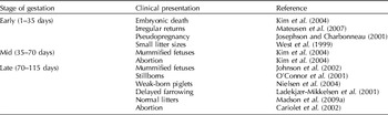
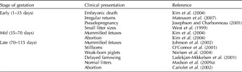
The clinical manifestation of gestational PCV2 infection is variable and is seemingly subject to timing of viral infection, antibody status and length of viremia in the dam. Table 1 summarizes the clinical presentation of PCV2-associated reproductive failure in affected dams related to the timing of fetal infection. The most consistent clinical presentation of PCV2-associated reproductive failure is increased stillborns and mummified fetuses at parturition (O'Connor et al., Reference O'Connor, Gauvreau, West, Bogdan, Ayroud, Clark, Konoby, Allan and Ellis2001). Clinical signs of PCV2 infection in the dam are typically absent or inapparent; however, a low percentage of females may abort due to systemic illness. Pyrexia and anorexia are frequently observed in aborting dams (Cariolet et al., Reference Cariolet, Blanchard, Le Dimna, Mahé, Jolly, de Boisséson, Truong, Ecobichon, Madec and Jestin2002; Park et al., Reference Park, Kim, Ha, Jung, Choi, Lim, Kim and Chae2005). Delayed farrowing (>118 days of gestation) (Ladekjær-Mikkelsen et al., Reference Ladekjær-Mikkelsen, Nielsen, Storgaard, Bøtner, Allan and McNeilly2001) or pseudopregnancy (Josephson and Charbonneau, Reference Josephson and Charbonneau2001) can be additional clinical features with PCV2-associated reproductive failure.
Gestational PCV2 infection in the dam
Occasionally, replacement gilts in off-site locations or in acclimation facilities can display clinical signs of systemic PCV2 infection and can plausibly indicate impending reproductive problems after their introduction into the breeding herd. During a recent outbreak of PCV2-associated reproductive failure, a small percentage of gilts in a developer unit that supplied the farm with gilts displayed clinical signs of pneumonia, diarrhea and systemic wasting (Pittman, Reference Pittman2008). Another report noted that 32.9% (31/94) of replacement gilts developed necrotizing dermatitis with enlarged kidneys similar to PDNS on entry into a sow farm (Mauch and Bilkei, Reference Mauch and Bilkei2004).
As with disease in growing swine, the clinical expression of PCV2 infection in the dam is highly variable. PCV2 should be a differential diagnosis in any instance when there is a reproductive problem occurring within the breeding herd.
PCV2 in breeding females: prevalence of PCV2-associated reproductive failure and in utero infection
The first description of PCV2-associated reproductive failure (West et al., Reference West, Bystrom, Wojnarowicz, Shantz, Jacobson, Allan, Haines, Clark, Krakowka, McNeilly, Konoby, Martin and Ellis1999) was reported in Canada and published 8 years following the initial recognition of PMWS (Harding and Clark, Reference Harding and Clark1997) in 1991. The failure to detect PCV2-associated reproductive failure prior to 1999 was likely due to lack of assays for the detection of PCV2 (Allan and Ellis, Reference Allan and Ellis2000) and to the lack of knowledge about the pathogenesis of viral infection as it relates to pregnant dams. Furthermore, clinical signs can be inapparent in the dam. Abortion is not a common feature and not all fetuses are infected in utero; thus, PCV2-associated reproductive failure was likely frequently misdiagnosed because of the similarities to PPV-associated reproductive failure.
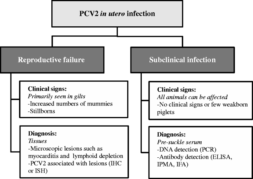
The authors believe there is a discrete distinction between PCV2-associated reproductive failure and subclinical PCV2 in utero infection, similar to the situation in growing pigs. Clinical PCVAD requires that there are clinical signs, microscopic lesions and demonstration of PCV2 antigen or DNA within the area of the microscopic change. We propose similar criteria for PCV2-associated reproductive failure. The following are needed for diagnosis: (1) clinical signs which can include early termination of pregnancy, increased numbers of mummified fetuses, stillborns or weak-born pigs, (2) +/−microscopic lesions within fetal tissues (heart or lymphoid tissues) and (3) PCV2 antigen or DNA within fetal tissues. In contrast, subclinical PCV2 in utero infection is identified by the detection of PCV2 DNA or antibodies in fetal tissues, presuckle serum or fetal thoracic fluid without the presence of microscopic lesions or indication of reproductive failure (Fig. 1). This presentation is likely related to timing of fetal PCV2 infection (late gestation) and the degree of PCV2 replication.
Outcomes of PCV2 in utero infection.

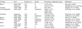
The prevalence of clinical and subclinical in utero PCV2 infection has been studied in many countries over the past decade (Table 2). Early reports indicated a low prevalence of PCV2, but increased numbers of in utero PCV2 infections have been noted in countries recently affected by severe PCVAD in the growing pig population. Shen et al. (Reference Shen, Wang, Madson and Opriessnig2010) evaluated presuckle serum samples obtained from healthy live-born piglets from conventional breeding farms located in the U.S.A. and in Mexico to determine the prevalence of in utero PCV2 infection. PCV2 viremia and specific anti-PCV2 antibodies were detected in 199/499 (39.9%) and 107/499 (21.4%) of the presuckle serum samples, respectively. Differences were apparent across breeding farms and ranged from 17 to 71% in utero fetal PCV2 infection. In utero infection with PCV2b was more common, but some piglets were also found to be dually infected with PCV2a and PCV2b (Shen et al., Reference Shen, Wang, Madson and Opriessnig2010). This finding is likely associated with introduction of different PCV2 subtypes circulating in these regions.
Prevalence of PCV2-associated reproductive failure and in utero fetal infection across different countries
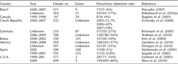
a Number of cases; the number of fetuses tested was not given.
Most studies investigating PCV2 prevalence were conducted prior to widespread vaccine availability and usage, and under those circumstances approximately 10–20% of the reproductive failure cases in the last decade were suggested to be associated with PCV2 in utero infection. To support this speculation, an infectious pathogen was identified in 501 cases of reproductive failure in swine submitted to the Iowa State University Veterinary Diagnostic Laboratory between 2003 and 2010; 12% of the cases (58/501) were diagnosed as having PCV2 involvement (data not shown). With the availability of vaccines, the strategy to vaccinate dams is important to prevent in utero PCV2 infection or associated reproductive failure prior to these animals entering the breeding population. Information on PCV2 vaccination protocols and their effect on dams and on prevention of fetal infection and reproductive failure is provided in a later section on PCV2 vaccination in breeding herds.
PCV2 in breeding females: impact of PCV2 isolates
Initial molecular comparisons of PCV2 isolates obtained from reproductive failure cases and other clinical manifestations (PDNS and PCVAD) showed genetic variation suggesting dissimilarities (Meehan et al., Reference Meehan, McNeilly, McNair, Walker, Ellis, Krakowka and Allan2001; Farnham et al., Reference Farnham, Choi, Goyal and Joo2003). When PCV2 isolates associated with reproductive failure were compared, characterization of the Open Reading Frame (ORF2) region demonstrated that the PCV2 isolates were 99% (nucleic acid sequence) and 97% (protein sequence) identical. When compared with a PDNS PCV2 isolate, the two reproductive failure associated PCV2 isolates were 96% (nucleic acid sequence) and 92% (protein sequence) identical (Meehan et al., Reference Meehan, McNeilly, McNair, Walker, Ellis, Krakowka and Allan2001). In a similar study, the ORF2 sequence homology of two North American PCV2-associated reproductive failure isolates was characterized and compared to 13 PCV2 sequences obtained from the GenBank and identified in Canada, France, China, Taiwan and the United Kingdom. The two PCV2 reproductive isolates varied at the nucleotide level from 90.6–98.7% when compared with the 10 PCV2a and 3 PCV2b isolates (Farnham et al., Reference Farnham, Choi, Goyal and Joo2003).
In vitro replication kinetics of PCV2 reproductive failure isolates have been compared with those of PCV2 isolates recovered from PDNS- and PMWS-affected pigs. A greater amount of PCV2 capsid protein was detected in the cytoplasm of PK-15 cells inoculated with PCV2 strains recovered from reproductive failures compared to cells inoculated with the other isolates; however, the production of new virus particles was slightly diminished when compared with a reference PMWS-associated PCV2 isolate. Nevertheless, the reproductive failure PCV2 isolates were able to infect a greater number of fetal myocardiocytes and pulmonary macrophages over a 72 h time period (Meerts et al., Reference Meerts, Misinzo, McNeilly and Nauwynck2005).
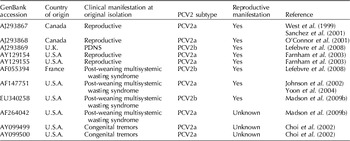
PCV2 fetal infection in pregnant sows has been experimentally reproduced using field PCV2 isolates from cases of reproductive failures (Nielsen et al., Reference Nielsen, Ladekjær-Hansen, Bille-Hansen, Lohse and Bøtner2004; Park et al., Reference Park, Kim, Ha, Jung, Choi, Lim, Kim and Chae2005; Lefebvre et al., Reference Lefebvre, Barbé, Atanasova and Nauwynck2008). Furthermore, PCV2-associated reproductive failure has been reproduced in naïve sows artificially inseminated with semen contaminated with a PCV2b isolate from a PMWS outbreak (Madson et al., Reference Madson, Patterson, Ramamoorthy, Pal, Meng and Opriessnig2009c). A recent study reported on the outcome of in utero inoculation of fetuses with different PCV2 isolates at 55 days of gestation. PCV2 isolates were obtained from a reproductive failure case (PCV2a), a PDNS case (PCV2b), and two isolates were from PMWS-affected pigs (one PCV2a and one PCV2b) (Lefebvre et al., Reference Lefebvre, Barbé, Atanasova and Nauwynck2008). Twenty-one days post-fetal inoculation, sows were euthanized. Gross lesions of PCV2 fetal infection and high PCV2 titers in tissues were noted across all PCV2 isolates used in the experimental study (Lefebvre et al., Reference Lefebvre, Barbé, Atanasova and Nauwynck2008). Similarly, PCV2 replication was observed after placement of a PCV2a PMWS isolate into fetal fluids by laparotomy or ultrasound-guided trans-abdominal inoculation (Johnson et al., Reference Johnson, Joo, Direksin, Yoon and Choi2002; Yoon et al., Reference Yoon, Jepsen, Pogranichniy, Sorden, Stammer and Evans2004). With the information known to date (Table 3), it becomes evident that ORF2 differences among PCV2 isolates cannot be used to predict different clinical manifestations of the virus. This information also indicates that any PCV2 isolate is capable of infecting intrauterine fetuses and causing PCV2-associated reproductive failure.
PCV2 isolates associated with or perceived to be linked with in utero infection

PCV2 in breeding females: commonly observed coinfections
Singular PCV2 in utero fetal infection can result in reproductive failure (West et al., Reference West, Bystrom, Wojnarowicz, Shantz, Jacobson, Allan, Haines, Clark, Krakowka, McNeilly, Konoby, Martin and Ellis1999; Pittman, Reference Pittman2008; Hansen et al., Reference Hansen, Hjulsager, Bille-Hansen, Haugegaard, Dupont, Hogedal, Kunstmann and Larsen2010). However, many field reports and retrospective studies have identified other pathogens in combination with PCV2 in cases of reproductive failure. Other pathogens recognized in fetal tissues include PPV (O'Connor et al., Reference O'Connor, Gauvreau, West, Bogdan, Ayroud, Clark, Konoby, Allan and Ellis2001; Kim et al., Reference Kim, Jung and Chae2004; Ritzmann et al., Reference Ritzmann, Wilhelm, Zimmermann, Etschmann, Bogner, Selbitz, Heinritzi and Truyen2005; Pescador, Reference Pescador2007; Sharma and Saikumar, Reference Sharma and Saikumar2010; Woods et al., Reference Woods, McDowell, Holtkamp, Gillespie and Pogranichniy2010), PRRSV (O'Connor et al., Reference O'Connor, Gauvreau, West, Bogdan, Ayroud, Clark, Konoby, Allan and Ellis2001; Farnham et al., Reference Farnham, Choi, Goyal and Joo2003; Kim et al., Reference Kim, Jung and Chae2004; Ritzmann et al., Reference Ritzmann, Wilhelm, Zimmermann, Etschmann, Bogner, Selbitz, Heinritzi and Truyen2005; Pescador, Reference Pescador2007), PCV1 (Pescador, Reference Pescador2007), porcine torque teno virus (Ritterbusch et al., Reference Ritterbusch, Sa Rocha, Mores, Simon, Zanella, Diniz-Mendes, Niel and Ciacci-Zanella2010b) and Escherichia coli (Pescador, Reference Pescador2007). The significance of coinfections in PCV2-associated reproductive failure is unknown, but concurrent infection by two or more pathogens within a herd is suspected to increase the risk of reproductive failure and decrease reproductive production parameters (López-Soria et al., Reference López-Soria, Maldonado, Riera, Nofrarías, Espinal, Valero, Blanchard, Jestin, Casal, Domingo, Artigas and Segalés2010). Moreover, additional infectious pressure likely leads to greater numbers of fetuses infected with PCV2 due to the added stress and the possible enhancement of PCV2 replication and shedding.
PCV2 in breeding females: impact of the anti-PCV2 antibody status of the dam at the time of PCV2 exposure
PCV2-associated reproductive failure outbreaks are typically reported in gilt-start-ups or new farms and likely related to seronegative populations (West et al., Reference West, Bystrom, Wojnarowicz, Shantz, Jacobson, Allan, Haines, Clark, Krakowka, McNeilly, Konoby, Martin and Ellis1999; Josephson and Charbonneau, Reference Josephson and Charbonneau2001; Ladekjær-Mikkelsen et al., Reference Ladekjær-Mikkelsen, Nielsen, Storgaard, Bøtner, Allan and McNeilly2001; O'Connor et al., Reference O'Connor, Gauvreau, West, Bogdan, Ayroud, Clark, Konoby, Allan and Ellis2001; Sanford, Reference Sanford2002; Mauch and Bilkei, Reference Mauch and Bilkei2004; Mikami et al., Reference Mikami, Nakajima, Kawashima, Yoshii and Nakajima2005; Brunborg et al., Reference Brunborg, Jonassen, Moldal, Bratberg, Lium, Koenen and Schonheit2007; Høgedal et al., Reference Høgedal, Ovesen, Kunstmann, Haugegaard and Bille-Hansen2008; Pittman, Reference Pittman2008; Hansen et al., Reference Hansen, Hjulsager, Bille-Hansen, Haugegaard, Dupont, Hogedal, Kunstmann and Larsen2010). The spread of PCV2 in a naïve population can, however, result in few clinical signs and only a portion of affected dams may develop abnormal litter characteristics (Ladekjær-Mikkelsen et al., Reference Ladekjær-Mikkelsen, Nielsen, Storgaard, Bøtner, Allan and McNeilly2001). Additionally PCV2-associated reproductive failure has been reported in seropositive dams (Janke, Reference Janke2000; Nielsen et al., Reference Nielsen, Ladekjær-Hansen, Bille-Hansen, Lohse and Bøtner2004). Janke (Reference Janke2000) reported increased numbers of non-viable piglets at parturition in both gilts and multiparous sows. The affected herd was well established with no new animal introductions for many years. The reason for the PCV2 circulation, or re-circulation, was not determined, but 3 months after the initial presentation the production in the herd was back to normal. In a report by Nielsen et al. (Reference Nielsen, Ladekjær-Hansen, Bille-Hansen, Lohse and Bøtner2004), PCV2 seropositive sows were experimentally inoculated intranasally with PCV2 in late gestation. One challenged sow delivered a mummified fetus and three weak-born piglets that were positive for PCV2 DNA by PCR.
Sixty-days after oro-nasally inoculating naïve, non-pregnant sows with PCV2, Rose et al. (Reference Rose, Blanchard, Cariolet, Grasland, Amenna, Oger, Durand, Balasch, Jestin and Madec2007) inseminated two sows with spiked semen containing the homologous PCV2 isolate. PCV2 viremia was not measured at insemination, but both sows were seropositive at 35 days post-oro-nasal inoculation. Both sows delivered PCV2-positive mummified fetuses varying in crown-to-rump length from 5 to 33 cm (Rose et al., Reference Rose, Blanchard, Cariolet, Grasland, Amenna, Oger, Durand, Balasch, Jestin and Madec2007). Conversely, Madson et al. (Reference Madson, Patterson, Ramamoorthy, Pal, Meng and Opriessnig2009a) found different results following oro-nasal challenge in three PCV2 naïve dams 120 days prior to artificial insemination (AI) with PCV2 spiked semen containing 5 ml of cell culture-propagated PCV2 (104.2 TCID50 per ml). Dams were not PCV2 viremic at insemination. Post-insemination viremia was detected in two of the three sows, but reproductive failure and PCV2 fetal infection were not observed (Madson et al., Reference Madson, Patterson, Ramamoorthy, Pal, Meng and Opriessnig2009a).
These reports suggest that dam-associated homologous anti-PCV2 antibodies are at least partially protective for in utero PCV2 infection and the development of reproductive failure. However, if anti-PCV2 antibody titers are low, re-circulation within the breeding herd is possible and can be associated with reproductive problems. In addition, introduction of a different PCV2 subtype and infection of dams could potentially be associated with reproductive problems, though the incidence of clinical disease is likely diminished because of partial immunological protection.
PCV2 in gilts/sows: detection of PCV2 in colostrum or milk
PCV2 DNA
Sow colostrum and milk can contain PCV2 DNA post-partum. The prevalence of PCV2 in sow milk was reported by Shibata et al. (Reference Shibata, Okuda, Kitajima and Asai2006) after investigating three Japanese swine herds. PCV2 DNA was found by PCR or isolated in 33/33 samples (Shibata et al., Reference Shibata, Okuda, Kitajima and Asai2006). PCV2 DNA was demonstrated in colostrum samples obtained 24 h after birth in PCV2 seronegative sows experimentally infected with PCV2 on day 93 of gestation. In another report sow milk samples collected on days 2 and 3 were negative by PCR, but PCV2 antigen was detected in macrophage-like cells within ductal lumina in mammary tissue 3 days post-partum (Park et al., Reference Park, Ha, Kwon, Cho, Lee and Chae2009). The duration of PCV2 DNA shedding in sow milk has been described to be at least 27 days post-partum following oro-nasal challenge in late gestation (Ha et al., Reference Ha, Ahn, Kim, Cho, Lee, Oh, Kim and Chae2009).
In colostrum or milk, PCV2 can be free or cell associated and is infectious as virus isolation has been successful on colostral samples (Gerber et al., Reference Gerber, Garrocho, Lana and Lobato2010). Vaccination will decrease the amount of PCV2 shedding in colostrum and milk but may not eliminate this route as a possible source of infection for newborn piglets (Madson et al., Reference Madson, Patterson, Ramamoorthy, Pal, Meng and Opriessnig2009a; Gerber et al., Reference Gerber, Garrocho, Lana and Lobato2010). The role and the infectious potential of PCV2 in colostrum/milk have not been elucidated. Piglet infection by this route is feasible, but since colostrum and milk also contain antibodies it is not known whether PCV2 is neutralized in the gastrointestinal tract prior to absorption. One could hypothesize that the virus likely is absorbed and sequestered in the piglet until maternal antibodies have waned, at which time there is increased replication and potential disease. Alternatively, after waning of passively acquired anti-PCV2 antibodies, piglets become infected with PCV2 through continuous exposure to infectious PCV2 in the environment or other animals.
Anti-PCV2 antibodies
Passively acquired, maternally derived colostral antibodies are important to protect neonates prior to full development of the immune system (Sangild, Reference Sangild2003). With regard to PCV2, most swine herds are seropositive (Allan and Ellis, Reference Allan and Ellis2000; Opriessnig et al., Reference Opriessnig, Meng and Halbur2007), and thus dams are likely to transfer PCV2 antibodies to offspring via the colostrum. Dam antibody levels vary among individual dams within a herd (Opriessnig et al., Reference Opriessnig, Yu, Thacker and Halbur2004; Calsamiglia et al., Reference Calsamiglia, Fraile, Espinal, Cuxart, Seminati, Martin, Mateu, Domingo and Segalés2007) and between herds (Opriessnig et al., Reference Opriessnig, Yu, Thacker and Halbur2004; Zhou et al., Reference Zhou, Chen, Ye, Shen, Chen and Shang2006), and piglet antibody uptake from the same dam can vary (Opriessnig et al., Reference Opriessnig, Yu, Thacker and Halbur2004; McKeown et al., Reference McKeown, Opriessnig, Thomas, Guenette, Elvinger, Fenaux, Halbur and Meng2005). PCV2 maternal antibody half-life in piglets is approximately 3 weeks (Opriessnig et al., Reference Opriessnig, Yu, Thacker and Halbur2004), and depending on the initial titer, may last up to 18 weeks of age (Opriessnig et al., Reference Opriessnig, Patterson, Madson, Pal, Rothschild, Kuhar, Lunney, Juhan, Meng and Halbur2009). This highly variable length of time for maternal antibody decay on a population level makes the exact timing of PCV2 infection within a population difficult to predict.
Passively acquired maternal antibodies, in general, protect the piglet from developing clinical disease associated with PCV2 (Allan et al., Reference Allan, McNeilly, McNair, Meehan, Marshall, Ellis, Lasagna, Boriosi, Krakowka, Reynaud, Boeuf-Tedeschi, Bublot and Charreyre2002; Harms et al., Reference Harms, Sorden, Halbur, Nawagitgul, Lager, Bolin and Paul2002; Ostanello et al., Reference Ostanello, Caprioli, Di, Battilani, Sala, Sarli, Mandrioli, McNeilly, Allan and Prosperi2005; Calsamiglia et al., Reference Calsamiglia, Fraile, Espinal, Cuxart, Seminati, Martin, Mateu, Domingo and Segalés2007), but do not protect against PCV2 infection (McKeown et al., Reference McKeown, Opriessnig, Thomas, Guenette, Elvinger, Fenaux, Halbur and Meng2005). Therefore, the environment of the piglets including the dam could serve as a potential source of initial infection under certain circumstances. Of note, one report found no significant protective effect of PCV2 passively derived antibodies on the development of PMWS (Hassing et al., Reference Hassing, Kristensen and Baekbo2003) indicating that clinical disease can develop in animals regardless of their level of passively acquired immunity.
The titer of anti-PCV2 antibodies in colostrum has only recently been evaluated (Madson et al., Reference Madson, Patterson, Ramamoorthy, Pal, Meng and Opriessnig2009a, Reference Madson, Patterson, Ramamoorthy, Pal, Meng and Opriessnigb; Gerber et al., Reference Gerber, Garrocho, Lana and Lobato2010). Colostrum and serum from experimentally PCV2-infected dams had similar anti-PCV2-IgG antibody levels as determined by an ORF2 capsid protein-based enzyme-linked immunosorbent assay. As a more accessible sample, dam sera could be screened to estimate the amount of colostral anti-PCV2 antibodies delivered to the offspring. In addition, PCV2 vaccination of the dam confers colostral antibody levels similar to those induced by natural infection of the dam (Madson et al., Reference Madson, Patterson, Ramamoorthy, Pal, Meng and Opriessnig2009a; Gerber et al., Reference Gerber, Garrocho, Lana and Lobato2010).
Fetal infection with PCV2
PCV2 in oocytes and embryos
Detection of PCV2 in oocytes and embryos
Viral pathogens can attach to the zona pellucida of developing swine oocytes and embryos (Singh and Thomas, Reference Singh and Thomas1987; Bielanski et al., Reference Bielanski, Larochelle and Magar2004b). PCV2 DNA can be demonstrated by PCR in follicular fluid, oviductal cells, uterine washes and oocytes (Bielanski et al., Reference Bielanski, Larochelle, Algire and Magar2004a). Thus, the reproductive tract environment can be a potential source of PCV2 contamination for a subset of oocytes prior to fertilization.
Ability of PCV2 to cross the zona pellucida
Fluorescent particles with a diameter of 20 and 26 nm can cross the zona pellucida and reach the embryo while 200 nm particles are deposited on the outer surface (Mateusen et al., Reference Mateusen, Sanchez, Van Soom, Meerts, Maes and Nauwynck2004). Previously, PPV with an approximate diameter of 20 nm was reported to pass through an intact zona pellucida after 48 h of incubation (Bane et al., Reference Bane, James, Gradil and Molitor1990). Virions of PCV2 are slightly smaller than those of PPV with an approximate diameter of 17 nm (Tischer et al., Reference Tischer, Gelderblom, Vettermann and Koch1982). When swine morulae with intact or absent zona pellucida (6-day-old embryos), early blastocysts (7-day-old embryos) and late blastocysts (8-day-old-embryos) were exposed to PCV2 for 48 h, only zona pellucida free morulae and blastocysts became infected with PCV2 (Mateusen et al., Reference Mateusen, Sanchez, Van Soom, Meerts, Maes and Nauwynck2004). Based on the relative size of PCV2, it could penetrate the zona pellucida; however, this was not observed. Incubation of zona pellucida – free morulae and early blastocysts with PCV2 for 48 h had no significant effect on development when compared with intact zona pellucida embryos during the incubation period (Mateusen et al., Reference Mateusen, Sanchez, Van Soom, Meerts, Maes and Nauwynck2004). In contrast, incubation of PCV2 with hatched blastocysts (7-day-old embryos) for 1 h resulted in increased numbers of non-viable embryos 21 days later, though development still occurred in some embryos exposed to PCV2 (Mateusen et al., Reference Mateusen, Maes, Van Soom, Lefebvre and Nauwynck2007). Free PCV2 virions within the uterus have the potential to be associated with early embryonic death and can be present due to contamination of semen with PCV2, using contaminated breeding equipment or circulating PCV2 within the dam.
Attempts to decontaminate embryos infected with PCV2
Embryo transfer is a growing field for genetic improvement. Decontamination protocols were tested on oocytes and embryos that were exposed to PCV2 in an attempt to provide clean specimens that may be used for embryo transfer (Bielanski et al., Reference Bielanski, Larochelle and Magar2004b; Bureau et al., Reference Bureau, Dea and Sirard2005). Similar protocols involving trypsin, hyaluronidase, DNase and RNase were used. Bielanski et al. (Reference Bielanski, Larochelle and Magar2004b) was unable to identify a protocol that removed PCV2 DNA from contaminated embryos; however, Bureau et al. (Reference Bureau, Dea and Sirard2005) showed that treatment with 0.1% hyaluronidase removed PCV2 DNA along with the DNA of other swine viruses.
PCV2 fetal infection: experimental inoculation
Experimental PCV2 fetal infection has been documented following exposure of the dam to PCV2 via oro-nasal infection (Nielsen et al., Reference Nielsen, Ladekjær-Hansen, Bille-Hansen, Lohse and Bøtner2004; Park et al., Reference Park, Kim, Ha, Jung, Choi, Lim, Kim and Chae2005) or by AI with PCV2-spiked semen (Madson et al., Reference Madson, Patterson, Ramamoorthy, Pal, Meng and Opriessnig2009c). PCV2 viremia in adult animals may last for several weeks (Madson et al., Reference Madson, Patterson, Ramamoorthy, Pal, Meng and Opriessnig2009c). During viremia in the dam, PCV2 crosses the placenta as free virus or cell-associated virus (Pensaert et al., Reference Pensaert, Sanchez, Ladekjær-Mikkelsen, Allan and Nauwynck2004). Fetal infection and death can occur at different stages of gestation resulting in variably sized mummified fetuses (crown-to-rump length) and stillborns at parturition (Josephson and Charbonneau, Reference Josephson and Charbonneau2001; Ladekjær-Mikkelsen et al., Reference Ladekjær-Mikkelsen, Nielsen, Storgaard, Bøtner, Allan and McNeilly2001; O'Connor et al., Reference O'Connor, Gauvreau, West, Bogdan, Ayroud, Clark, Konoby, Allan and Ellis2001; Cariolet et al., Reference Cariolet, Blanchard, Le Dimna, Mahé, Jolly, de Boisséson, Truong, Ecobichon, Madec and Jestin2002; Pittman, Reference Pittman2008; Madson et al., Reference Madson, Patterson, Ramamoorthy, Pal, Meng and Opriessnig2009c). Timing of PCV2 fetal infection will also result in differences in gross lesions and distribution of PCV2 (Sanchez et al., Reference Sanchez, Nauwynck, McNeilly, Allan and Pensaert2001, Reference Sanchez, Meerts, Nauwynck and Pensaert2003, Reference Sanchez, Meerts, Nauwynck, Ellis and Pensaert2004; Johnson et al., Reference Johnson, Joo, Direksin, Yoon and Choi2002). After fetal infection, PCV2 was capable of spreading to other fetuses (Yoon et al., Reference Yoon, Jepsen, Pogranichniy, Sorden, Stammer and Evans2004). Furthermore, PCV2 was detected by PCR in early-aborted fetuses less than 30 days of gestation, implicating PCV2 in early embryonic fetal death (Kim et al., Reference Kim, Jung and Chae2004).
PCV2 fetal infection: PCV2 distribution and replication
After fetal infection is established, PCV2 can be detected in many tissues. In one experimental study, in utero fetuses were infected at 57, 75 and 92 days of gestation by injecting PCV2 into the amniotic fluid (Sanchez et al., Reference Sanchez, Nauwynck, McNeilly, Allan and Pensaert2001). All fetuses were retrieved 21 days later. Gross lesions of edema, hepatomegaly and congestion were observed in fetuses inoculated at 57 days, but not in fetuses inoculated at 75 or 92 days of gestation. Inoculation at 57 days of gestation resulted in significantly higher amounts of PCV2 in tissues that indicate systemic organ distribution, including heart, lung, spleen, kidney, lymph nodes and brain. Myocardium and liver contained the highest amounts of infectious virus. PCV2 inoculation at 75 and 90 days of gestation resulted in a restricted range of organ infection which included myocardium and to a lesser degree lymphoid tissue (spleen and lymph node), lung and liver (Sanchez et al., Reference Sanchez, Nauwynck, McNeilly, Allan and Pensaert2001). The distribution of PCV2 antigen was similar to what had been previously reported in field cases of PCV2-associated reproductive failure, indicating that the myocardium is likely the preferred site of fetal PCV2 replication (Sanchez et al., Reference Sanchez, Meerts, Nauwynck and Pensaert2003).
PCV2 DNA or antigen has also been detected in sera (Sanchez et al., Reference Sanchez, Nauwynck, McNeilly, Allan and Pensaert2001; Farnham et al., Reference Farnham, Choi, Goyal and Joo2003; Madson et al., Reference Madson, Patterson, Ramamoorthy, Pal, Meng and Opriessnig2009c), thymus (Park et al., Reference Park, Kim, Ha, Jung, Choi, Lim, Kim and Chae2005), tonsil (Park et al., Reference Park, Kim, Ha, Jung, Choi, Lim, Kim and Chae2005; Madson et al., Reference Madson, Patterson, Ramamoorthy, Pal, Meng and Opriessnig2009c), thoracic fluid (Sanchez et al., Reference Sanchez, Nauwynck, McNeilly, Allan and Pensaert2001; Yoon et al., Reference Yoon, Jepsen, Pogranichniy, Sorden, Stammer and Evans2004), small intestine (Johnson et al., Reference Johnson, Joo, Direksin, Yoon and Choi2002), colon (Pescador, Reference Pescador2007) and ear notch skin (Madson et al., Reference Madson, Patterson, Ramamoorthy, Pal, Meng and Opriessnig2009c) of in utero PCV2-infected fetuses. Myocardial PCV2 replication diminishes with increased fetal age. This associated shift is related to myocardial development (Sanchez et al., Reference Sanchez, Meerts, Nauwynck and Pensaert2003). As fetuses near full development, PCV2 replication appears to take place in cells of the monocyte-macrophage lineage (Sanchez et al., Reference Sanchez, Meerts, Nauwynck and Pensaert2003, Reference Sanchez, Meerts, Nauwynck, Ellis and Pensaert2004).
PCV2 fetal infection: gross lesions
PCV2 affected litters are commonly composed of increased numbers of non-viable fetuses (mummified and stillborn) at parturition. Mummified fetuses can vary in crown-to-rump length with the smallest fetuses around 6–7 cm (O'Connor et al., Reference O'Connor, Gauvreau, West, Bogdan, Ayroud, Clark, Konoby, Allan and Ellis2001; Madson et al., Reference Madson, Patterson, Ramamoorthy, Pal, Meng and Opriessnig2009c). Live-born or weak-born piglets can also be delivered and display ill-thrift (West et al., Reference West, Bystrom, Wojnarowicz, Shantz, Jacobson, Allan, Haines, Clark, Krakowka, McNeilly, Konoby, Martin and Ellis1999; O'Connor et al., Reference O'Connor, Gauvreau, West, Bogdan, Ayroud, Clark, Konoby, Allan and Ellis2001; Cariolet et al., Reference Cariolet, Blanchard, Le Dimna, Mahé, Jolly, de Boisséson, Truong, Ecobichon, Madec and Jestin2002; Nielsen et al., Reference Nielsen, Ladekjær-Hansen, Bille-Hansen, Lohse and Bøtner2004; Mikami et al., Reference Mikami, Nakajima, Kawashima, Yoshii and Nakajima2005; Park et al., Reference Park, Kim, Ha, Jung, Choi, Lim, Kim and Chae2005; Madson et al., Reference Madson, Patterson, Ramamoorthy, Pal, Meng and Opriessnig2009c). PCV2 fetal infection can occur prior to fetal bone mineralization (Kim et al., Reference Kim, Jung and Chae2004), resulting in early embryonic death with fetal reabsorption, low litter sizes or possibly irregular returns to estrus of the dam if all embryos are lost. Fetal gross lesions are not always present following in utero PCV2 infection (Madson et al., Reference Madson, Patterson, Ramamoorthy, Pal, Meng and Opriessnig2009a, Reference Madson, Patterson, Ramamoorthy, Pal, Meng and Opriessnigb). Frequently, the only indication of in utero infection are increased numbers of stillborn and mummified fetuses at parturition. When present, gross lesions can be observed in live-born pigs, stillborn piglets or late-term mummified fetuses and are generally associated with myocardial failure. The lesions include dilated cardiomyopathy with regions of pallor, pulmonary edema, hepatomegaly with an accentuated lobular pattern (congestion), hydrothorax, ascites and subcutaneous edema (West et al., Reference West, Bystrom, Wojnarowicz, Shantz, Jacobson, Allan, Haines, Clark, Krakowka, McNeilly, Konoby, Martin and Ellis1999; O'Connor et al., Reference O'Connor, Gauvreau, West, Bogdan, Ayroud, Clark, Konoby, Allan and Ellis2001; Cariolet et al., Reference Cariolet, Blanchard, Le Dimna, Mahé, Jolly, de Boisséson, Truong, Ecobichon, Madec and Jestin2002; Park et al., Reference Park, Kim, Ha, Jung, Choi, Lim, Kim and Chae2005; Brunborg et al., Reference Brunborg, Jonassen, Moldal, Bratberg, Lium, Koenen and Schonheit2007; Madson et al., Reference Madson, Patterson, Ramamoorthy, Pal, Meng and Opriessnig2009c). Other infrequently observed gross lesions include lymphadenopathy (Johnson et al., Reference Johnson, Joo, Direksin, Yoon and Choi2002; Lefebvre et al., Reference Lefebvre, Barbé, Atanasova and Nauwynck2008), thymic atrophy (Nielsen et al., Reference Nielsen, Ladekjær-Hansen, Bille-Hansen, Lohse and Bøtner2004; Madson et al., Reference Madson, Patterson, Ramamoorthy, Pal, Meng and Opriessnig2009c), perirenal edema (Madson et al., Reference Madson, Patterson, Ramamoorthy, Pal, Meng and Opriessnig2009c), mesocolonic edema (Pescador, Reference Pescador2007; Madson et al., Reference Madson, Patterson, Ramamoorthy, Pal, Meng and Opriessnig2009c) and cerebral and splenic petechiation (Brunborg et al., Reference Brunborg, Jonassen, Moldal, Bratberg, Lium, Koenen and Schonheit2007; Lefebvre et al., Reference Lefebvre, Barbé, Atanasova and Nauwynck2008; Madson et al., Reference Madson, Patterson, Ramamoorthy, Pal, Meng and Opriessnig2009c).
PCV2 fetal infection: clinical signs
In utero PCV2 infection has been linked with congenital tremors in newborn piglets in the U.S.A. (Stevenson et al., Reference Stevenson, Kiupel, Mittal, Choi, Latimer and Kanitz2001). PCV2 DNA and antigen were demonstrated in the brain and spinal cord of piglets less than 2 days old displaying congenital tremors. The piglets were born to gilts; however, no other gross lesions or clinical characteristics of PCV2-associated reproductive failure were noted in affected herds (Stevenson et al., Reference Stevenson, Kiupel, Mittal, Choi, Latimer and Kanitz2001). The PCV2 isolates obtained from affected piglets were characterized as PCV2a isolates and shared 99% nucleotide homology (Table 3). The congenital tremor isolates were compared with known PMWS isolates and 99% genetic homology was reported (Choi et al., Reference Choi, Stevenson, Kiupel, Harrach, Anothayanontha, Kanitz and Mittal2002). In other countries, PCV2 association with congenital tremor has not been confirmed following the examination of 95 piglet cases (Kennedy et al., Reference Kennedy, Segalés, Rovira, Scholes, Domingo, Moffett, Meehan, O'Neill, McNeilly and Allan2003; Ha et al., Reference Ha, Jung and Chae2005; Brunborg et al., Reference Brunborg, Jonassen, Moldal, Bratberg, Lium, Koenen and Schonheit2007). A clear association of congenital tremors has been previously documented with several swine viruses (Harding et al., Reference Harding, Done and Darbyshire1966, Reference Harding, Done, Harbourne, Randall and Gilbert1973; Mare and Kluge, Reference Mare and Kluge1974), but there is no substantial evidence of PCV2 in utero infection leading to congenital tremors.
PCV2 fetal infection: microscopic lesions
Microscopic lesions associated with PCV2 in fetuses are most consistently present in the myocardium of mummified, stillborn and live-born piglets (West et al., Reference West, Bystrom, Wojnarowicz, Shantz, Jacobson, Allan, Haines, Clark, Krakowka, McNeilly, Konoby, Martin and Ellis1999; O'Connor et al., Reference O'Connor, Gauvreau, West, Bogdan, Ayroud, Clark, Konoby, Allan and Ellis2001; Madson et al., Reference Madson, Patterson, Ramamoorthy, Pal, Meng and Opriessnig2009c). Myocardiocytes are often degenerate, necrotic or lost and replaced by variable amounts of fibrous connective tissue and mineralization with infiltration of inflammatory cells composed of lymphocytes, plasma cells, macrophages and, to a lesser extent, multinucleated giant cells. Occasionally, botyroid intranuclear inclusion bodies are present within cardiomyocytes (O'Connor et al., Reference O'Connor, Gauvreau, West, Bogdan, Ayroud, Clark, Konoby, Allan and Ellis2001; Pescador, Reference Pescador2007; Madson et al., Reference Madson, Patterson, Ramamoorthy, Pal, Meng and Opriessnig2009c). Other microscopic changes may include mild multifocal non-suppurative interstitial pneumonia (Kim et al., Reference Kim, Jung and Chae2004; Park et al., Reference Park, Kim, Ha, Jung, Choi, Lim, Kim and Chae2005; Pescador, Reference Pescador2007), bronchopneumonia (Yoon et al., Reference Yoon, Jepsen, Pogranichniy, Sorden, Stammer and Evans2004; Brunborg et al., Reference Brunborg, Jonassen, Moldal, Bratberg, Lium, Koenen and Schonheit2007), hepatic congestion with hepatocellular loss (West et al., Reference West, Bystrom, Wojnarowicz, Shantz, Jacobson, Allan, Haines, Clark, Krakowka, McNeilly, Konoby, Martin and Ellis1999), non-suppurative hepatitis with periacinar necrosis (Brunborg et al., Reference Brunborg, Jonassen, Moldal, Bratberg, Lium, Koenen and Schonheit2007), lymph node and splenic lymphocyte depletion with occasional multinucleated giant cells (Mikami et al., Reference Mikami, Nakajima, Kawashima, Yoshii and Nakajima2005; Brunborg et al., Reference Brunborg, Jonassen, Moldal, Bratberg, Lium, Koenen and Schonheit2007; Madson et al., Reference Madson, Patterson, Ramamoorthy, Pal, Meng and Opriessnig2009c) or lymph node follicular hyperplasia (Brunborg et al., Reference Brunborg, Jonassen, Moldal, Bratberg, Lium, Koenen and Schonheit2007).
PCV2 fetal infection: diagnosis
Clinical signs
PCV2 infection should be suspected in cases with increased numbers of non-viable piglets at parturition or in cases of abortion; however, several other infectious and non-infectious causes of reproductive failure in swine can be associated with similar clinical presentations (Almond et al., Reference Almond, Flowers, Batista, D'Allaire, Straw, Zimmerman, D'Allaire and Taylor2006). The diagnostic success rate can depend on the timing of PCV2 infection, length of viremia in the dam, the number of fetuses infected and number of fetuses sampled. Pregnant dams infrequently show clinical signs of infection, are usually seropositive and can be PCV2 viremic or non-viremic at farrowing (Madson et al., Reference Madson, Ramamoorthy, Kuster, Pal, Meng, Halbur and Opriessnig2008). Therefore, interpreting fetal infection based on sow diagnostics is extremely difficult and should be avoided. Interpretation of sow serology is frequently complicated by the ubiquitous nature of PCV2 and viremia does not always correlate with fetal infection (Madson et al., Reference Madson, Patterson, Ramamoorthy, Pal, Meng and Opriessnig2009a).
Number of fetuses that need to be examined
Intrauterine PCV2 exposure may result in all fetuses becoming infected (Madson et al., Reference Madson, Patterson, Ramamoorthy, Pal, Meng and Opriessnig2009c). Alternatively, as little as one piglet in a litter of 10 clinically normal piglets may have an active infection at birth following gestational viremia in dams (Madson et al., Reference Madson, Patterson, Ramamoorthy, Pal, Meng and Opriessnig2009b). Fetuses can also clear active PCV2 infection prior to farrowing if the primary infection occurs in the post-immunocompetent phase (approximately 70 days of gestation) (Madson et al., Reference Madson, Patterson, Ramamoorthy, Pal, Meng and Opriessnig2009b). In trying to apply experimentally generated information to field scenarios, the authors speculate that most mummified fetuses will contain PCV2 antigen or DNA if PCV2 is the underlying cause of the fetal death. However, in situations where viral fetal infection occurs later in gestation, the authors speculate that approximately 50% of fetuses may contain viral particles. Therefore, sampling 4–6 fetuses per litter is recommended to improve the chances of detecting PCV2.
Demonstration of PCV2 antigen in tissues
Several diagnostic assays have been used for the detection of PCV2 in fetal samples. Similar to PCVAD in growing pigs, it is beneficial to find PCV2 DNA or antigen associated with affected fetal tissue. Immunochemistry (IHC) (Ellis et al., Reference Ellis, Hassard, Clark, Harding, Allan, Willson, Strokappe, Martin, McNeilly, Meehan, Todd and Haines1998; Sorden et al., Reference Sorden, Harms, Nawagitgul, Cavanaugh and Paul1999) and in situ hybridization (ISH) (Kim et al., Reference Kim, Han, Choi and Chae2003) are considered to be the gold standard for the detection of PCV2 antigen or DNA in formalin fixed, paraffin embedded tissues from fetuses suspected to be infected with PCV2. The most consistent tissue for the detection and diagnosis of in utero PCV2 infection is myocardium (Sanchez et al., Reference Sanchez, Nauwynck, McNeilly, Allan and Pensaert2001; Brunborg et al., Reference Brunborg, Jonassen, Moldal, Bratberg, Lium, Koenen and Schonheit2007; Madson et al., Reference Madson, Patterson, Ramamoorthy, Pal, Meng and Opriessnig2009c). Myocardium contains PCV2 antigen in the cytoplasm and the nucleus of infected cells stained by IHC. PCV2 antigen or DNA can also be present in thymus, spleen, tonsil and lung tissue.
Demonstration of PCV2 DNA
Detection of PCV2 DNA in fetal tissues or presuckle sera by PCR (Ellis et al., Reference Ellis, Krakowka, Lairmore, Haines, Bratanich, Clark, Allan, Konoby, Hassard, Meehan, Martin, Harding, Kennedy and McNeilly1999; Opriessnig et al., Reference Opriessnig, Yu, Gallup, Evans, Fenaux, Pallares, Thacker, Brockus, Ackermann, Thomas, Meng and Halbur2003) or by virus isolation (Ellis et al., Reference Ellis, Hassard, Clark, Harding, Allan, Willson, Strokappe, Martin, McNeilly, Meehan, Todd and Haines1998; Kim and Chae, Reference Kim and Chae2004) is another method for diagnosing PCV2 in utero infection. A PCR method allows for the detection of small amounts of virus, and tissue pooling can alleviate problems with timing of in utero infection and the distribution of virus. Myocardium should always be a part of the tissue pool. PCV2 detection by PCR has been reported to be successful in fetal thoracic fluid following direct fetal inoculation (Yoon et al., Reference Yoon, Jepsen, Pogranichniy, Sorden, Stammer and Evans2004); however, the sensitivity of the assay with the described sample is unknown. In field cases of PCV2-associated reproductive failure, Brunborg et al. (Reference Brunborg, Jonassen, Moldal, Bratberg, Lium, Koenen and Schonheit2007) detected PCV2 DNA in heart, liver, kidney, spleen, lymph node and brain tissues of non-viable fetuses (mummified and stillborn) and live-born pigs at farrowing using quantitative real-time PCR. The amount of PCV2 DNA in examined tissues ranged from 107 to 1012 genomic copies per 500 ng of DNA in non-viable fetuses. Myocardial and liver samples contained the highest amounts of DNA. The amount of PCV2 DNA in live-born piglets was lower and ranged from 102 to 1010 genomic copies per 500 ng DNA. Myocardium, liver and splenic tissues were positive for PCV2 DNA in all live-born piglets (Brunborg et al., Reference Brunborg, Jonassen, Moldal, Bratberg, Lium, Koenen and Schonheit2007).
Demonstration of anti-PCV2 antibodies
In utero PCV2 infection can also be diagnosed by demonstrating anti-PCV2-antibodies in fetal or live-born pre-suckle serum samples (Farnham et al., Reference Farnham, Choi, Goyal and Joo2003; Madson et al., Reference Madson, Patterson, Ramamoorthy, Pal, Meng and Opriessnig2009c; Shen et al., Reference Shen, Wang, Madson and Opriessnig2010). PCV2 antibodies in pre-suckle serum samples have been detected using a PCV2 recombinant capsid protein-based ELISA (Nawagitgul et al., Reference Nawagitgul, Harms, Morozov, Thacker, Sorden, Lekcharoensuk and Paul2002) on specimens in live-born piglets (Madson et al., Reference Madson, Patterson, Ramamoorthy, Pal, Meng and Opriessnig2009b; Shen et al., Reference Shen, Wang, Madson and Opriessnig2010). In an experimental sow challenge model, anti-PCV2 antibodies were the only indication of fetal infection (Madson et al., Reference Madson, Patterson, Ramamoorthy, Pal, Meng and Opriessnig2009b). Other serological tests include indirect fluorescent antibody and immunoperoxidase monolayer assay (IPMA) (Pogranichniy et al., Reference Pogranichniy, Yoon, Harms, Swenson, Zimmerman and Sorden2000). Anti-PCV2 antibodies in fetal thoracic fluid were also detected by Ladekjær-Mikkelsen et al. (Reference Ladekjær-Mikkelsen, Nielsen, Storgaard, Bøtner, Allan and McNeilly2001) using an IPMA technique.
PCV2 virus isolation
Virus isolation is more difficult and less sensitive than antigen, antibody and DNA detection. Following experimental trans-uterine inoculation of fetuses, PCV2 was isolated from the major organs including heart, lung, liver, spleen, brain, lymph node pools, serum and body fluids and successful isolation was age dependent (Sanchez et al., Reference Sanchez, Nauwynck, McNeilly, Allan and Pensaert2001). PCV2 was isolated from myocardium of all fetuses 21 days after in utero inoculation at 57, 75 and 92 days of gestation (Sanchez et al., Reference Sanchez, Nauwynck, McNeilly, Allan and Pensaert2001). Others were also successful in isolating PCV2 from fetuses that were experimentally infected in late gestation (86, 92 and 93 days of gestation) via laparotomies (Johnson et al., Reference Johnson, Joo, Direksin, Yoon and Choi2002). At farrowing PCV2 infection was confirmed in 12/12, 10/15 and 4/9 fetuses from their respective litters. Positive PCV2 isolation was confirmed in 11 spleens, 10 lymph nodes, 6 livers, 6 lungs and 3 intestinal samples from 34 fetuses. PCV2 was not isolated from any of the 34 kidney samples (Johnson et al., Reference Johnson, Joo, Direksin, Yoon and Choi2002).
Definitive diagnosis of PCV2-associated reproductive failure
Clinical abortion or increased numbers of non-viable fetuses at parturition should be observed and PCV2 DNA or antigen must be detected in tissues of affected fetuses. Detection of PCV2 DNA or antibodies in pre-suckle serum or thoracic fluid from fetuses only indicates in utero infection.
PCV2 fetal infection: sequelae for surviving pigs
It has been speculated that there may be differences in dam or litter susceptibility for the development of PCVAD in growing pigs (Madec et al., Reference Madec, Eveno, Morvan, Hamon, Blanchard, Cariolet, Amenna, Morvan, Truong, Mahé, Albina and Jestin2000). The authors suggested that dams transferred infection, but in utero PCV2 infection was not determined in the epidemiological study (Madec et al., Reference Madec, Eveno, Morvan, Hamon, Blanchard, Cariolet, Amenna, Morvan, Truong, Mahé, Albina and Jestin2000). Further examination of a possible litter effect and association with the development of PMWS in growing pigs was conducted by Calsamiglia et al. (Reference Calsamiglia, Fraile, Espinal, Cuxart, Seminati, Martin, Mateu, Domingo and Segalés2007). Sow viremia and antibody titer at parturition were documented and litter mates were followed into the growing phase. Piglets derived from dams with low PCV2 antibody titers or with viremia had higher morbidity and mortality rates associated with PMWS (Calsamiglia et al., Reference Calsamiglia, Fraile, Espinal, Cuxart, Seminati, Martin, Mateu, Domingo and Segalés2007). This information confirms a litter effect and implies that in utero fetal infection can be a contributing factor in the development of PMWS.
In one study, Jung et al. (Reference Jung, Kim, Ha, Choi and Chae2006) infected PCV2 naïve sows oro-nasally at 93 days of gestation and subsequently infected PCV2-positive live-born piglets at 3 days of age with porcine epidemic diarrhea virus (PEDV). Piglets from sows exposed to PCV2 and challenged with PEDV at 3 days of age contained higher numbers of PEDV-infected intestinal cells 24 h post-challenge compared with piglets infected with PEDV only (Jung et al., Reference Jung, Kim, Ha, Choi and Chae2006). Similarly, Ha et al. (Reference Ha, Lee, Ahn, Kim and Chae2008) inoculated PCV2 naïve dams oro-nasally in late gestation (3 weeks prior to expected farrowing) and challenged PCV2-positive live-born piglets at 28 days of age with PPV or stimulated their immune systems with keyhole limpet hemocyanin mixed with incomplete Freund's adjuvant (KLH/ICFA). All PCV2 in utero infected live-born pigs that were challenged or immunostimulated developed PMWS by 63 days of age. PMWS lesions were also observed in pigs infected after birth with PCV2 and PPV (Ha et al., Reference Ha, Lee, Ahn, Kim and Chae2008). Non-stimulated or coinfected PCV2-positive live-born piglets in these studies did not develop PMWS (Jung et al., Reference Jung, Kim, Ha, Choi and Chae2006; Ha et al., Reference Ha, Lee, Ahn, Kim and Chae2008). PCV2 in utero infection likely makes pigs more susceptible to coinfections with other pathogens and therefore may be associated with PMWS in the growing pig.
PCV2 infection in mature boars
PCV2 and boars: importance of AI
AI is a standard practice commonly implemented by most large swine production systems worldwide. In North America, nearly 60% of the swine herds are estimated to be practicing AI (Singleton, Reference Singleton2001). This percentage increased from the previous decade when estimates were around 5% (Flowers and Esbenshade, Reference Flowers and Esbenshade1993). Estimates in Western European countries approach 90% or greater (Madsen, Reference Madsen2005; Maes et al., Reference Maes, Nauwynck, Rijsselaere, Mateusen, Vyt, de Kruif and Van Soom2008). The main advantage of AI is genetic improvement (Gerrits et al., Reference Gerrits, Lunney, Johnson, Pursel, Kraeling, Rohrer and Dobrinsky2005). Although AI has many benefits in swine production, introduction of boar semen to sow herds from outside sources (AI centers) has been implicated in disease transmission (Yaeger et al., Reference Yaeger, Prieve, Collins, Christopher-Hennings, Nelson and Benfield1993). A recent review summarized swine viruses that can be detected in boar semen and included PCV2 (Guerin and Pozzi, Reference Guerin and Pozzi2005).
PCV2 and boars: clinical signs and gross lesions
Mature boars infected with PCV2 generally lack clinical signs and gross lesions associated with disease (Larochelle et al., Reference Larochelle, Bielanski, Muller and Magar2000; Madson et al., Reference Madson, Ramamoorthy, Kuster, Pal, Meng, Halbur and Opriessnig2008). However, Opriessnig et al. (Reference Opriessnig, Kuster and Halbur2006) reported clinical disease in an 11-month-old PCV2-positive boar coinfected with Mycoplasma hyopneumoniae and Pasteurella multocida. Semen morphology, motility or quality is typically not affected by natural or experimental PCV2 infection (McIntosh et al., Reference McIntosh, Harding, Parker, Ellis and Appleyard2006; Ciacci-Zanella et al., Reference Ciacci-Zanella, Zanella, Locatelli, Brambatti, Simon and Coldebella2007; Madson et al., Reference Madson, Ramamoorthy, Kuster, Pal, Meng, Halbur and Opriessnig2008).
PCV2 and boars: detection of PCV2
Detection of PCV2 in semen
In the acute phase of infection, PCV2 DNA can be detected in serum and semen at 2 and 5 days post-inoculation, respectively (Larochelle et al., Reference Larochelle, Bielanski, Muller and Magar2000; Madson et al., Reference Madson, Ramamoorthy, Kuster, Pal, Meng, Halbur and Opriessnig2008). Detection of PCV2 viremia commonly precedes the detection of semen-associated virus (Madson et al., Reference Madson, Ramamoorthy, Kuster, Pal, Meng, Halbur and Opriessnig2008), but shedding in semen has been reported in the absence of viremia (Larochelle et al., Reference Larochelle, Bielanski, Muller and Magar2000). PCV2 antibodies typically develop by 2 weeks post-inoculation (Larochelle et al., Reference Larochelle, Bielanski, Muller and Magar2000; Madson et al., Reference Madson, Ramamoorthy, Kuster, Pal, Meng, Halbur and Opriessnig2008). Following intranasal inoculation of four PCV2 negative boars, Larochelle et al. (Reference Larochelle, Bielanski, Muller and Magar2000) documented intermittent shedding of PCV2 DNA in semen over a 47-day observation period. Intermittent shedding in semen was confirmed by Grasland et al. (Reference Grasland, Blanchard, Jan, Oger, Rose, Madec, Jestin and Cariolet2008) after testing semen samples for 8 weeks obtained from four inoculated boars. In contrast, Madson et al. (Reference Madson, Ramamoorthy, Kuster, Pal, Meng, Halbur and Opriessnig2008) found continuous shedding of the virus in semen over 90 days following intranasal and intramuscular inoculation in 12 Landrace boars. Naturally infected boars sporadically shed PCV2 DNA in semen for up to 27.3 weeks in a PCV2-positive boar stud (McIntosh et al., Reference McIntosh, Harding, Parker, Ellis and Appleyard2006). Peak PCV2 shedding in semen occurs between 9 and 20 days post-inoculation and ranges from 105.1 (Madson et al., Reference Madson, Ramamoorthy, Kuster, Pal, Meng, Halbur and Opriessnig2008) to 106.0 (Grasland et al., Reference Grasland, Blanchard, Jan, Oger, Rose, Madec, Jestin and Cariolet2008) PCV2 genomic copies/ml. PCV2 PCR-positive results in serum or blood swabs are not correlated with semen PCR results, and therefore not predictive of the presence of PCV2 DNA in semen (Larochelle et al., Reference Larochelle, Bielanski, Muller and Magar2000; Reicks et al., Reference Reicks, Davies and Rossow2007; Madson et al., Reference Madson, Ramamoorthy, Kuster, Pal, Meng, Halbur and Opriessnig2008); however, there is correlation and predictability of serum PCR in the acute stage of infection in naïve boars (Madson et al., Reference Madson, Ramamoorthy, Kuster, Pal, Meng, Halbur and Opriessnig2008).
Detection of PCV2 in blood swabs
Experimentally inoculated boars can be viremic for at least 90 days and blood swabs can be positive for DNA at least 47 days after cell-free viremia was last detected (Madson et al., Reference Madson, Ramamoorthy, Kuster, Pal, Meng, Halbur and Opriessnig2008). This phenomenon is thought to be due to cell-associated virus in circulating peripheral blood mononuclear cells, which has previously been described in pregnant dams (Pensaert et al., Reference Pensaert, Sanchez, Ladekjær-Mikkelsen, Allan and Nauwynck2004).
Detection of PCV2 in boar tissues
Sex organs and accessory sex glands of PCV2-infected boars can contain PCV2 DNA or antigen. In non-clinical and clinically diseased boars, PCV2 antigen and DNA have been demonstrated by IHC and ISH in bulbourethral glands, testes, epididymis, prostate and seminal vesicles (Opriessnig et al., Reference Opriessnig, Kuster and Halbur2006; Ciacci-Zanella et al., Reference Ciacci-Zanella, Zanella, Locatelli, Brambatti, Simon and Coldebella2007; Ciacci-Zanella et al., Reference Ciacci-Zanella, Zanella, Morés, Neide, Kelen, Giseli, Almiro and Gava2008; Gava et al., Reference Gava, Zanella, Morés and Ciacci-Zanella2008). Abundant IHC staining of PCV2 antigen was identified within interstitial macrophages and fibroblast-like cells of seminal vesicles in a clinically affected, naturally infected boar from a commercial boar stud (Opriessnig et al., Reference Opriessnig, Kuster and Halbur2006). PCV2 was identified at a higher frequency in epididymis and bulbourethral gland sections compared to other accessory sex glands of naturally infected boars (Ciacci-Zanella et al., Reference Ciacci-Zanella, Zanella, Morés, Neide, Kelen, Giseli, Almiro and Gava2008). PCV2 DNA in testes, bulbourethral glands, epididymis, prostate and seminal vesicles has been demonstrated by quantitative real-time PCR in experimentally infected boars. Similar amounts were detected between accessory sex glands (Madson et al., Reference Madson, Ramamoorthy, Kuster, Pal, Meng, Halbur and Opriessnig2008). PCV2 DNA was also detected in penile tissue of naturally infected boars (Gava et al., Reference Gava, Zanella, Morés and Ciacci-Zanella2008).
PCV2 and boars: association of AI and PCV2 transmission
Several investigators have evaluated the risk of boar semen as a point source of PCV2 dissemination. A survey of 25 PCV2 seropositive Ontario swine farms using purchased semen or natural mating found no correlation with semen and the incidence of PMWS in growing pigs (Cottrell et al., Reference Cottrell, Friendship, Dewey, Allan, Walker and McNeilly1999). An epidemiological assessment of affected herds in Sweden concluded that dissemination via infected semen was not occurring (Wallgren et al., Reference Wallgren, Hasslung, Bergström, Linder, Belak, Hård af Segerstad, Stampe, Molander, Björnberg Kallay, Nörregård, Ehlorsson, Törnquist, Fossum, Allan and Robertsson2004). Similarly, Lawton et al. (Reference Lawton, Morris and King2004) investigated the spread of PMWS in New Zealand swine herds and determined that imported boar semen could not explain the spatial distribution of affected farms. In 2005–2006, North American swine populations saw an increase in PMWS associated with PCV2b isolates. One group investigated multiple PCV2b PMWS finishing sites in Kansas. The affected farms were sourced from two sow herds which were supplied with semen from a single boar stud. Evaluation and PCV2 sequencing showed that boars within AI centers supplying semen were infected with PCV2a. There was no correlation between the boar stud and the downstream PWMS associated with PCV2b (Horlen et al., Reference Horlen, Schneider, Anderson, Nietfeld, Henry, Tokach and Rowland2007). A more thorough risk analysis was conducted on 149 French swine farms to determine the association of boar semen with the development of PMWS. The investigators evaluated 59 farrow-to-finish farms clinically affected by PMWS, 55 farms that had never experienced PMWS and 35 farms that previously experienced PMWS and recovered. No correlation between the purchase of boar semen and the development of PMWS was demonstrated; however, an increased risk of PMWS in downstream growing pigs was positively correlated with on-farm semen collection (Rose et al., Reference Rose, Larour, Le, Eveno, Jolly, Blanchard, Oger, Le, Jestin and Madec2003).
PCV2 and boars: field investigations on prevalence of PCV2 DNA in semen
Detection of PCV2 DNA in boar semen is variable and depends on age (McIntosh et al., Reference McIntosh, Harding, Parker, Ellis and Appleyard2006; Reicks et al., Reference Reicks, Davies and Rossow2007; Schmoll et al., Reference Schmoll, Lang, Steinrigl, Schulze and Kauffold2008) and breed (McIntosh et al., Reference McIntosh, Harding, Parker, Ellis and Appleyard2006). Table 4 summarizes reported prevalence of PCV2 semen shedding in various geographic locations. With age and breed differences aside, collectively, the literature supports that approximately 20% of individual boars within a stud are shedding PCV2 DNA in semen at any particular time point if preventive vaccination is not being implemented.
Prevalence of shedding of PCV2 in boar semen

PCV2 and boars: semen infectivity
Virus isolation
Numerous bacteria and viruses can be present in boar semen and have been shown to be transmitted to breeding sows (Maes et al., Reference Maes, Nauwynck, Rijsselaere, Mateusen, Vyt, de Kruif and Van Soom2008). Cell culture assays for PCV2 are difficult to conduct and evaluate because semen is cytotoxic to cell lines (Medveczky and Szabo, Reference Medveczky and Szabo1981; Schultz et al., Reference Schultz, Adams, Letchworth, Sheffy, Manning and Bean1982; Weiblen et al., Reference Weiblen, Kreutz, Canabarro, Schuch and Rebelatto1992) and because PCV2 is a non-cytopathic virus (Tischer et al., Reference Tischer, Peters, Rasch and Pociuli1987; Allan and Ellis, Reference Allan and Ellis2000). However, successful PCV2 virus isolation from raw semen samples has been reported by Kim et al. (Reference Kim, Han, Choi and Chae2001, Reference Kim, Han, Choi and Chae2003).
Bioassay and AI
Recently, Madson et al. (Reference Madson, Patterson, Ramamoorthy, Pal, Meng and Opriessnig2009b) used a swine bioassay model to demonstrate infectivity of both PCV2a and PCV2b in semen from acutely infected boars injected intraperitoneally into 3-week-old PCV2 naïve pigs. When the same semen that contained PCV2a or PCV2b was extended and used for AI in PCV2 naïve gilts, transmission of PCV2 did not occur to inseminated gilts or gilt progeny (Madson et al., Reference Madson, Ramamoorthy, Kuster, Pal, Meng, Halbur and Opriessnig2009d). Other groups have also failed to infect PCV2 negative gilts or their offspring following AI with PCV2 contaminated semen (Grasland et al., Reference Grasland, Blanchard, Jan, Oger, Rose, Madec, Jestin and Cariolet2008). Semen extender used for AI has little effect on PCV2 viability in semen (Madson et al., Reference Madson, Ramamoorthy, Kuster, Pal, Meng, Halbur and Opriessnig2009d). The lack of PCV2 transmission in the AI model using contaminated semen from PCV2-infected boars suggests that infectivity of PCV2 via AI is dose dependent (Madson et al., Reference Madson, Ramamoorthy, Kuster, Pal, Meng, Halbur and Opriessnig2009d). In support, PCV2-associated reproductive failure was experimentally reproduced in PCV2 naïve sows following AI with extended semen spiked with 5 ml of cell culture-propagated PCV2 (104.2 TCID50 per ml) (Madson et al., Reference Madson, Patterson, Ramamoorthy, Pal, Meng and Opriessnig2009c). The reproducibility of in utero fetal infection was also demonstrated in seropositive sows using a PCV2-spiked semen model (Madson et al., Reference Madson, Patterson, Ramamoorthy, Pal, Meng and Opriessnig2009a). Seropositive sows were viremic for a short duration (1–2 weeks), had no clinical signs of disease, but delivered PCV2 viremic live-born piglets in the absence of increased numbers of non-viable piglets.
Combining what is known on semen epidemiology, infectivity of PCV2 in semen and breeding studies using PCV2-positive semen, we conclude that there is a potential for PCV2 transmission to occur via semen. Because of the low amount of semen that is delivered after extension, this risk is extremely low. Adding to the low risk is the high seroprevalance of PCV2 as most herds are seropositive for PCV2. Increased risk of transmission of PCV2 by semen may exist for PCV2 naïve animals or for herds where natural mating is practiced. In naïve herds or animal breeding scenarios testing of semen is recommended to negate the potential risk of transmission.
PCV2 and boars: detection of PCV2 in semen
Aside from virus isolation techniques, PCR methodologies, which are more sensitive, are routinely used to demonstrate that PCV2 DNA is in semen. Detection of PCV2 DNA in semen has been demonstrated by nested-PCR (Larochelle et al., Reference Larochelle, Bielanski, Muller and Magar2000; Kim et al., Reference Kim, Han, Choi and Chae2001, Reference Kim, Han, Choi and Chae2003; McIntosh et al., Reference McIntosh, Harding, Parker, Ellis and Appleyard2006) and quantitative real-time PCR (Pal et al., Reference Pal, Huang, Madson, Kuster, Meng, Halbur and Opriessnig2008) for both naturally exposed and experimentally infected boars. In one report, 60 raw semen samples were centrifuged and separated into seminal plasma, non-sperm cell fraction and sperm head fraction. Twenty-eight semen samples (46.7%) were positive for PCV2 DNA. PCV2 DNA was detected in all 28 seminal plasma samples, but only 11 of the non-sperm cell and 4 of the sperm head fractions contained PCV2 DNA (Kim et al., Reference Kim, Han, Choi and Chae2001). This same group also tested 98 semen fractions for the presence of PCV1, PCV2 and PPV using a multiplex nested PCR. Twenty-six of 98 whole semen samples (26.5%) were found to contain PCV2 DNA. When whole positive semen was fractionated and tested; 26/26 seminal plasma fractions, 9/26 non-sperm cells fractions and 3/26 sperm head fractions were positive for PCV2 DNA (Kim et al., Reference Kim, Han, Choi and Chae2003). More recently, Pal et al. (Reference Pal, Huang, Madson, Kuster, Meng, Halbur and Opriessnig2008) used a quantitative real-time PCR with an internal positive control and found semen cell fractions to contain more DNA than seminal fluid. Furthermore, quantitative analysis in raw semen was comparable to cell fraction samples for the detection of PCV2 DNA (Pal et al., Reference Pal, Huang, Madson, Kuster, Meng, Halbur and Opriessnig2008). Previous work has clearly demonstrated that PCV2 DNA can be detected in all fractions of raw semen. The seminal plasma fraction appears to contain higher amounts of PCV2 DNA when nested PCR techniques are applied. To date, raw semen has been established as the sample of choice using quantitative real-time PCR assays. Real-time PCR on raw semen can be used not only as a screening tool but also as a diagnostic assay confirming PCV2 shedding in boar semen.
Control of PCV2-associated disease in breeding herds and boar studs
Managing and controlling PCVAD in affected herds has evolved in recent years with the introduction of a number of commercial killed PCV2 vaccines. Prior to the availability of commercial PCV2 vaccines, good production practices with elimination of stressful pig-associated events and coinfections were promoted for controlling disease (Madec et al., Reference Madec, Eveno, Morvan, Hamon, Morvan, Albina, Truong, Hutet, Cariolet, Arnauld and Jestin1999).
PCV2 vaccination in breeding herds
There is only one vaccine currently marketed for sows (Circovac®, Merial, Lyon, France); however, other vaccines have been used in sows and are generally labeled for animals older than 3 weeks of age (Opriessnig et al., Reference Opriessnig, Madson, Shen, Brockmeier, Beach and Meng2010b). The two-dose sow vaccine is labeled for use to induce passive immunization of piglets via dam colostrum. Dam vaccination is recommended to be done 2–4 weeks prior to farrowing and will induce PCV2 specific antibodies in naïve gilts (Charreyre et al., Reference Charreyre, Bésème, Brun, Bublot, Joisel, Lapostolle, Sierra and Vaganay2005). A significant increase in production parameters has been observed in many herds in different geographical locations in Europe after implementing using Circovac® vaccine prior to farrowing.
In Denmark, sow PCV2 vaccination increased the number of live-born piglets, total born pigs and litters/sow/year while decreasing sow mortality, stillborn fetuses and dam non-productive days in many herds (Schøning et al., Reference Schøning, Nielsen and Lau2008; Bech and Kunstmann, Reference Bech and Kunstmann2008; Kunstmann and Lau, Reference Kunstmann and Lau2008). Moreover, in over 100 French herds, Circovac® sow vaccination resulted in similar production benefits (Hérin et al., Reference Hérin, Fily, Longo and Joisel2007; Delisle et al., Reference Delisle, Delisle, Bridoux, Thibault, Longo and Joisel2008), which was also confirmed in 233 (Joisel et al., Reference Joisel, Brune, Schade, Longo and Charreyre2007a, Reference Joisel, Brune, Schade, Longo and Charreyreb) and 277 (Joisel et al., Reference Joisel, Bune, Schade, Longo and Charreyre2008) German sow farms.
There are few experimental studies evaluating PCV2 vaccination followed by PCV2 challenge in mature sows. Madson et al. (Reference Madson, Patterson, Ramamoorthy, Pal, Meng and Opriessnig2009a, Reference Madson, Patterson, Ramamoorthy, Pal, Meng and Opriessnigb, Reference Madson, Patterson, Ramamoorthy, Pal, Meng and Opriessnigc, Reference Madson, Ramamoorthy, Kuster, Pal, Meng, Halbur and Opriessnigd) vaccinated PCV2 naïve pregnant dams at 28 days of gestation using a 1-dose baculovirus-expressed PCV2 vaccine (Ingelvac®, CircoFLEX™, Boehringer Ingelheim, Vetmedica Inc.) followed by oro-nasal PCV2 challenge at 56 days of gestation. Vaccinated dams developed neutralizing antibodies against PCV2 prior to challenge and PCV2 viremia was not detected in these dams during gestation. However, PCV2 in utero infection was detected at parturition in 10% (5/50) of the piglets born to vaccinated sows (Madson et al., Reference Madson, Patterson, Ramamoorthy, Pal, Meng and Opriessnig2009b). Similarly, in utero infection was observed in the spiked semen model when PCV2 naïve dams were vaccinated (Ingelvac®, CircoFLEX™) 8 weeks prior to AI (Madson et al., Reference Madson, Patterson, Ramamoorthy, Pal, Meng and Opriessnig2009a).
PCV2 vaccination at the breeding herd level benefits the dam which is actively vaccinated and protected, in utero fetuses which benefit by a reduction of PCV2 viremia in their dam, and piglets which receive higher amounts of anti-PCV2 antibodies via colostral intake. The timing of vaccination is critical and depends on the objectives of the management. If the situation in the breeding farm requires prevention of intrauterine infection and increase in production parameters, sow vaccination has the most benefit if given to the dam post-farrowing or pre-breeding. Vaccination at this point will protect the dam and the fetuses throughout the entire gestational period. If the goal is to protect the piglets in the earlier stages of the growing period, then vaccination prior to farrowing is suggested as higher levels of anti-PCV2 antibodies will be delivered in colostrum.
Another critical management step to be assessed in breeding herds is the need for PCV2 vaccination of replacement gilts that are entering the herd. In North America, most replacement gilts are PCV2 vaccinated as nursery pigs and the duration of immunity, commonly assumed to last for approximately 6 months, may not protect these animals as they enter the breeding facility where PCV2 circulation can occur. Therefore, boosting the immune response in replacement gilts 2–3 weeks prior to entering the breeding facility is highly recommended.
PCV2 vaccination in boar studs
Since the introduction of commercial PCV2 vaccines, three studies have evaluated vaccine efficacy in boars. In one study, the authors vaccinated 15 of 30 boars entering a boar stud isolation unit with a single dose of PCV2 vaccine (Suvaxyn® PCV2 One Dose, Fort Dodge Animal Health, Inc.). All boars were seropositive for anti-PCV2 antibodies upon entry into the isolation facility and 23 of 30 were either PCV2 viremic (12/23) and or shedding PCV2 DNA in feces (23/23) upon entry into the isolation facility. Vaccinated boars had a higher average ELISA sample-to-positive ratio of anti-PCV2 antibodies compared to non-vaccinates in the study, but significant differences were not found between groups for fecal shedding and PCV2 viremia during the 84 days of the study. Boar vaccination did not reduce semen shedding after entry into the stud on day 30 (Erlandson et al., Reference Erlandson, Connor and Groth2007). The second boar vaccine study involved vaccination of 8/16 PCV2 naïve landrace boars at 5 months of age with a single-dose PCV2 vaccine (Ingelvac®, CircoFLEX™). All boars were inoculated with PCV2 35 days later. Post-challenge, vaccinated boars became viremic and were positive for PCV2 DNA in semen at 4, 7, 14 and 21 days. Compared to non-vaccinated boars, vaccinated boars had lower quantities of PCV2 DNA in serum and semen after 4 days post-challenge (Reicks and Leuwerke, Reference Reicks and Leuwerke2008). PCV2 vaccination of boars under experimental conditions was evaluated in a PCV2 and M. hyopneumoniae coinfection model. One half of the PCV2 naïve mature boars were vaccinated with a single-dose PCV2 vaccine (Suvaxyn® PCV2) 35 days and 21 days prior to PCV2 and M. hyopneumoniae inoculation, respectively. Semen and serum samples were collected from all boars weekly for 5 weeks following PCV2 inoculation and tested for PCV2 DNA. Results indicated coinfected boars and non-vaccinated boars shed significantly higher amounts of PCV2 DNA in semen compared to singly infected and vaccinated boars (Opriessnig et al., Reference Opriessnig, Madson, Shen, Brockmeier, Beach and Meng2010a).
With limited information in the literature concerning boar vaccination, speculations on efficacy, timing, dose, number of vaccinations or type of vaccine, it is difficult to make recommendations. However, experimental data implies that boar vaccination will significantly reduce shedding of the virus in semen. In many ways, stud operations are similar to breeding farms with potential PCV2 circulation occurring. Thus, vaccinating or boosting the immune response in incoming boars prior to entry into the stud farm is recommended. This strategy has been adopted recently by the American Association of Swine Veterinarians, Boar Stud Biosecurity Committee (personal communication, Dr Chris Kuster).
Conclusions
Although PCV2-associated reproductive failure was recognized more than a decade ago, the exact pathogenesis of this manifestation of PCV2 infection and specifics on transmission are still poorly understood. For example, the minimal dose of PCV2 required for successful vertical transmission is unknown. Similarly, the minimal dose of PCV2 that needs to be present in semen for successful transmission into breeding herds is unknown. Regardless of transmission route, fetal myocardium appears to be the preferred site of viral replication although PCV2 can be found in many tissues. Outcomes of fetal PCV2 infection include fetal death, resolution of in utero infection and virus clearance or subclinical infection present at parturition with the latter being a more common event. In utero infected piglets can be a source of PCV2 transmission to other pigs and can succumb to PCV2-associated disease during the growing period.
PCV2 has been circulating in the global swine population at a subclinical level for several decades resulting in unrecognized economic losses to the industry. The recent advent of killed commercial PCV2 vaccines and the widespread implementation of PCV2 vaccination in growing pigs have resulted in marked improvement in previously unforeseen production losses, thereby highlighting the true cost of PCV2 infection, especially in clinically affected herds. Economic assessment of PCV2 circulation within a breeding herd has not been conducted to date. This is likely due to lack of PCV2 research in breeding herds and the lack of availability of an approved sow vaccine in the U.S. Preliminary data from Europe indicate economic benefit in vaccinating sows. Sows vaccinated with two doses of a killed vaccine (Circovac®) had significantly higher numbers of weaned pigs/sow/year and more live-born pigs per litter. Additionally, farrowing rate, return to estrus intervals and total born pigs were improved compared to periods prior to use of a vaccine (Vila et al., Reference Vila, Autret, Callen, Bridoux, Brune, Charreyre, Hérin, Joisel, Larour, Lau, Longo, Lorenzo, Shade and Smits2009). Vaccination data are promising for the prevention of PCV2 in utero fetal infection, but future assessments are needed to fully understand the significance and the economic impact of PCV2 circulation within breeding herds.