Introduction
Warthin's tumours are the second most common type of salivary gland tumour; they are benign neoplasms that occur almost exclusively within the parotid gland.Reference Goldenberg and Goldstein1 They present as slow-growing painless masses arising from the tail of the parotid gland, but occasionally present as rapidly growing and/or painful lesions outside the parotid gland, suggesting malignancy. The latter can pose a diagnostic and management problem.
A rare case of multifocal extraparotid Warthin's tumour is described. Based on clinical, radiological and cytological findings, it was considered to represent a metastatic squamous cell carcinoma (SCC) from an unknown upper aerodigestive tract primary tumour and treated by total conservative parotidectomy and modified radical neck dissection.
Case report
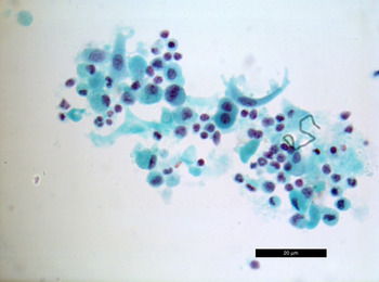
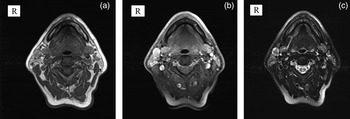
A 57-year-old male smoker was referred to the Otolaryngology Department with a rapidly growing right submandibular swelling. He admitted to having had a lump at that site for two years but had noted a recent, significant increase in size. He also complained of pain at the back of his tongue. Examination revealed a 2 cm mobile posterior right submandibular (level 1B) lump; the remainder of the ENT and neck examination was normal. Fine needle aspiration (FNA) of the neck lump was performed in the clinic; cytology was reported as suspicious for metastatic non-small carcinoma (Figure 1). Magnetic resonance imaging (MRI) findings were consistent with multiple necrotic nodes (intra- and extraparotid) and suspicious for malignancy (Figure 2).
Fine needle aspiration cytology showing small lymphocytes along with acellular and necrotic debris and large epithelioid cells with some nuclear irregularity and pleomorphism with slightly prominent nuclei. (Papanicolaou staining, ×630; scale bar, 20 µm)

(a) T1-weighted axial, (b) T1-weighted with fat suppression post-contrast axial and (c) T2-weighted with fat suppression axial magnetic resonance images showing a well-defined lesion (1.5 × 1.7 cm) lying anterior to the right parotid tail and displaying a partially intact fat plane and heterogenous appearances consistent with a necrotic node. Further lesions noted within the right parotid gland display unusual imaging characteristics compatible with necrotic intra-parotid lymph nodes.

The Head and Neck Oncology multidisciplinary team (MDT) concluded that findings were consistent with metastatic carcinoma in the right level 1 neck node, with an unknown primary or primary parotid tumour. Positron electron tomography (PET) scanning and ultrasound-guided FNA of the mass were recommended.
Further investigations
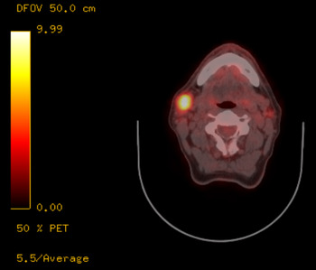
PET findings are shown in Figure 3. No primary site was identified, and PET was suggestive of metastatic cancer. Ultrasound-guided FNA of two of the intraparotid lesions were consistent with metastatic SCC. Examination under anaesthesia of the upper aerodigestive tract and directed biopsies including bilateral tonsillectomy were performed in an attempt to identify a primary site. The resultant histology findings revealed no evidence of malignancy. Evaluation of the left parotid nodule (identified by PET) with ultrasound-guided FNA cytology (FNAC) revealed a benign adenolymphoma (i.e. Warthin's tumour).
Positron electron tomography displaying a high standardised uptake value (SUV; 11.9) in the extraparotid lesion in the right side of the neck. This had increased in size from 1.7 cm to 3.1 cm since the previous magnetic resonance imaging scan. It also showed low-grade uptake in the lesion within the right parotid (maximum SUV 3.9) and a 0.6 cm sized nodule with low-grade uptake (maximum SUV 3.4) in the opposite left parotid gland.

The MDT concluded that investigations had identified a growing metastatic SCC of the upper right neck and parotid nodes with an unknown, possibly primary, parotid SCC. Subsequently, right total conservative parotidectomy and right-sided modified radical neck dissection were performed to provide definitive histology findings for diagnosis and treatment.
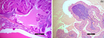
During surgery, multiple necrotic tumours were found within the inferior parotid gland and upper neck, including one within the deep parotid lobe and another in the submandibular triangle. Final histology findings showed multifocal benign Warthin's tumours (see Figure 4). A total of eight separate Warthin's tumour lesions were identified, with two in a completely extraparotid position and a further two attached to the parotid gland but almost completely exophytic. SCC was not identified.
Final histology findings (haematoxylin and eosin staining) showing a Warthin's tumour at (a) ×25 (scale bar 500 µm) and (b) ×200 (scale bar 50 µm) magnification, reported as lymphoid tissue and showing double-layered epithelium with tall columnar, eosinophilic and granular epithelial cells lining cystic cavities and forming papillary projections.

Discussion
Warthin's tumours account for 4–15 per cent of all salivary gland tumours.Reference Goldenberg and Goldstein1–Reference Cawson, Gleeson and Eveson3 They are more common in men and in the seventh decade of life; they almost exclusively occur within the parotid gland, where they can be multicentric and/or bilateral in 4–10 per cent of cases.Reference Goldenberg and Goldstein1, Reference Watkinson and Gilbert2, Reference Roland, McRae and McCombe4–Reference Van der Wal, Davids and Van der Waal7 The tumours usually manifest as a slow-growing, painless mass for 2 years on average, although sometimes for as much as 20 years, before presentation. However, they can also present with rapid growth, sometimes with pain and occasionally fluctuating in size.Reference Cawson, Gleeson and Eveson3 A small proportion (2.7–12.0 per cent) present as lesions outside the parotid gland; in a large series reported by the British Salivary Gland Tumour Panel, only 2 out of 335 cases of Warthin's tumours occurred outside the parotid gland.Reference Cawson, Gleeson and Eveson3, Reference Chae, Sohn, Shin, Choi and Kim8 Multifocal extraparotid Warthin's tumours are even rarer.Reference Naujoks, Sproll, Singh, Heikaus, Depprich and Kubler5, Reference Van der Wal, Davids and Van der Waal7–Reference Snyderman, Johnson and Barnes9
Warthin's tumour histology is unique and characteristic, showing varying pathognomonic proportions of lymphoid tissue and epithelium (and thereby accounting for its other names, i.e. adenolymphoma and papillary cystadenoma lymphomatosum).Reference Cawson, Gleeson and Eveson3, Reference Gnepp10 Foci of mucous or squamous metaplasia may sometimes be seen; rarely, such squamous metaplasia foci may be abundant.Reference Gnepp10 These foci are unlikely to lead to difficulties in histological diagnosis, although they may do so upon cytological analysis.
A cytological diagnosis of Warthin's tumour aims to identify three characteristic components: oncocytic cells, lymphoid stroma and acellular granular debris.Reference Klijanienko and Vielh11 Problems in cytological diagnosis arise when squamous metaplasia is present because such cells can lead to the suspicion, and sometimes the false-positive diagnosis, of a muco-epidermoid carcinoma or SCC. When such suspicious cytology findings arise from an intraparotid lesion, experienced clinicians and cytologists will recognise that the differential diagnosis includes a benign Warthin's tumour with squamous metaplasia. However, diagnostic problems are reported to arise when such cytology findings derive from an extraparotid lesion.Reference Naujoks, Sproll, Singh, Heikaus, Depprich and Kubler5, Reference Van den Brekel, Risse, Tiwari and Stel12
The distinct histology and clinical presentation of Warthin's tumours are believed to reflect the origin of this unusual category of tumour. One popular theory assumes that the tumours result from the mixing of heterotopic salivary and lymphoid tissue during development, leading to nests of mucosal epithelial cells becoming entrapped within lymph nodes.Reference Naujoks, Sproll, Singh, Heikaus, Depprich and Kubler5 In addition, the parotid capsule forms late during embryological development (late within the first trimester), which permits lymphatic inclusions to form within the parotid capsule. Both of these events lead to mucosal salivary tissue and lymphoid tissue mixing together within a capsule, which may then present as spectrum of lymphoepithelial inclusions such as Warthin's tumours, lymphoepithelial parotid cysts or branchial cysts. Similar mechanisms may explain the presence of aberrant salivary tissue in the lower neck or even the upper mediastinum.Reference Naujoks, Sproll, Singh, Heikaus, Depprich and Kubler5 Therefore, the Warthin's tumour size fluctuations that are sometimes observed clinically may be caused by mucosal proliferation secondary to local lymphokine release; when exaggerated and prolific, such mucosal proliferation would result in epithelial metaplasia resembling malignant proliferation (i.e. pseudo-neoplasia). Rapid cellular proliferation that outstrips the vascular supply can lead to necrosis and infarction, which can also mimic malignancy. Moreover, rapid Warthin's tumour growth in the absence of signs of inflammation will raise a clinical suspicion of malignancy. Rapid tumour growth has been observed following exogenous infection (e.g. a dental infection) or trauma, although specific case series are not well documented, except in association with FNA.Reference Taylor, Cozens and Robinson13
Warthin's tumours are therefore difficult to diagnose because they potentially mimic an aggressive malignancy, particularly if situated in an extraparotid position. In our patient, a diagnosis of metastatic lymphadenopathy was supported by clinical presentation, cytology and imaging, as described above.
Metastatic lymphadenopathy is a common presentation of head and neck cancer;Reference Roland, McRae and McCombe4 it usually presents with enlarged extraparotid neck nodes, but can include intraparotid nodes. In our case, since there was evidence of an intraparotid abnormality, the differential diagnosis included the rare primary parotid SCC, which represents 0.3–1.5 per cent of primary parotid cancers.Reference Gleeson, Cawson and Gleeson6
It is possible to explain the clinical presentation of our patient's benign tumour as a pseudomalignancy. As discussed, the literature on Warthin's tumours attests to their potential for rapid growth in response both to infection and FNA. Our patient showed evidence of post-operative lower right dental inflammation and possible pre-operative inflammation. In addition, diagnostic FNAC may have also contributed to tumour growth.
• Extraparotid multifocal Warthin's tumours are rare
• They may present as rapidly enlarging masses, suggesting malignancy
• Squamous metaplasia may confuse cytological reporting and lead to a false-positive diagnosis of malignancy
• Careful cytological analysis and frozen section examination at surgery may aid diagnosis and prevent unnecessary morbidity
Extraparotid Warthin's tumours remain a diagnostic challenge. This case highlighted some of the issues that should alert clinicians to the need to include this condition in the differential diagnosis of suspected metastatic upper neck lymphadenopathy. As discussed, cytological analysis can be problematic. Certain MRI protocols have been proposed to aid diagnosis but their specificity remains low.Reference Ikeda, Motoori, Hanazawa, Naqai, Yamamoto and Ueda14–Reference Yabuuchi, Matsuo, Kamitani, Setoquchi, Okafuji and Soeda16 Frozen section examination at surgery may therefore be necessary to guide management.
Conclusion
Experience in and knowledge of the unusual presentations and diagnostic difficulties of Warthin's tumours should help prevent unnecessary treatments that lead to morbidity. A rare case of multifocal Warthin's tumour is presented in which several extraparotid components suggest malignancy. A definitive diagnosis was based only on the final histology findings, despite an extensive radiological and cytological investigation. Therefore, cytological material should be carefully assessed and frozen section examination considered when doubts regarding diagnosis exist.
Acknowledgements
Grateful thanks to Dr E Loney, Bradford Royal Infirmary, for providing and interpreting radiological images.