I. INTRODUCTION
The binary Cu–Ni alloys are promising materials for a variety of applications such as heat exchangers and condenser tubes and thermoseeds because of their interesting properties including corrosion resistance, excellent strength, thermal conductivity, and antimicrobial. In a certain composition Cu (30–33%)–Ni (66–70%) alloy has a Curie temperature in the range of 41–46 °C which is appropriate for the production of ferromagnetic thermoseeds with a biodegradable overcoat such as an organic carbon layer. Reference Lilly, Brezovich and Atkinson1 Amorphous carbon films containing a mixture of sp 2 and sp 3 bonded carbon atoms have superior properties such as biocompatibility, chemical inertness, high hardness, wear resistance, low friction coefficient, high electrical resistivity for modifying surface properties of metal substrates used in medical implants and tools, storage devices, and micro-electromechanical devices. Reference Robertson2–Reference Ahmad, Roy, Rahman, Okpalugo, Maguire and McLaughlin11 Although several studies have been done to investigate properties of amorphous carbon overcoats on metal substrates (Cu–Ni alloy, Ni, stainless steel, Monel), the differences in physical properties of substrates, deposition methods (magnetron sputtering, chemical vapor deposition and etc.), and deposition parameters make more investigations necessary. Reference Yari, Larijani, Afshar, Eshghabadi and Shokouhy8–Reference Meigooni, Yoe-Sein, Al-Otoom and Sowards15 The ion beam sputtering deposition (IBSD) method that was contrived by Aisenberg and Chabot in 1971 Reference Aisenberg and Chabot4 for deposition of amorphous carbon is a widely used deposition technique for production of overcoats with good adhesion to a substrate because of simultaneous deposition by ion implantation. Reference Donnet and Erdemir16 Thus, a gradual strain transition from substrate to deposited film can be created and less built-in strain in the deposited film can be realized, which leads to a much more durable adhesion of the film to the substrate. Reference Bai, Zhang, Wu, Wang and Yan6 The possibility of independent control of deposition parameters including ion beam energy, substrate temperature, and ion current density is the most important issue of IBSD. Reference Tang, Li, Yang and Hirose5
The requirement of deposition at low temperatures for prevention of variation in the magnetic properties (Curie temperature) of Ni–Cu alloy during deposition process and the possible use of higher average particle energies in the ion beam deposition technique for creating a high durable amorphous carbon (a-C) coating lead to the selection of IBSD method for coating of Ni–Cu alloy in this study. Reference Manova, Gerlach and Mändl17 A large number of previous research results have indicated that the structure and physical properties of the amorphous carbon films strongly depend on the deposition process parameters, for instance, deposition temperature, kinetic energy of the atoms, deposition pressure. Among these parameters, the kinetic energy of the carbon atoms is the most effective one in a-C film deposition. Reference Pauleau18,Reference Bai, Zhang, Wu, Wang and Yan19 Nevertheless, various surveys have been performed in this context, most of them designed to investigate the effect of ion beam energy on the mechanical, Reference Bai, Zhang, Wu, Wang and Yan19 tribological, Reference Donnet and Erdemir16 corrosion properties Reference Chiang, Yang, Wei and Coulter20 in the intermediate ion energy ranges. Reference Tang, Li, Yang and Hirose5,Reference Ensinger21 In addition, most of them have been done on hydrogenated amorphous carbon deposited on other substrates. Reference Tang, Li, Yang and Hirose5,Reference Ahmad, Roy, Rahman, Okpalugo, Maguire and McLaughlin11 The free surface energy is an important parameter in controlling catalytic activity, adsorption, and adhesion to the surfaces that depends on the amount and nature of surface bonds. This parameter can be determined by the wetting behavior of a surface. Reference Banerjee, Mukherjee and Chattopadhyay22
In this paper that follows our previous work, Reference Mohagheghpour, Rajabi, Gholamipour, Larijani and Sheibani23 we investigate the effects of a variation in the ion beam energy from an intermediate value of 2 keV up to high value of 5 keV on the structural, optical, and wetting behavior of amorphous carbon films deposited on Ni–Cu alloy by the ion beam sputtering deposition method. The surface roughness, optical band gap, sp 3/sp 2 content, the wetting behavior, and the surface energy dependence on the argon ion beam energy and the existing relations are determined systematically.
II. EXPERIMENT
a-C thin films were deposited on Ni–Cu alloy substrate by ion beam sputtering deposition technique. The Ni (70.4%)–Cu (29.6%) alloy specimens were prepared by a vacuum arc remelting (VAR) furnace and homogenized with 4 times remelting. The specimens after rolling were cut out with an approximate size of 10 × 20 mm and 0.5 mm thickness, and then were mechanically wet ground, polished, and smoothed with Al2O3 to get a mirror finish. These substrates were cleaned ultrasonically with acetone, ethanol, and deionized water prior to film deposition. The experimental details of a-C deposition were published in our previous work. Reference Mohagheghpour, Rajabi, Gholamipour, Larijani and Sheibani23 The chamber was evacuated to a base pressure of 7–9 × 10⁻⁶ Torr and depositions were made in the constant temperature of water-cooling system. The deposition time and the ion current were 30 min and 25 mA, respectively. Five sets of a-C films were obtained by variation of the argon ion beam energy from 2 to 5 keV, while the other parameters include substrate temperature and pressure, and deposition time was kept constant. The thickness and band gap energy of the a-C films were obtained by using an optical method with simultaneous deposition of film on glass substrate (microscope slides).
The thickness and morphology of the films were determined by reflection measurement (AvaSpec-2048) and atomic force microscope (AFM), respectively. The AFM operated in a tapping mode (DME dual scope DS 95, Danish Micro Engineering, Herlev, Denmark) with a scan rate of approximately 3 Hz and 0.1 nN force in a typical scan area equal to 1 × 1 μm2. The surface roughness of Ni–Cu alloy substrates was measured by the portable surface roughness tester (TR 200, Time Instruments, Beijing, China). The optical properties were investigated from the reflectance and transmittance data using a UV/visible spectrophotometer (Cary 500, Varian, Palo Alto, California). The Raman spectra were measured using the 532 nm excitation wavelength of Nd:YLF laser with a power of 30 mW. The accuracy of Raman instrument is 4 cm−1. The samples for the X-ray photoelectron spectroscopy (XPS) measurements were prepared in a multi-chamber UHV evaporation tool (BESTEC GmbH, Berlin, Germany) having a base pressure of 0.75 × 10⁻¹⁰ Torr. Monochromatic Al Kα X-rays (1486.6 eV) were used as the excitation source operated at 10 kV. The wetting test was carried out at room temperature by using a contact angle measuring device (CAM). The contact angle (CA) was determined from digital images taken by a DFK23 U618 USB3.0 color industrial camera with the help of an a2X lens. An open source image processing software of ImageJ1.46v was used to measure contact angles.
III. RESULTS AND DISCUSSION
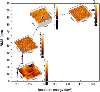
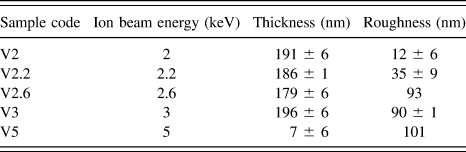
The surface roughness is an important parameter in determining the quality, the physical properties, and the growth mechanisms of amorphous carbon thin films. So, the surface morphology of the samples was measured to investigate the correlation between the ion beam energy and the growth mechanism of a-C on the Ni–Cu alloy substrate. The average surface roughness of the Ni–Cu alloy substrates before coating was 272 ± 20 nm. Figure 1 shows the variation of surface roughness of amorphous carbon thin films deposited on these substrates versus the deposition ion beam energy. The corresponding data are presented in Table I.
The evolution of roughness during the growth process of the a-C film as a function of ion beam energy.

The argon ion beam energy, thickness, and roughness of amorphous carbon films deposited on Ni–Cu alloy substrates, with a deposition time of 30 min.

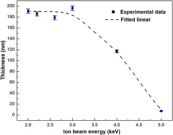
Figure 2 shows the variation of film thickness as a function of the sputtering ion beam energy. The film thickness is nearly constant in the ion beam energy range from 2 to 3 keV, while at higher energies it drops sharply from 188 nm to 7 nm (Table I).
The thickness of amorphous carbon films versus the argon ion beam energy with constant deposition time of 30 min.

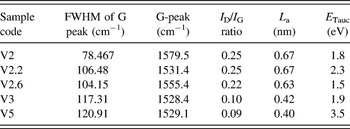
Figure 3 shows the Raman spectra of a-C films prepared at 2–5 keV ion beam energy. The Raman spectra of the carbon film are mainly composed of two broad peaks. The G-peak places in the range of 1528–1580 cm−1 as it appears in aromatic (rings) and olefinic (chains) molecules and the D peak around 1370 cm−1 is a breathing mode. This mode is forbidden in perfect graphite and only becomes active in the presence of the disorder and its intensity is strictly connected to the presence of 6-fold aromatic rings, so, the I D/I G ratio falls as the number of rings per cluster reduces and the fraction of chain groups elevates. Reference Robertson2,Reference Shin, Lee, Lee and Eun32 To extract the information from Raman spectra to determine the I D/I G ratio, as well as D and G peak positions, the spectra were deconvoluted with Gaussian functions. The data extracted from the Raman deconvolution versus ion beam energy are reported in Table II. From the deconvoluted spectra, it is observed that the integrated intensity ratio I D/I G has a little variation from 2 keV to 2.6 keV (Table II). This could be mainly due to a little change of sp 2 content in the a-C thin film with the low variation in the ion energy. While as the ion energy increases from 2.6 keV to 5 keV, the peak position of the G band shifts from 1555 to 1529 cm−1, the integrated intensity ratio I D/I G decreases from 0.22 to 0.09, and the FWHM of the G peak increases from 104 cm−1 to 121 cm−1. The size of the graphite cluster (L a) is calculated according to the following relation Reference Robertson2 :
 $${{{I_{\rm{D}}}} \over {{I_{\rm{G}}}}} = cL_{\rm{a}}^2\quad ,$$
where c is equal to ∼0.0055 (L a is in Angstrom), Reference Robertson2,Reference Paulmier, Bell and Fredericks24 decreases from 0.67 nm to 0.4 nm (Table II). These results imply a reduction in the sp 2 content of a-C thin films deposited with the higher energies.
Raman spectra of a-C films deposited on Ni–Cu alloy by different ion beam energies of (a) 2 keV, (b) 2.2 keV, (c) 2.6 keV, (d) 3 keV, and (e) 5 keV (
 $$
ion beam energy,
 $$
Gaussian fit peak, and
 $$
deconvoluted peaks).

The data extracted from Gaussian deconvolution of Raman spectra and the calculated values of the optical bandgap.

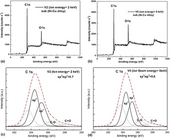
The XPS survey scans of the a-C samples V2 and V5 that were carried out over the 0–1200 eV binding energy range with 1.5 eV steps indicates clearly the contributions of C 1s (∼285 eV) and O 1s (∼532 eV) peak as shown in Figs. 4(a) and 4(b). To determine the sp 3 to sp 2 ratio of the amorphous carbon films, the C 1s peak position was deconvoluted into five subpeaks as illustrated in Figs. 4(c) and 4(d). The carbon film is mainly composed of disordered graphite at 283.4 eV, graphite (C sp 2 peak at 284.4 eV), oxygen bonded to carbon through hydroxyl or the other group (C–O at 286 eV), and carbonyl groups (C=O at 287 eV). Reference Paulmier, Bell and Fredericks24 It is noteworthy that two peaks at about 286–287 eV are attributed to some C/O contamination formed on the surface of the samples due to air exposure. Reference Pauleau25,Reference Merel, Tabbal, Chaker, Moisa and Margot26 After subtracting the background and fitting the sp 2 and sp 3 peaks, the sp 3/sp 2 ratio of the a-C films is evaluated which increases from 0.7 to 0.8 by increasing the ion beam energy from 2 keV to 5 keV. These data have been obtained by fitting the experimental spectrum with several Gaussian functions.
Analysis of the XPS C 1s peak of a-C films deposited on Ni–Cu alloy by different ion beam energies of (a) V2, (b) V5, (c) deconvoluted C 1s for V2, (d) deconvoluted C 1s for V5 and their sp 3/sp 2 ratio were depicted outside the spectra.

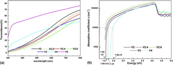
Figure 5 shows the optical transmission and absorption spectra of a-C films deposited on a glass substrate at the same condition of Ni–Cu substrates. The values of E 04 are determined in Fig. 5(b). The calculated data of Tauc band gap are presented in Table II. Reference Mohagheghpour, Rajabi, Gholamipour, Larijani and Sheibani23
(a) Optical transmission and (b) the absorption coefficients of a-C films deposited on glass substrates. The values of E 04 gap are also determined in the diagram.

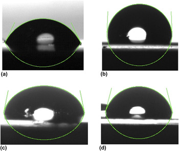
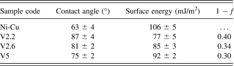
The surface energy is an important parameter for determining the adsorption, wetting, and adhesion properties of the a-C structure that depend on surface properties such as roughness and the surface content of carbon bonds (sp 3/sp 2 ratio). Reference Banerjee, Mukherjee and Chattopadhyay22 Here we investigate the variation of surface energy as a function of argon ion beam energies and the resultant observed change in physical properties of a-C thin films. Figure 6 shows the profile of the liquid distilled water drop and its contact angle on Ni–Cu uncoated substrate and coated with an amorphous carbon layer as a function of argon ion beam energy. The corresponding values of the contact angle are presented in Table III. The contact angle of the uncoated substrate is around 62° [Fig. 6(a)], so it is hydrophilic in nature. By a-C coating of the substrates, the values of the contact angle vary in the range of 87°–75° by increasing the argon ion beam energy that indicates the hydrophobic nature of amorphous carbon films. Reference Vogler27 According to our results, surfaces deposited at lower ion beam energies have higher contact angle and more hydrophobicity.
Profiles of the liquid distilled water droplet on (a) Ni–Cu alloy substrate and amorphous carbon films deposited by different ion beam energies of (b) 2.2 keV, (c) 2.6 keV, and (d) 5 keV.

The contact angle (CA), surface energy, and porosity of a-C film coated Ni–Cu alloy with different ion beam energies.

The surface free energy can be calculated by using the Young–Dupre equation Reference Pauleau25
 $$E = \gamma (1 + \cos \,\theta )\quad ,$$
where γ is the surface tension of water at 20 °C (γ = 72.8 mJ/m2) and θ is the contact angle (°).
The CA on a rough surface can be described by two different models: Wenzel’s law that is applicable if there is no air entrapped in the pits of the rough surface below the liquid and the equation proposed by Cassie and Baxter that describes the CA at a heterogeneous surface composed of two different materials (solid and air trapped in the hollows of the rough surface). Reference Banerjee, Mukherjee and Chattopadhyay22,Reference Peng and Chen28,Reference Zhou, Wang, Song, Li, Li, Zhao and Yan29 Since air could be easily trapped into a micro-porous surface, and then water cannot intrude into the interspaces among the microstructures, Reference Zhou, Wang, Song, Li, Li, Zhao and Yan29 we used Cassie and Baxter’s model for calculation of the surface porosity of the thin films by the following equation Reference Banerjee, Mukherjee and Chattopadhyay22 :
 $$\cos \,{\theta ^*} = f\,\cos \,\theta - \left( {1 - f} \right)\quad ,$$
where
 ${{\rm{\theta }}^{\rm{*}}}$
is the contact angle,
 ${\rm{\theta }}$
is the contact angle for an equivalent flat surface, and f is the liquid–solid contact fraction. For a suitable choice of
 ${\rm{\theta }}$
, according to the study of Zhou et al.
Reference Zhou, Wang, Song, Li, Li, Zhao and Yan29
that reported the CA on the smooth surface created with the magnetron sputtering method when the wetting angle is 40°, does not change with time, we have taken
 ${\rm{\theta }}$
equal to 40° for the measurement of porosity. This value of
 ${\rm{\theta }}$
was also used by D. Banerjee et al. for a-C thin films deposited by using plasma enhanced chemical vapor deposition (PECVD) method.
Reference Banerjee, Mukherjee and Chattopadhyay22
Comparing the results reveals that the surface roughness of layers increases with the enhancement in the ion beam energy, while the surface porosity decreases. The roughness measurement of Ni–Cu alloy substrates shows that the surface of the deposited films is smoother than the substrate even at the highest ion beam energy of 5 keV. The enhancement of surface roughness and the reduction of film thickness by increasing the argon ion beam energy in the range of 2–3 keV have been reported by L. Bai and coworkers. Reference Bai, Zhang, Wu, Wang and Yan19
It has been widely reported that the sp 3/sp 2 ratio is closely related to the I D/I G ratio in the amorphous carbon thin films. Reference Robertson2,Reference Tang, Li, Yang and Hirose5,Reference Pauleau25,Reference Ferrari30,Reference Ruso, Soga, Jimbo, Umen and Sharon31 Since the Raman results depend only on the ordering of the sp 2 sites and cluster size, Reference Shin, Lee, Lee and Eun32 the I D/I G ratio decreases as the number of rings per cluster decreases and the amount of chain groups increases; thus the possibility of creating sp 3 bonds increases. Therefore, it is evident that by the reduction in the I D/I G ratio due to increasing ion beam energy, sp 3/sp 2 ratio increases. This result is in good agreement with our XPS measurements.
According to the amorphization trajectory proposed by Ferrari and Robertson, Reference Ferrari and Robertson33 when the I D/I G ratio is very low, there exists a simple relation between cluster size and optical band gap
 $${{{I_{\rm{D}}}} \over {{I_{\rm{G}}}}} = {{C''} \over {E_{\rm{g}}^2}}\quad .$$
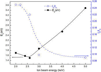
Since the size of graphite clusters is lower than 2 nm, according to the cluster model of Robertson, the I D/I G ratio is proportional to L a [Eq. (1)]. This model proposes the optical band gap of a-C depends on the sp 2 cluster size. Reference Robertson2 So, the increase of the optical band gap of a-C thin films due to the reduction of the I D/I G ratio by the increase in ion beam energy is reasonable.
As illustrated in Fig. 7, the value of the I D/I G ratio decreases by applying higher ion beam energies, while the optical band gap of thin films changes in the range of 1.5–3.5 eV (Table II). In addition, according to the previously reported data, an enhancement in the optical band gap by increasing the sp 3/sp 2 bond ratio should be expected. Reference Adamopoulos, Robertson, Morrison and Godet34,Reference Vasin, Matveeva and Kutsa35 So, the sp 2 bonds as well as the sp 2 cluster size decrease with the increasing ion beam energy and as a result of sp 3 enhancement.
The relation existing between optical bandgap and I D/I G ratio as a function of the argon ion beam energy.

Furthermore, the effect of the resputtering phenomena observed in the evaluation of optical properties of the a-C thin films that deposited on the glass substrate under the same deposition conditions on Ni–Cu alloy [Figs. 5(a) and 5(b) and Table I]. Since the film thickness has a sharp reduction due to resputtering phenomena, it can be concluded that the enhancement of transmittance in the deposited a-C thin film by 5 keV ion beam energy is due to a sharp reduction in the film thickness versus a little reduction of free volume. As mentioned in Table III the surface free energy is enhanced by increasing the ion beam energy that leads to higher surface roughness and lower I D/I G ratio. In fact, the contact angles of about 35 and 78° were reported for natural diamond and graphite, respectively. Reference Piazza and Morell36,Reference Ostrovskaya37 This phenomenon is attributed to the difference in the free surface energy between the basic crystallographic faces of diamond and graphite and proposed that the low surface energy of graphite planes resulted from the absence of reaction centers (dangling bonds) on the surface leading to hydrophobicity of the graphite surface. Reference Ostrovskaya37 Also, it has been shown that a decrease in surface energy is related to an increase in sp 2 bonding content and formation of nano crystalline graphite. Reference Yan, Xu, Yue, Liu, Xue and Yang38,Reference ModabberAsl, Kameli, Ranjbar, Salamati and Jannesari39 The CA could be correlated with the density of a-C films and increases with the decrease of the density of films due to the decreasing of sp 3/sp 2 ratio. Reference Robertson2,Reference ModabberAsl, Kameli, Ranjbar, Salamati and Jannesari39 Since the porosity decreases with increasing the density of thin films and according to the reported results of J. Kaban, Reference Kaban, Nowak, Bruzda, Xi, Sobczak, Eckert and Giebeler40 there exists a tendency of a better wetting of electrographite compared to glassy carbon due to a larger density, as a consequence of stronger van der Waals interactions. Thus, it can be suggested that the reduction of CA of a-C films with increasing deposition ion beam energy may be ascribed to the reduction of the sp 2 content, as revealed by XPS and Raman studies and porosity. The correlation between density, structural properties, and wettability of a-C thin films that were reported in the other studies confirms our results. Reference Robertson2,Reference Banerjee, Mukherjee and Chattopadhyay22,Reference Zhou, Wang, Song, Li, Li, Zhao and Yan29,Reference Piazza and Morell36,Reference Kaban, Nowak, Bruzda, Xi, Sobczak, Eckert and Giebeler40
According to the presented results and subplantation model, we propose a growth mechanism for the deposition of amorphous carbon film on Ni–Cu alloy with the argon ion beam energies of 2–5 keV. First, for the energy of 2 keV the carbon atoms and ions have enough energy to penetrate the subsurface layer and reduce the magnitude of voids in the film. Thus, a relatively smoother layer can be obtained on the rough surface of Ni–Cu alloy substrate. Second, by increasing the ion beam energy from 2.2 to 3.0 keV the sputtered C atoms and ions have increased mobility to agglomerate on energetically favorable sites of the substrate surface which leads to higher surface roughness. At energies higher than 3.0 keV, the high mobility and the resputtering effect lead to the reduction of film thickness and the roughness enhancement. The energy transferred to the structure by the impinging particle (thermal spike model) can give rise to the enhancement of the sp 3 bond content observed in the structural study.
IV. CONCLUSION
The ion beam energy enhancement from 2 keV to 5 keV increases the surface roughness sharply because of the resputtering effect. The surface free energy is reduced by creating surface height perturbation. Variation in structural and optical properties of the a-C thin films can be divided into two stages. The optical band gap and the I D/I G ratio have little change in the relatively low energy region (2–2.6 keV), after that the optical band gap begins to increase while the I D/I G ratio and G peak position decrease as the ion beam energy increases. These results imply a reduction in the values of porosity calculated by using Cassie and Baxter’s model, water contact angle on the surface, and the sp 2 content of the a-C thin films by increasing ion beam energy that is confirmed by the observed enhancement of the sp 3/sp 2 ratio obtained from XPS measurements. It should be noted that the highest optical transparency (in 1.55–6.2 eV) is observed in the case of the a-C thin films deposited by 5 keV ion beam energy with an optical band gap equal to 3.5 eV.