Introduction
Historically, Bulinus globosus was the only known snail host for Schistosoma haematobium in Lake Malaŵi. This snail typically inhabits inland, slow-moving waters and limited areas within the lake that are protected and, therefore, it was suspected that S. haematobium transmission in Lake Malaŵi was limited to such habitats (Stauffer et al., Reference Stauffer, Arnegard, Cetron, Sullivan, Chitsulo, Turner, Chiotha and McKaye1997). Beginning in the mid-1980s, reports of schistosomiasis among tourists suggested that, for the first time, transmission was occurring in the open waters of Lake Malaŵi (Harries et al., Reference Harries, Fryatt, Walker, Chiodini and Bryceson1986; Whitworth, Reference Whitworth1993; Pollner et al., Reference Pollner, Schwartz, Kobrine and Parenti1994; Cetron et al., Reference Cetron, Chitsulo, Sullivan, Pilcher, Wilson, Noh, Tsang, Hightower and Addiss1996). It was clear that the epidemiology of S. haematobium in Lake Malaŵi was changing, and the open waters of Lake Malaŵi were no longer free from urinary schistosomes.
Madsen et al. (Reference Madsen, Bloch, Kristensen and Furu2001) discovered an endemic snail, Bulinus nyassanus, infected with human schistosomes in the open waters of Nankumba Peninsula in southern Lake Malaŵi. Extensive sampling efforts (Stauffer et al., Reference Stauffer, Madsen, McKaye, Konings, Bloch, Fereri, Likongwe and Makaula2006) yielded no infected B. nyassanus outside of Nankumba Peninsula; thus, we believed that open-water transmission was limited to this area. The school-aged children residing in Chembe Village (Nankumba Peninsula) had a very high prevalence of urinary schistosomiasis (>87% in 1998; Madsen et al., Reference Madsen, Bloch, Kristensen and Furu2001) and currently the highest found in villages along Lake Malaŵi (Stauffer et al., Reference Stauffer, Madsen, McKaye, Konings, Bloch, Fereri, Likongwe and Makaula2006). Therefore, we postulated that the current high prevalence of human infection at Chembe Village is linked to B. nyassanus being an intermediate host in this portion of Lake Malaŵi.
The host specificity of S. haematobium can be divided as follows: strains most closely adapted to B. truncatus, a tetraploid member of the B. truncatus/tropicus species complex (North Africa and West Africa), strains most closely adapted to members of the B. africanus species group (West Africa, East Africa and southern Africa), and strains most closely adapted to the B. forskalii species group (Brown, Reference Brown1994; Southgate et al., Reference Southgate, de-Clercq, Sene, Rollinson, Ly and Vercruysse2000; Rollinson et al., Reference Rollinson, Stothard and Southgate2001). The finding of B. nyassanus, endemic to Lake Malaŵi and a diploid member of the B. tropicus/truncatus species group, as intermediate host in Lake Malaŵi was therefore unexpected.
We compare the genetic variation between S. haematobium from the southern part of the lake (Cape Maclear), where both B. globosus and B. nyassanus are intermediate hosts, and from the northern part (Likoma Island), and investigate the compatibility of the S. haematobium from Likoma Island with B. nyassanus from Chembe Village. Because infected B. nyassanus were collected only around Nankumba Peninsula in Lake Malaŵi, we concluded that B. globosus was the only intermediate host at Likoma Island (Stauffer et al., Reference Stauffer, Madsen, McKaye, Konings, Bloch, Fereri, Likongwe and Makaula2006).
Methods
Bulinus globosus and B. nyassanus were collected from Cape Maclear (Nankumba Peninsula). Snails were placed in 6 ml of water in 12.5 ml beakers and exposed to light to stimulate cercarial shedding. Cercariae shed from each snail were collected and preserved in 99% ethanol for DNA extraction.
DNA extraction, DNA sequencing and phylogenetic analysis
Cercariae collected from three snails of each species were used for molecular analysis. The ethanol-preserved cercariae were centrifuged in a 1.5-ml Eppendorf tube at 1300 rpm for 3 min to collect the cercariae at the bottom of the tube. Most of the ethanol was pipetted from the sample, which was then left to air dry for 10–30 min, allowing any residual ethanol to evaporate from the sample. Once dry, total genomic DNA was extracted from each sample using the DNeasyTM Tissue Kit (Qiagen, West Sussex, UK) according to the manufacturer's protocol.
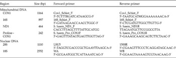
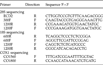
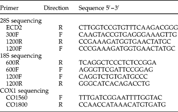
To identify any genetic variation between the samples, two nuclear DNA and four mitochondrial DNA partial regions were amplified by polymerase chain reaction (PCR), as shown in table 1. Amplifications were performed in a total reaction volume of 25 μ l using Ready-to-go PCR Beads (Amersham Pharmacia Biotech, Buckinghamshire, UK), each containing 1.5 units DNA Taq polymerase, 10 mm Tris–HCl (pH 9), 50 mm KCl, 1.5 mm MgCl2, 200 μ m of each dNTP and stabilizers, including bovine serum albumin (BSA), 0.4 μm of each primer and 2 μl of DNA (~10 ng). Thermal cycling in a Perkin Elmer 9600 Thermal Cycler used the following PCR conditions: 5 min denaturing at 95°C: 40 cycles of 30 s at 95°C, 30 s at 40–55°C, 1–2 min at 72°C; followed by a final extension period of 7 min at 72°C. PCR products were visualized on a 0.8% ethidium bromide agarose gel and then purified using Qiagen PCR Purification Kits (Qiagen) according to the manufacturer's protocol. Sequencing was performed using Fluorescent Dye Terminator Sequencing Kits (Applied Biosystems, Foster City, USA) and the sequencing reactions run on either an Applied Biosystems 377 or a 373A automated sequencer. PCR products were sequenced directly using the original PCR primers; and for COX1, 28S and 18S, internal sequencing primers were also used to obtain the full sequence of each fragment from both strands (table 2). The sequences were assembled and manually edited using Sequencher version 4.5 (GeneCodes Corp., Ann Abor, USA) and submitted to EMBL/GenBank. Each DNA region from each sample was aligned using Sequencher and sequence differences were checked by visualization of the sequence chromatograms.
DNA regions utilized in this study and the primers used to amplify them. COX1, 16S, 28S and 18S regions were amplified as they have proved useful in phylogenetic studies, and the variable regions ND1 and a small section between Proline and COX1 were amplified as a result of preliminary data from studies by Littlewood, Webster & Huyse (pers. comm.) on Schistosoma haematobium mitochondrial genomes.

Internal sequencing primers.
The 28S and COX1 sequences were aligned by eye in Maclade 4.05 (Sinauer Associates, Sunderland, Connecticut, USA) to sequence alignments of several other Schistosoma species and strains. Phylogenetic analyses were conducted separately using a neighbour-joining (NJ) algorithm to examine the evolutionary distance relationships among the samples.
Compatibility testing
Schistosoma haematobium eggs were obtained from urine samples from 10–15 infected children from Cape Maclear and Likoma Island. Urine samples were washed three times in saline water and once in fresh water (filtered lake water; 20 μm filter). After sedimentation in conical flasks for 25 min in the dark, the supernatant in the absence of light was discarded and the flask filled with more washing medium (saline or filtered lake water). After washing in filtered lake water, the eggs were transferred to Petri dishes and more filtered lake water added. Hatching was stimulated by exposing the Petri dishes to direct sunlight. Miracidia were captured with a tapering glass pipette and each snail was exposed to 5–20 miracidia. The uninfected B. nyassanus and B. globosus that were exposed came from Chembe Village. We exposed 25 B. globosus and 25 B. nyassanus to miracidia from eggs collected from children at Chembe Village and 25 B. globosus to miracidia from eggs from children at Likoma Island. Subsequently, we repeated the exposure of 25 B. nyassanus from Chembe Village to miracidia from eggs captured from Likoma Island children. The exposed snails were kept in separate beakers under light for 6 h to facilitate infection, and maintained in aquaria (Madsen et al., Reference Madsen, Bloch, Kristensen and Furu2001). From 5 to 8 weeks after exposure, snails were checked individually once a week for cercarial release: snails were transferred to beakers as described above and exposed to indirect sunlight for 2 h. The water in each beaker was visually checked for the presence of cercariae.
Results
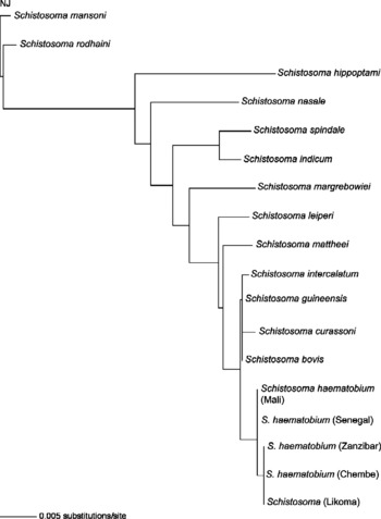
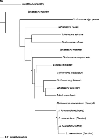
The 28S and 18S sequences from all the Lake Malaŵi isolates recovered from snails were identical, showing no nuclear genetic variation. The phylogenetic analysis of the 28S sequences showed that there is very little variation between the S. haematobium isolates incorporated into the tree, and the isolates from Lake Malaŵi match the Zanzibar strain of S. haematobium (fig. 1).
Neighbour joining (distance tree); 28S (nuclear DNA).

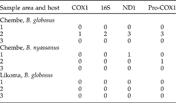
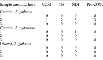
In all mitochondrial DNA sequences, there was very little variation between the Lake Malaŵi isolates (table 3). All nucleotide differences were also non-synonymous, suggesting that there is no genetic difference between the different isolates. The phylogenetic analysis of the COX1 sequences shows that there is very little variation between the different S. haematobium isolates incorporated into the tree. The Malaŵi isolates clearly group with other S. haematobium strains, but there is not enough genetic variation between the isolates to give clear resolution (fig. 2).
Mitochondrial DNA nucleotide differences of S. haematobium isolates from different intermediate hosts and localities (number of single nucleotide polymorphisms) (accession numbers EU567124–EU567144) compared to the Likoma isolate.
Neighbour joining (distance tree); COX1 (mitochondrial DNA).

In the first exposure experiment, 13 B. globosus were infected from eggs that were captured from Chembe Village children, 10 B. globosus were infected from eggs that were captured from Likoma Island, and no B. nyassanus were infected. When we repeated the exposure of B. nyassanus to miracidia from eggs captured from Likoma Island children, one snail was infected.
Discussion
The lack of genetic variation between the S. haematobium isolates from the two different intermediate snail hosts was surprising, especially due to the fact that B. nyassanus (B. truncatus/tropicus complex) is an unusual intermediate host for this parasite. Within the B. truncatus/tropicus complex, most diploid species seem to be resistant to infection in nature (Brown, Reference Brown1994), although B. liratus on the island of Madagascar appears to be an exception (Stothard et al., Reference Stothard, Bremond, Andriamaro, Sellin, Sellin and Rollinson2001). The fact that B. nyassanus is also susceptible is therefore another interesting exception to the rule, because it is diploid (Madsen et al., Reference Madsen, Bloch, Kristensen and Furu2001), and is a species within the B. truncatus/tropicus complex (Jørgensen et al., Reference Jørgensen, Jørgensen, Kristensen, Madsen and Stothard2007). Both B. tropicus and B. truncatus are known to occur in other parts of Malaŵi and, given the infectivity of Lake Malaŵi strains of S. haematobium to B. nyassanus, it is recommended that further compatibility studies are conducted to assess the possible risk of the spread of schistosomiasis transmission to other areas.
Preliminary mitochondrial genome data from different strains of S. haematobium have suggested that surprisingly little genetic variation occurs between different strains and even between strains from very distant geographical areas (Littlewood, Webster & Huyse, pers. comm.). In this study, we amplified, together with some conserved regions, the most variable regions of the mitochondrial genomes, aiming to capture any genetic variation that might be occurring between the isolates. We postulated that the miracidia collected from children in Chembe Village would infect both B. globosus and B. nyassanus, since we have collected infected individuals of both snail species on Nankumba Peninsula. Although we have examined some 700 B. nyassanus (3.0–9.1 m depth) from sites on Likoma Island and sites along the mainland shore in the northern part of the lake for cercarial shedding, we have not found a single individual that was infected. Based on these data, we concluded that all of the urinary schistosomiasis found in inhabitants of Likoma Island originated from cercariae shed from B. globosus. Therefore, it is significant that we were able to infect one B. nyassanus with eggs that originated from children from Likoma Island. The fact that we exposed 50 B. nyassanus collected from Chembe Village, and only had one infected snail is consistent with the field observations that showed that of 24,775 B. nyassanus collected, only 87 (0.4%) were infected (Stauffer et al., Reference Stauffer, Madsen, McKaye, Konings, Bloch, Fereri, Likongwe and Makaula2006). The fact that the S. haematobium from Lake Malaŵi can utilize Bulinus species from both the B. truncatus/tropicus and the B. africanus group is somewhat unique. Furthermore, the fact that miracidia isolated from children at Likoma Island, where the only infected snails found were B. globosus, suggests that the Lake Malaŵi S. haematobium always had the potential to infect both B. globosus and B. nyassanus.
Certainly, along the lake shore, where both B. globosus and B. nyassanus shed cercariae, the prevalence of infection in school-aged children is 2–3 times higher than where only B. globosus is a host (Stauffer et al., Reference Stauffer, Madsen, McKaye, Konings, Bloch, Fereri, Likongwe and Makaula2006). Our current studies suggest that only in the waters of Nankumba Peninsula have population numbers of B. nyassanus reached a critical level in shallow waters, enabling it to become involved in the schistosome life cycle.
Acknowledgements
Partial funding was provided by the National Science Foundation (USA)/National Institutes of Health (USA) joint programme in ecology of infectious diseases (DEB0224958).