Introduction
It is widely accepted that individuals who attempt suicide or die by suicide have a predisposition to this behavior (Mann, Reference Mann2003; Turecki et al. Reference Turecki, Ernst, Jollant, Labonte and Mechawar2012). According to this model, suicidal behaviors result from a complex interplay between vulnerability and contextual factors, including stressful proximal events, acute mental disorders such as major depression, and alcohol consumption or physical pain (Mann, Reference Mann2003). This stress–vulnerability model has been borne out by clinical, cellular, molecular and genetic studies (Mann, Reference Mann2003) and, more recently, by neuropsychological (Richard-Devantoy et al. Reference Richard-Devantoy, Gorwood, Annweiler, Olie, Le Gall and Beauchet2012a , Reference Richard-Devantoy, Berlim and Jollant2014a ) and neuroimaging studies (Jollant et al. Reference Jollant, Lawrence, Olie, Guillaume and Courtet2011). Neurocognitive deficits may represent relevant factors of vulnerability to suicide.
Impaired cognitive control, which is found across ages in suicide attempters (Keilp et al. Reference Keilp, Sackeim, Brodsky, Oquendo, Malone and Mann2001, Reference Keilp, Gorlyn, Oquendo, Burke and Mann2008; Richard-Devantoy et al. Reference Richard-Devantoy, Jollant, Kefi, Turecki, Olie, Annweiler, Beauchet and Le Gall2012b , Reference Richard-Devantoy, Szanto, Butters, Kalkus and Dombrovski2015), appears to be a promising avenue of investigation. Cognitive control is a general term underlying performance on tests measuring cognitive inhibition, task switching, error detection, response conflict and cognitive flexibility (Miller & Cohen, Reference Miller and Cohen2001). Cognitive control makes it possible to flexibly adapt one's behavior to meet current demands (Barch et al. Reference Barch, Braver, Carter, Poldrack and Robbins2009), especially in the face of ambiguous, complex and changing environments (Botvinick et al. Reference Botvinick, Braver, Barch, Carter and Cohen2001). Thus, deficient cognitive control is said to reduce one's ability to respond adaptively to stressors. Cognitive inhibition – a major component of cognitive control – refers to active suppression mechanisms that limit the processing of irrelevant stimuli for the ongoing task (Shallice & Burgess, Reference Shallice and Burgess1991). Cognitive inhibition deficits among suicide attempters may underlie inadequate regulation of emotional and cognitive responses (Jollant et al. Reference Jollant, Lawrence, Olie, Guillaume and Courtet2011). We need to improve our understanding of the mechanisms underlying these deficits among suicide attempters.
Although we previously proposed a model suggesting a role for the dorsomedial (including the anterior cingulate) and dorsolateral prefrontal cortices in cognitive control deficits among suicide attempters (Jollant et al. Reference Jollant, Lawrence, Olie, Guillaume and Courtet2011), the exact neural basis underlying cognitive inhibition deficits in this population is largely unknown. Only one neuroimaging study among adolescents has been conducted to date. Using a Go/No-Go task, it found greater activity in the right anterior cingulate gyrus and left insula among non-attempters compared with suicide attempters, but similar activation between attempters and healthy controls (Pan et al. Reference Pan, Batezati-Alves, Almeida, Segreti, Akkal, Hassel, Lakdawala, Brent and Phillips2011).
Here, we used functional magnetic resonance imaging (MRI) to measure brain activation in response to a cognitive inhibition paradigm among suicide attempters in comparison with controls. An a priori design was implemented with the specific aim of investigating vulnerability to suicidal behavior independently of co-morbid disorders. Unmedicated male and female depressed suicide attempters were compared with depressed individuals with no personal or family history of suicidal behavior and with healthy controls. The well-validated Go/No-Go task (Simmonds et al. Reference Simmonds, Pekar and Mostofsky2008) was used as a classical measure of cognitive and response inhibition. Deficits on the Go/No-Go task have previously been found in suicide attempters. Among the elderly, Richard-Devantoy et al. (Reference Richard-Devantoy, Jollant, Kefi, Turecki, Olie, Annweiler, Beauchet and Le Gall2012b ) reported greater impairment in elderly depressed suicide attempters than in patient and healthy controls. Raust et al. (Reference Raust, Slama, Mathieu, Roy, Chenu, Koncke, Fouques, Jollant, Jouvent, Courtet, Leboyer and Bellivier2007) found a trend toward more commission errors in middle-aged suicide attempters than in healthy controls, while Keilp et al. (Reference Keilp, Gorlyn, Russell, Oquendo, Burke, Harkavy-Friedman and Mann2013) did not find any differences.
We hypothesized that suicide attempters would show (i) deficits in Go/No-Go performances in comparison with the control groups; and that (ii) these deficits would be related to impaired activation of the dorsomedial prefrontal cortex.
Method
Population
Three groups of participants aged 18–55 years were recruited: (1) 25 currently depressed patients with a personal history of attempted suicide (suicide attempters); (2) 22 currently depressed patient controls without a personal or first- or second-degree family history of suicidal behavior (patient controls); and (3) 27 healthy controls with no personal or first- or second-degree family history of suicidal behavior (healthy controls).
All participants were right-handed as confirmed by the Edinburgh Handedness Inventory (Oldfield, Reference Oldfield1971). All suicide attempters and patient controls were depressed at the time of scanning, as determined by a Hamilton Depression Rating Scale (HAM-D) score higher than 20 (Hamilton, Reference Hamilton1960), and all presented with a diagnosis of major depressive episode according to the Structured Clinical Interview for DSM-IV Axis I Disorders (SCID-I) (First et al. Reference First, Spitzer, Gibbon and Williams2002). Only patients with a major depressive disorder were recruited. None of the participants was medicated at the time of the scanning. All participants were English- or French-speaking natives of Québec. Informed written consent was obtained from all participants. This study was conducted at the Douglas Mental Health University Institute in Montréal and approved by the local ethics committee. Participants received 100 Canadian dollars for their time.
Suicide attempts were defined as any acts carried out with the intent to die and thus did not include non-suicidal self-injuries (Mann, Reference Mann2003). Furthermore, in order to reduce possible heterogeneity and eliminate acts with a low suicidal drive, we excluded dubious or low-intent attempts on the basis of a Suicide Intent Scale (SIS) score below 15/30 (Beck et al. Reference Beck, Schuyler, Herman, Beck, HLP Resnik and Lettieri1974). Exclusion criteria included a lifetime history of schizophrenia or bipolar disorder, a history of alcohol/substance abuse or dependence spanning the previous 6 months, a major general medical condition requiring ongoing pharmacological treatment, a lifetime history of severe head trauma or central nervous system disorder, and contraindication to MRI.
No previous studies have been published on this population.
Clinical evaluation
Clinical assessment
Diagnoses were made using the SCID-I (First et al. Reference First, Spitzer, Gibbon and Williams2002) and SCID-II (First et al. Reference First, Gibbon, Spitzer, Williams and Benjamin1997). Level of depression was rated using the 24-item HAM-D (HAM-D-24) (Hamilton, Reference Hamilton1960). Level of anxiety was assessed using the Hamilton Rating Scale for Anxiety (HAM-A) (Hamilton, Reference Hamilton1959) and the Spielberger Anxiety State Trait Inventory (STAI) (Spielberger, 1983), and level of functioning with the Clinical Global Impressions (CGI) Scale (Guy, Reference Guy2000). An analog scale measured current level of psychological pain, as this scale has been shown to discriminate suicide attempters from depressed non-attempters and to be correlated with suicidal ideas (Olie et al. Reference Olie, Guillaume, Jaussent, Courtet and Jollant2010).
The Buss–Durkee Hostility Inventory (BDHI) (Buss & Durkee, Reference Buss and Durkee1957), the Brown–Goodwin Assessment of Lifetime History of Aggression (BGLHA) (Brown & Goodwin, Reference Brown and Goodwin1986), and Barratt's Impulsivity Scale (BIS-11) (Barratt, Reference Barratt1965) were used to assess traits of hostility, aggression and impulsivity, respectively.
Suicidal history was assessed using the Colombia Suicide History Form (Posner et al. Reference Posner, Oquendo, Gould, Stanley and Davies2007), while suicide intent and current ideation were assessed, respectively, using the SIS (Beck et al. Reference Beck, Schuyler, Herman, Beck, HLP Resnik and Lettieri1974) and the Scale for Suicidal Ideation (SSI) (Beck et al. Reference Beck, Kovacs and Weissman1979).
Neuropsychological assessment
Cognitive inhibition was assessed using the Stroop Color Test [Stroop, Reference Stroop1935; Godefroy & La GREFEX (Groupe de Réflexion pour l'Evaluation des Fonctions Exécutives), Reference Godefroy2008], the Trail Making Test [Godefroy & La GREFEX (Groupe de Réflexion pour l'Evaluation des Fonctions Exécutives), Reference Godefroy2008] and the Hayling Sentence Completion Test (Burgess & Shallice, Reference Burgess and Shallice1996). The Iowa Gambling Task (IGT) was used to assess decision-making (Bechara et al. Reference Bechara, Damasio, Damasio and Lee1999), the FAS Verbal Fluency Test [Godefroy & La GREFEX (Groupe de Réflexion pour l'Evaluation des Fonctions Exécutives), Reference Godefroy2008] to assess verbal fluency, the Wechsler Adult Intelligence Scale (WAIS)-IV Digit Span Test (forward and backward) (Wechsler, Reference Wechsler2008) to assess working memory, and the National Adult Reading Test (NART) (Beardsall & Brayne, Reference Beardsall and Brayne1990; Mackinnon & Mulligan, Reference Mackinnon and Mulligan2005) to assess verbal intelligence quotient. The order of the tasks was randomized.
Statistical analyses
For continuous variables, distributions were tested with the Shapiro–Wilk test and showed a deviation from normality for most of the scores. Non-parametric tests were therefore used. Comparisons of quantitative values among groups were performed using the Kruskal–Wallis test (for three-group comparisons) or Mann–Whitney U test (for two-group comparisons). A χ 2 test was used to compare qualitative values. Spearman's correlations were used to assess the link between quantitative measures.
An α threshold of 0.05 was set a priori with Bonferroni corrections applied for multiple comparisons. SPSS 21.0 (SPSS, USA) was used.
Functional neuroimaging
Image acquisition
The functional neuroimaging scans were carried out on the same day as the clinical and neuropsychological assessment. The scans were conducted at the Douglas Mental Health University Institute's Cerebral Imaging Centre using a Siemens Magnetom Trio (Tim System 3T, MR B17) MRI scanner with a 12-channel head coil. For the blood oxygen-level dependent (BOLD) functional scans, 175 volumes consisting of 38 contiguous 3.5-mm transversal slices were acquired with a T2-weighted gradient echo-planar imaging sequence (repetition time 2.09 ms; echo time 30 ms; field of view 24 mm; base resolution 64 × 64; in-plane resolution 3.5 × 3.5 mm2; GRAPPA acceleration 2; descending sequential acquisition). A structural sequence was also acquired consisting of a high-resolution, whole-brain T1-weighted acquisition using a magnetization prepared rapid gradient echo (MPRAGE) sequence with repetition time/echo time/flip angle = 2300/2.98 ms/9°, and a base resolution of 256 × 256, with 1 mm3 isotropic voxels resulting in acquisition time of 9.25 min.
Go/No-Go task
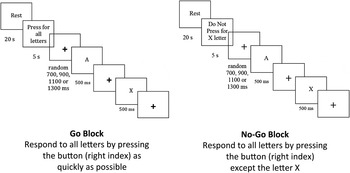
A classical version of the Go/No-Go task (Simmonds et al. Reference Simmonds, Pekar and Mostofsky2008) was implemented in E-Prime 2.0.10.182 (USA) as a measure of cognitive inhibition. Stimuli were displayed in an MRI-compatible liquid crystal display at the rear of the scanning bore, viewable via a mirror by the participant. Each task was composed of six blocks: three Go and three No-Go blocks were presented in an ABBAAB order interleaved with 20-s blank-screen resting periods and 5 s instructions prior to the start of each block. In the Go block, participants were instructed to respond to all letters (black letters on a white screen) indiscriminately by pressing the button with their right index finger as quickly as possible. In the No-Go block, participants were instructed to respond by pressing a button corresponding only to target letters (i.e. letters other than X) but not to an equally frequent non-target letter (letter X). See Fig. 1.
Representation of the Go/No-Go paradigm used in this study.

Each block (be it a Go or No-Go block) lasted 62 s and consisted of 24 trials. Each trial was composed of a fixation cross followed by a letter (target or non-target letter). The duration of the fixation cross varied between 700, 900, 1100 or 1300 ms, randomized to prevent habituation, six trials of each duration resulting in an average fixation cross duration of 1000 ms across the entire block and experiment. The target/non-target letters were always displayed for 500 ms. All blocks had 12 predetermined pseudo-randomly distributed target letters (50%) and 12 non-target letters (50%).
In all sequences, reaction times were recorded, as were omission errors (i.e. not pressing a target letter) in both conditions and commission errors (i.e. responding to a non-target letter) in the No-Go conditions. Omission scores are usually interpreted as reflecting attention abilities, while commission scores measure inhibitory processes.
Image analyses
MRI data were analysed with SPM12b (Wellcome Department of Imaging Neuroscience, UK) implemented in Matlab 2013b (Mathworks, Inc., USA). A standard indirect normalization preprocessing routine was performed: slice-timing correction for the functional time-series using sinc function, realignment of functional data to its first volume within each individual, co-registration of functional and structural images, segmentation of structural image to produce forward map Template-O-Matic (TOM) Montreal Neurological Institute (MNI) template space, spatially normalizing to MNI template, and smoothing with an isotropic 8 mm full-width half maximum Gaussian kernel. Low-frequency temporal drifts in functional MRI signal were removed by applying a high-pass filter with a cut-off of 128 s.
A first-level fixed-effect block-design model was constructed for each individual subject, including eight regressors composed of two block types (Go and No-Go). Canonical hemodynamic response function and its temporal and dispersion derivatives were used in the model. The following contrasts were conducted for the first-level fixed-effect model: Go v. No-Go, and No-Go v. baseline.
Then, we conducted three-group comparisons followed by direct pairwise comparisons by constructing separate second-level random-effect analyses (Friston et al. Reference Friston, Frith, Frackowiak and Turner1995). As these analyses yielded mixed results, we conducted a regression analysis with two factors: depression (both patient groups = 1, healthy controls = 0) and suicidal acts (suicide attempters = 1; both control groups = 0). For all analyses, statistical parametric maps were thresholded at an uncorrected voxel-wise p value of 0.001, with a minimum extent threshold of 10 voxels for exploration and visualization purposes. We then considered any clusters with family-wise error-corrected threshold of p < 0.05 as statistically significant. All results are reported using an MNI coordinate system.
Results
Clinical data (Table 1)
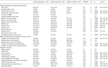
The three groups were similar in terms of gender. Healthy controls were younger than the two patient groups. As expected, they also showed lower levels of functional impairment at the CGI, lower depression (HAM-D) and anxiety (HAM-A) scores, and lower levels of impulsivity (BIS) and hostility (BDHI) than both patient groups, as well as a more infrequent history of aggression (BGLHA) and lower trait and state anxiety scores (STAI) than suicide attempters.
Comparison of sociodemographic and clinical and neuropsychological variables between suicide attempters, patient controls and healthy controls

Data are given as mean (standard deviation) unless otherwise indicated.
KW, Kruskal–Wallis; U, Mann–Whitney U; df, degrees of freedom; HC, healthy controls; SA, suicide attempters; PC, patient controls; CGI-E, Clinical Global Impressions scale, severity; HAM-D-24, 24-item Hamilton Rating Scale for Depression; MDE, major depressive episode; HAM-A, Hamilton Rating Scale for Anxiety; BIS-11, Barratt's Impulsivity Scale; BGLHA, Brown–Goodwin Assessment of Lifetime History of Aggression; BDHI, Buss–Durkee Hostility Inventory; STAI-A, Spielberger Trait Anger Inventory – State; STAI-B, Spielberger Trait Anger Inventory – Trait; SSI, Scale for Suicide Ideation; SIS, Suicidal Intent Scale; NART, National Adult Reading Test; IGT, Iowa Gambling Task; TMT, Trail Making Test.
a Analyses in 25 healthy controls.
The two patient groups did not differ significantly in terms of the age at onset of mood disorder, number of previous depressive episodes, current levels of depressive or anxiety symptoms or suicidal ideation, impulsivity trait, past psychotropic exposure and burden of medical illness. Suicide attempters had higher levels of past suicidal ideas than patient controls but no difference in terms of psychological or physical pain levels (currently or over the past 15 days).
Eight patient controls and 11 suicide attempters had never received an antidepressant medication before starting the study. For those who previously received an antidepressant just before starting the study, the washout period was 8.4 (s.d. = 2.7) days for patient controls and 6.2 (s.d. = 3.3) days for suicide attempters. None of them used fluoxetine and lithium previously. All healthy controls had no previous exposure to medications.
Suicide attempters made a mean of 1.1 (s.d. = 1.3) suicide attempts, mainly using non-violent methods: overdose medication (n = 20; 80%); drowning (n = 1; 4%); jumping (n = 1; 4%); and wrist cutting (n = 3; 12%). None necessitated surgery or intensive unit care. Suicide ideation level (SSI score) was moderate for the most severe attempt (median = 16, on a maximum score of 38). The overall level of intent for the previous attempt was moderate (total SIS median score = 22.2, on a maximum score of 42), with moderate planning scores (planning SIS median score = 5.5, on a maximum score of 16).
Cognitive performance (Table 1)
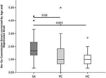
Compared with healthy and patient controls, suicide attempters made a higher number of commission errors during the Go/No-Go task with age as a covariate (Fig. 2). Reaction times were similar between groups. The mean number of omission errors during the Go and No-Go blocks was lower in healthy controls compared with both suicide attempters and patient controls, with no difference between the two patient groups. Healthy controls performed better than the two patient groups on all other cognitive measures, with no difference between suicide attempters and patient controls. The three groups had similar memory capacities and NART scores.
Number of commission errors on the Go/No-Go task between the three groups. SA, Suicide attempters; PC, patient controls; HC, healthy controls. Values are means. Boxes are number of commission errors, error bars are standard deviations and circles are outliers; each point represents a participant.

Functional imaging
Within-group analyses
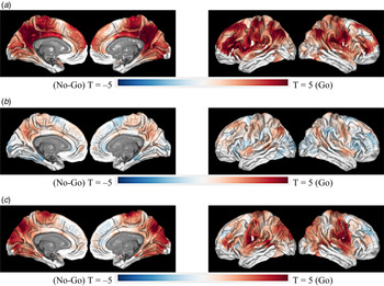
In healthy controls, contrast between Go v. No-Go conditions showed higher activation in a large network of multiple interconnected regions, including the precuneus, superior temporal gyrus, precentral gyrus, cingulate gyrus, insula and inferior frontal gyrus (Fig. 3a ), which survived whole-brain correction. The greatest peak activational differences within that large network were observed in the right medial precuneus region (p corrected < 0.001, 12297 voxels, peak Z = 5.51 at 6, −52, 14). Another cluster in the right cerebellum region also survived correction. We did not observe any significant differences in the No-Go v. Go contrast, even at a liberal threshold (p uncorrected < 0.001).
Within-group analyses for the Go v. No-Go contrasts: (a) healthy controls; (b) patient controls; (c) suicide attempters.

In patient controls, no cluster survived whole-brain correction in either the Go v. No-Go or No-Go v. Go contrast (Fig. 3b ).
In suicide attempters, the Go v. No-Go contrast showed increased activation difference in two large and four small clusters, surviving whole-brain corrections. The largest two clusters of activations were located in the precuneus/posterior cingulate gyrus (p corrected < 0.001, 1466 voxels, peak Z = 5.15 at −3, −70, 32) and the right post-central gyrus/superior temporal gyrus region (p corrected < 0.001, 730 voxels, peak Z = 4.95 at 63, −4, 32) (Fig. 3c ). The smaller clusters covered the left temporal cortex (p corrected = 0.005, 381 voxels, peak Z = 4.98 at −60, −16, 5), medial supplementary motor area (p corrected < 0.05, 307 voxels, peak Z = 4.33 at 3, −13, 56), right cerebellum (p corrected = 0.18, 266 voxels, peak Z = 4.54 at 39, −61, −37) and left putamen (p corrected < 0.05, 201 voxels, peak Z = 3.68 at −33, −16, −7). There were no significant differences in the No-Go v. Go contrasts in the suicide attempter group.
Brain activation associated with depression and suicidal acts
Direct group comparisons yielded mixed results (detailed in the online Supplementary material). Briefly, in the whole-brain Go v. No-Go contrast, suicide attempters showed reduced activation in the precuneus and posterior cingulate cortex in comparison with healthy controls. However, there was no group difference between suicide attempters and patient controls, or between patient controls and healthy controls. Yet, online Supplementary Fig. S1b suggests a subthreshold difference in brain activation between patient controls and healthy controls, with no difference between suicide attempters and patient controls, which was confirmed by the following regression analyses.
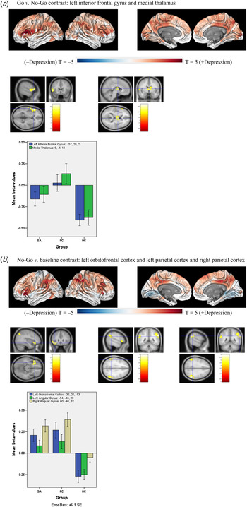
For Go v. Go-No, we observed a positive association with depression ( Fig. 4a ) in the left inferior frontal gyrus (Brodmann area 45; p corrected < 0.05, 292 voxels, peak Z = 4.59 at −57, 20, 2) and the medial thalamus (p corrected < 0.05, 196 voxels, peak Z = 4.21 at 6, −4, 11). There was no association with suicide attempt.
Brain regions showing a significant association with depression. (a) Between-group comparisons for the Go v. No-Go contrasts. (b) Between-group comparisons for the No-Go v. baseline contrasts. SA, Suicide attempters; PC, patient controls; HC, healthy controls. Values are means, with standard errors (SE) represented by vertical bars.

For No-Go v. baseline, we found a positive association with depression (Fig. 4b ) in a cluster encompassing bilateral inferior parietal lobules/angular gyrus/supramarginal regions (left: p corrected < 0.05, 200 voxels, peak Z = 3.70 at −54, −46, 29; right: p corrected < 0.05, 450 voxels, peak Z = 4.65 at 60, −46, 32) and in the left orbital part of the inferior frontal gyrus (Brodmann area 47; p corrected < 0.05, 198 voxels, peak Z = 4.08 at −36, 26, −13). Again, there was no association with suicide attempt.
Clinical correlation
For the Go v. No-Go contrast, brain activation in the left inferior frontal gyrus was correlated with levels of psychological pain (current: rs = 0.31, p < 10–2; last 15 days: rs = 0.30, p < 10–2; worst over last 15 days: rs = 0.33, p < 10–2) and CGI score (rs = 0.28, p < 0.05). Activation in the medial thalamus was correlated with levels of psychological pain (current: rs = 0.31, p < 10–2; last 15 days: rs = 0.40, p < 10–3; worst over last 15 days: r s = 0.35, p < 10–2), CGI score (rs = 0.32, p < 10–2) and current SIS score (rs = 0.26, p < 0.05).
For No-Go v. baseline, the left orbitofrontal cortex, and right and left angular gyri were both correlated with psychological pain (rs between 0.33 and 0.49, all p < 0.01) and CGI score (rs = 0.44 and 0.50, p < 10–3).
Discussion
In this study, we explored cognitive inhibition, as measured by the Go/No-Go task, in relation to suicidal vulnerability among unmedicated depressed patients. First, we found that suicide attempters exhibited an elevated number of commission errors in comparison with both control groups. However, other cognitive inhibition measures showed lower performance between both patient groups and healthy controls, but not between suicide attempters and patient controls. These latter results were confirmed by neuroimaging analyses showing a significant association between depression and brain activation during the Go/No-Go task in the inferior frontal gyrus, medial thalamus, orbitofrontal cortex and parietal cortex, but no significant association with a personal history of suicidal act. All these clusters were significantly correlated with current levels of psychological pain, suicidal ideas and global functioning. Moreover, suicide attempters did not differ from patient controls in terms of activation for any contrast at the whole-brain corrected level. Overall, these findings therefore suggest that deficits in cognitive inhibition are associated with the depressive state more than vulnerability to the suicidal act.
Efforts to interpret our findings must take into account several difficulties, notably: (1) the complexity of cognitive inhibition, what it is and how it works; (2) the complexity of the neural network underlying this function; and (3) the complexity of the Go/No-Go task itself and the processes it mobilizes. Cognitive inhibition is a generic term that encompasses a series of interactive processes. These include conflict monitoring (Garavan et al. Reference Garavan, Ross, Murphy, Roche and Stein2002; Graf et al. Reference Graf, Abler, Freudenmann, Beschoner, Schaeffeler, Spitzer, Schwab and Gron2011), error detection (Simoes-Franklin et al. Reference Simoes-Franklin, Hester, Shpaner, Foxe and Garavan2010), attention (Duann et al. Reference Duann, Ide, Luo and Li2009; Hampshire et al. Reference Hampshire, Chamberlain, Monti, Duncan and Owen2010), working memory (Mostofsky & Simmonds, Reference Mostofsky and Simmonds2008; Simmonds et al. Reference Simmonds, Pekar and Mostofsky2008), response selection and inhibition (Mostofsky & Simmonds, Reference Mostofsky and Simmonds2008; Simmonds et al. Reference Simmonds, Pekar and Mostofsky2008), task setting (Vallesi et al. Reference Vallesi, McIntosh, Alexander and Stuss2009) and the integration of bottom-up sensory information with top-down response-related information (Dodds et al. Reference Dodds, Morein-Zamir and Robbins2011). Not surprisingly, a considerable number of brain structures are involved in inhibitory control (Chikazoe, Reference Chikazoe2010; Swick et al. Reference Swick, Ashley and Turken2011). More specifically regarding the Go/No-Go task, a recent meta-analysis (Criaud & Boulinguez, Reference Criaud and Boulinguez2013) highlighted the implication of the temporo-parietal regions and the inferior, middle and superior prefrontal gyri during the No-Go condition. Many of these regions are reported in the present study. This meta-analysis also underscored the fact that, beyond response inhibition, attention and working memory are likely to play a significant role during the No-Go condition through the activation of the dorsolateral prefrontal cortex, inferior frontal gyrus and parietal regions. It is therefore not excluded that deficits reported in the present study relate less to deficits in inhibition per se, than to deficits in working memory or attention.
While we found more commission errors in suicide attempters v. both patient groups at the Go/No-Go task, we could not identify specific impairments on other cognitive inhibition tests comparing suicide attempters with patient controls. This is in line with findings from a recent meta-analysis (Richard-Devantoy et al. Reference Richard-Devantoy, Berlim and Jollant2014a ) (tests included the Hayling Test and Trail Making Test in the present study; the Trail Making Test, Wisconsin Card Sorting Test, or the Continuous Performance Task in the meta-analysis cited above). Moreover, our neuroimaging analyses suggest that several brain regions differentially activated during response inhibition may be more associated with the depressive state than with suicidal vulnerability. These regions – the inferior frontal gyrus, thalamus, orbitofrontal cortex and parietal cortex – have previously been associated with depression (see the meta-analysis by Graham et al. Reference Graham, Salimi-Khorshidi, Hagan, Walsh, Goodyer, Lennox and Suckling2013).
One hypothesis may be that, while vulnerability to suicidal acts is associated with a series of long-term (and possibly heritable) cognitive deficits, notably disadvantageous value-based decision-making and to a lesser extent a higher sensitivity to interference (as measured by the Stroop test), the acute depressive state may give rise to its own dysfunctional cognitive load, including deficits in cognitive inhibition, attention and memory (Jollant et al. Reference Jollant, Bellivier, Leboyer, Astruc, Torres, Verdier, Castelnau, Malafosse and Courtet2005; Richard-Devantoy et al. Reference Richard-Devantoy, Berlim and Jollant2014a , Reference Richard-Devantoy, Berlim and Jollant b ). The cognitive deficits accompanying the depressive state would be associated with increased risk of psychological pain (a measure correlated with suicidal ideas; Olie et al. Reference Olie, Guillaume, Jaussent, Courtet and Jollant2010), suicidal ideas and impaired functioning. These deficits would add to the cognitive deficits already present as part of the long-term vulnerability, leading to conditions of increased risk of suicidal acts. This is somewhat supported by recent findings showing risky decision-making but normal cognitive control and memory in non-depressed relatives of suicide completers who never attempted suicide (Hoehne et al. Reference Hoehne, Richard-Devantoy, Ding, Turecki and Jollant2015).
Several limitations should be underlined. First, although the number of participants was large for a neuropsychological/neuroimaging study, the size of the groups and the use of non-parametric tests – which are more robust but less sensitive than parametric tests – served to limit the statistical power of some comparisons. Replication in larger groups is required to validate these results. Moreover, while participants were not medicated, many patients had stopped taking their previous medication 1 week prior to the study, which may have modified the response in some brain structures. Finally, because suicide attempters and patient controls represent heterogeneous groups, there is a risk of variable findings based on sample selection. For instance, our sample of suicide attempters did not use violent suicidal means, and level of suicidal intent were scored moderate. This may explain the lack of difference in IGT performance between suicide attempters and patient controls. Findings may be different in different subgroups of attempters as it has been shown with risky decision-making (Gorlyn et al. Reference Gorlyn, Keilp, Oquendo, Burke and Mann2013).
In conclusion, our findings suggest that deficits in cognitive inhibition, related to the inferior frontal gyrus, thalamus, orbitofrontal cortex and parietal cortex, are associated with the depressive state more than vulnerability to suicidal behavior. Deficits in cognitive inhibition may nonetheless add up to trait-like cognitive alterations to increase the risk of suicidal acts. Future research should therefore identify state and trait cognitive alterations, as improvements to these two kinds of deficits may necessitate different strategies.
Supplementary material
For supplementary material accompanying this paper visit http://dx.doi.org/10.1017/S0033291715002421
Acknowledgements
This study was funded thanks to an operating grant received by F.J. from the Canadian Institutes of Health Research (CIHR). S.R.-D. received a CIHR fellowship grant during the course of this study. F.J. held a ‘chercheur-boursier clinicien’ salary grant from the Fond de Recherche du Québec – Santé (FRQS) and Y.D. a FRQS Ph.D. grant at the time of the study.
Declaration of Interest
None.