Liver transplantation is an effective therapy for liver dysfunction caused by Alagille syndrome.Reference Cardona, Houssin, Gauthier, Devictor, Losay, Hadchouel and Bernard1 Echocardiography often detects bilateral pulmonary arteries stenosis and sometimes an atrial septal defect in these patients.Reference Acosta Martínez, López-Herrera Rodríguez, González Rubio and López Romero2 During liver transplantation, a lot of blood influx the right atrium, resulting in a temporary increase in pressure of right atrium, causing embolism.Reference Starzl, Schneck and Mazzoni3 Recent studies show that a small atrial septal defect does not increase the risk of cerebral embolism during liver transplantation significantly.Reference Werlang, Palmer and Boyd4 However, in patients with bilateral pulmonary arteries stenosis and atrial septal defect, right atrium pressure may increase and excess left atrium pressure unexpectedly, thereby increasing the risk of embolism during reperfusion of the donor liver graft.
Case presentation
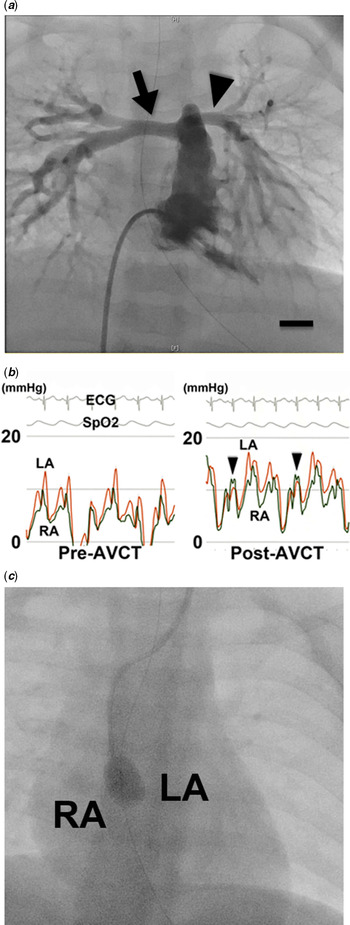
A 9-month-old boy diagnosed with Alagille syndrome was referred to our hospital for liver transplantation. Pre-operative echocardiography revealed that he had peripheral pulmonary artery stenosis with a 4.0 mm atrial septal defect and good right ventricular function. Angiography revealed bilateral peripheral pulmonary stenosis as shown in Figure 1a. Assessed by a cardiac catheter examination, right atrium and ventricular pressures values were measured as right atrium mean pressure 4 mmHg, right ventricular systolic pressure 50 mmHg, and right ventricular end-diastolic pressure 8 mmHg. These congenital heart conditions were not indicative of cardiac surgery before liver transplantation. Although the small defect is not contraindication for liver transplantation, the combination of atrial septal defect and bilateral peripheral pulmonary artery stenosis called into question the safety of liver transplantation. This is because right atrium pressure easily exceeds left atrium pressure, and the excess pressure may lead to systemic embolism. Therefore, we performed cardiac catheter examinations to compare the right and left atrium pressures simultaneously. At rest under anaesthesia, the pressure in the right atrium was lower than that in the left atrium. However, after an acute volume challenge test with natural saline (15 mL/kg/dose), there was a short time when right atrium pressure exceeded left atrium pressureReference Tan, Markowitz and Montgomery5 (Fig 1b). Therefore, temporal atrial septal defect occlusion was considered during reperfusion of donor liver. A 4 Fr balloon catheter was used for the defect occlusion (Fig 1c). The occlusion was performed just before reperfusion of the donor graft liver and maintained until the elevated central venous pressure fell to a level which it was. After liver transplantation, no neurological symptom and sign was detected in physical, laboratory, and echo examinations.
(a) A contrast images of the right ventricle and pulmonary arteries. Arrows indicate right and left pulmonary arteries. Bar: 10 mm. (b) Right atrium and left atrium pressure traces before (left panel) and after (right panel) acute volume challenge test. Arrowheads indicate the moments when the right atrium pressure is markedly higher than left atrium pressure. RA pressure curve: black, LA pressure curve: red. ECG: electrocardiogram, SpO2: saturation of percutaneous oxygen. (c) Temporal atrial septal defect occlusion was performed using a balloon catheter.

Discussion
Recently, liver transplantation has become more common in young children; thus, atrial septal defect is sometimes detected by pre-operative echocardiography.Reference Acosta Martínez, López-Herrera Rodríguez, González Rubio and López Romero2 Although some previous papers mentioned risks of brain infarction and other vascular embolism for patients with atrial septal defect during liver transplantation,Reference Starzl, Schneck and Mazzoni3 recent reports reveal that the defect did not increase the risk at all.Reference Werlang, Palmer and Boyd4 However, atrial septal defect patients with bilateral peripheral pulmonary artery stenosis tend to have elevated right atrium pressure, making right to left atrium shunts more likely. To these patients, atrial septal defect closure and/or bilateral pulmonary artery plasty were recommended precede to liver transplantation.Reference Harris, Cao, Waight and Hijazi6 But, for patients without operative indications, there is no evidence-based protocol for liver transplantation. To validate the occurrence of a right atrium–left atrium shunt during liver transplantation, we performed the acute volume challenge test for the patient.Reference Tan, Markowitz and Montgomery5 During the test, there were short moments in which right atrium pressure exceeded left atrium pressure. This could increase the risk of embolism. Therefore, temporal atrial septal defect occlusion using a balloon catheter was performed during liver transplantation. Successful defect closure did not interfere with the operation and neither patient suffered embolism.
In conclusion, temporal atrial septal defect occlusion during liver transplantation is a simple protocol for avoiding embolism for patients with atrial septal defect and bilateral peripheral pulmonary artery stenosis.
Financial support
This research received no specific grant from any funding agency, commercial or not-for-profit sectors.
Conflicts of interest
None.
Ethical standards
Helsinki’s declaration was compliant.