Protein synthesis requires protein synthesis machinery components and availability of amino acids (AAs) as well as the supply of energy (Bionaz et al., Reference Bionaz, Hurley, Loor and Hurley2012). The availability of AA is important for protein synthesis (Griinari et al., Reference Griinari, McGuire, Dwyer, Bauman, Barbano and House1997; Doepel et al., Reference Doepel, Pacheco, Kennelly, Hanigan, Lopez and Lapierre2004). AAs are anabolic factors that can induce protein gain by stimulating gene expression and mRNA translation (Lu et al., Reference Lu, Li, Huang and Gao2013) through mTOR pathway (Bionaz et al., Reference Bionaz, Hurley, Loor and Hurley2012). mTOR is a serine/threonine protein kinase which is a central regulator of cellular metabolism, growth and survival in response to hormones, growth factors, nutrients, energy and stress signals, that ultimately affects the expression of genes related to milk protein synthesis (Bruhat et al., Reference Bruhat, Jousse, Wang, Ron, Ferrara and Fafournoux1997; Deval et al., Reference Deval, Chaveroux, Maurin, Cherasse, Parry, Carraro, Milenkovic, Ferrara, Bruhat, Jousse and Fafournoux2009). Additionally, AAs are ‘building blocks’ of proteins. Many past studies have shown that AA can directly regulate the expression of genes related to protein synthesis through nutrient/gene interaction (Jousse et al., Reference Jousse, Averous, Bruhat, Carraro, Mordier and Fafournoux2004; Deval et al., Reference Deval, Chaveroux, Maurin, Cherasse, Parry, Carraro, Milenkovic, Ferrara, Bruhat, Jousse and Fafournoux2009). Nan et al. (Reference Nan, Bu, Li, Wang, Wei, Hu, Zhou and Loor2014) have found that adding supplemental Met and Lys into bovine mammary epithelial cells using in vitro experiment design has a strong nutrigenomic effect by significantly increasing expression of genes coding for major milk protein components (i.e. casein) as well as genes related to protein translation (ELF5, MTOR) and JAK-STAT signaling (JAK2, STAT5).
In general, Met is considered as a limiting AA for protein production in lactating dairy cows (National Research Council, 2001). Met is also an essential AA that has a major role in protein synthesis and several other biochemical and cellular processes (Orellana et al., Reference Orellana, Jeyapalan, Escobar, Frank, Nguyen, Suryawan and Davis2007; Metayer et al., Reference Metayer, Seiliez, Collin, Duchene, Mercier, Geraert and Tesseraud2008).
In addition, acetate is the chief source of energy for mammary gland of ruminant animals (Sabine and Johnson, Reference Sabine and Johnson1963). The positive effect of dietary energy on protein synthesis is partly due to the production of intracellular energy transfer molecules such as ATP, GTP, NADH, and NADPH in the presence of energetic precursors (Appuhamy et al., Reference Appuhamy, Knoebel, Nayananjalie, Escobar and Hanigan2012; Bionaz et al., Reference Bionaz, Hurley, Loor and Hurley2012). Another important function of energy, specifically glucose, is through increased secretion of insulin due to greater dietary energy. Schmidt (Reference Schmidt1966) has shown a positive role of insulin in mammalian protein synthesis.
N-Acetyl-L-Methionine (CMA) is a rumen-protected methionine in which L-Met is conjugated with acetate. It can be separated into L-Met and acetate in small intestine or liver. It has been shown that CMA itself is more effective than L-Met alone in mammalian synthesis because CMA has higher bioavailability in ruminants after being broken down in the intestine and in the liver (Fagundes et al., Reference Fagundes, Blaser, Yang, Eun and Moon2016; Toledo et al., Reference Toledo, Baez, Garcia-Guerra, Lobos, Guenther and Trevisol2017). In general, rumen-protected methionine (RPM) has been developed as a product that can significantly increase protein content in milk (Rulquin and Delaby, Reference Rulquin and Delaby1997; Socha et al., Reference Socha, Putnam, Garthwaite, Whitehouse, Kierstead, Schwab, Ducharme and Robert2005). In most studies, protein content is increased when RPM is supplemented (Chilliard and Doreau, Reference Chilliard and Doreau1997; Rulquin and Delaby, Reference Rulquin and Delaby1997; Kowalski et al., Reference Kowalski, Pisulewski and Gorgulu2003; Leonardi et al., Reference Leonardi, Stevenson and Armentano2003; Berthiaume et al., Reference Berthiaume, Thivierge, Patton, Dubreull, Stevenson, McBride and Lapierre2006). However, CMA and non-conjugated L-Met + Acetate (NMA) as digested forms of CMA have not been yet studied for their milk protein synthesis efficiency in mammary epithelial cells and regulation of mechanism on milk protein synthesis. We hypothesized that the addition of limiting amino acid L-Met together with the energy source, acetate, may cause the increase of protein synthesis by stimulating genes and proteins involved in the protein synthesis process.
Therefore, the objectives of this study were: (1) to compare the efficacy of L-Met, conjugated L-Met and acetate (CMA) and non-conjugated form (NMA; digested form of CMA), on protein synthesis using immortalized bovine mammary epithelial cell line (MAC-T cells), and (2) to elucidate the mechanism of milk protein synthesis (i.e. CSN2).
Materials and methods
Amino acid dosage and sampling time
Immortalized mammary epithelial cell line (MAC-T cells) (McGill University, Canada) were grown in 10-cm dishes (TPP, Trasadingen, Switzerland). These cells were then seeded into BD Falcon™ 6-well multiwall plate (Franklin Lakes, NJ, USA). They were cultured in DMEM/F12 basic medium (Thermo Scientific, South Logan, UT, USA) supplemented with 10% fetal bovine serum (FBS), 100 units/ml penicillin/streptomycin (Thermo Scientific, South Logan, UT, USA), 5 μg/ml insulin, 1 μg/ml hydrocortisone, and 50 μg/ml gentamycin (Sigma-Aldrich Corp., St. Louis, MO, USA) at 37 °C in a 5% CO2 incubator (Wang et al., Reference Wang, Lim, Bok, Kim, Kang, Lee, Hwang, Lee, Kang, Choi, Kim and Lee2014, Reference Wang, Lee, Hwang, Lim, Jung, Kim, Kang, Choi, Lee, Roh and Lee2015).
Lactogenic DMEM/F12 differentiation medium (without FBS) was used to differentiate MAC-T cells into CSN2 secreting cells for 72 h. This medium contained 5 μg/ml bovine insulin, 1 μg/ml hydrocortisone, 100 units/ml penicillin/streptomycin, 50 μg/ml gentamycin, and 5 μg/ml prolactin (Sigma-Aldrich Corp., St. Louis, MO, USA) (Wang et al., Reference Wang, Lim, Bok, Kim, Kang, Lee, Hwang, Lee, Kang, Choi, Kim and Lee2014, Reference Wang, Lee, Hwang, Lim, Jung, Kim, Kang, Choi, Lee, Roh and Lee2015). The AA profile of this medium was as follows: Arg, Cys, Gln, Gly, His, Ile, Leu, Lys, Met, Phe, Ser, Thr, Trp, Tyr, and Val at 0.70, 0.10, 2.5, 0.25, 0.15, 0.42, 0.45, 0.50, 0.12, 0.22, 0.25, 0.45, 0.04, 0.21, and 0.45 mm, respectively. When MAC-T cells reached 90% confluence, DMEM/F12 basic medium was replaced with lactogenic DMEM/F12 differentiation medium.
A preliminary study was conducted to determine the effects of different concentrations of supplemental Met and culture time on CSN2 mRNA expression. Additional L-Met (0 as control, 0.3, 0.6, 0.9 mm) was added to lactogenic medium without FBS. Cells were harvested at 24 h after treatment. To evaluate the effect of culture time of supplementing methionine on CSN2 secretion of MAC-T cells, the cells were harvested at 0, 12, 24, 36, 48, 72, 96, and 120 h after incubated in lactogenic medium. Each treatment was performed with six replicates. Resultant data were used to determine the best concentration and culture time for supplemental Met treatments. After the preliminary study, the determined best dosage and time was used uniformly for all treatments: L-Met conjugated L-Met and acetate (CMA) (CJ CheilJedang Corp., Suwon, Republic of Korea) and non-conjugated L-Met and acetate (NMA) (Sigma-Aldrich Corp., St. Louis, MO, USA). Each treatment was assigned with six replicates.
RNA extraction and cDNA synthesis
Total RNA was extracted from MAC-T cells using TRIzol® (Life Technologies Corporation, Carlsbad, CA, USA). RNA quality and quantity were measured using NanoDrop 1000® Spectrophotometer with RNA-40 module (Thermo Fisher Scientific, Wilmington, DE, USA). Then cDNA was prepared using iScript cDNA synthesis kit (Bio-Rad Laboratories, Inc., Foster City, CA, USA) according to the manufacturer's instructions. After incubating at 25 °C for 5 min, 42 °C for 30 min, and 85 °C for 5 min, the cDNA was quantified by using ssDNA-33 module of Thermo NanoDrop 1000® Spectrophotometer (Thermo Fisher Scientific, Wilmington, DE, USA).
Real-time PCR
Real-Time PCR (RT-PCR) analysis was performed using T100™ Thermal Cycler System. ACTB was used as a reference gene. Validated RT-PCR oligonucleotide primer sequences of forward and reverse primers specific for target genes were as follows: CSN2 Forward, 5′-AAATCTGCACCTTCCTCTGC-3′; CSN2 Reverse, 5′-GAACAGGCAGGACTTTGGAC-3′; ACTB Forward, 5′-GCATGGAATCCTGCGGC-3′; ACTB Reverse, 5′-GTAGAGGTCCTTGCGGATGT-3′. RT-PCR reactions were performed by initial incubation at 95 °C for 3 min followed by 50 cycles of denaturation at 95 °C for 10 s, annealing at specific temperature for 15 s (bovine CSN2 at 55 °C), and extension at 72 °C for 30 s. RT-PCR analysis was computed using the threshold cycle (2-ΔΔCT method, Livak and Schmittgen, Reference Livak and Schmittgen2001) as a convenient way to analyze relative gene expression changes from real-time quantitative PCR experiments. Relative quantification of expression levels of target genes in a treatment group was compared to the untreated group.
Protein extraction and quantification
After incubation in the treatment medium (lactogenic medium plus L-Met), conjugated L-Met and acetate (CMA) (CJ CheilJedang Corp., Suwon, Republic of Korea) and non-conjugated L-Met and acetate (NMA) (Sigma-Aldrich Corp., St. Louis, MO, USA) for 72 h, the culture medium was collected from adherent cells for protein quantity determination. Cells were washed twice with ice cold 1 × PBS and then 200 μl cell lysis buffer containing 10 mm Tris/HCl, pH 8.3, 8 m urea, 5 mm EDTA, 4% CHAPS, and 1 × protease inhibitors cocktail (GE Healthcare, Piscataway, NJ, USA) were added. The cell lysates were incubated at room temperature for 30 min and centrifuged at 21 952 × g/30 min at 20 °C. Also, the culture medium centrifuged at 300 × g/5 min at 4 °C. The supernatant of cell and medium was transferred to a new tube for protein quantification using Pierce BCA Protein Assay Kit (Pierce Biotechnology, Inc., Rockford, IL, USA) according to the manufacturer's instruction. Amino acid profile was analyzed via UPLC/Q-TOF-MS. LC performed using the Acquity UPLC system (Waters) with an Acquity HSS T3 column (1.8 μm, 2.1 × 150 mm; Waters).
Proteomic analysis
Upon completion of cell culture, cellular proteins were extracted by using cell lysis buffer containing 20 mm Tris, 10 mm KCl, 1.5 mm MgCl2, 0.5 mm EDTA, 0.1% SDS, protease inhibitor (Complete EDTA-free, Roche) after washing twice with ice cold 1 × PBS. The cell lysates were incubated at 4 °C for 30 min and centrifuged at 13 000 × g/10 min at 4 °C. In proteome analysis, 100 μg of cell lysate proteins were resuspended in 0.1% SDS in 50 mm triethyl ammonium bicarbonate (TEABC, pH 8.0) according to the procedure described by Ishihama, et al. (Reference Ishihama, Oda, Tabata, Sato, Nagasu, Rappsilber and Mann2005). Digested peptides were desalted and concentrated and then subjected to liquid chromatography (LC)–tandem mass spectrometry (MS/MS) analysis. Total peptides were analyzed by nano-ultra-performance LC-MS/electrospray ionization quadrupole time-of-flight (UPLC-MS/ESI-Q-TOF) (Waters, Manchester, UK). LC peptide separation was performed using the nano Acquity system equipped with a Symmetry C18 5 μm, 5 mm × 300 μm pre-column and CSH C18 1.7 μm, 25 cm × 75 μm analytical column (Waters).
Metabolomic analysis
After finishing the cell culture, the culture medium was removed from the adherent cells, and the cells were used for metabolite quantity determination as described by Park et al. (Reference Park, Yun, Lee, Park and Lee2012).
Statistical analysis
Statistical analysis (protein quantification data, n = 6; proteomics and metabolomics data, n = 3; quantitative real-time PCR data, n = 6) were tested for significance using SPSS statistic software (SPSS Inc., Chicago, IL, USA). Data were analyzed using one-way analysis of variance (one-way ANOVA) and differences among treatments were determined using Duncan Multiple Range Test (DMRT). Mean difference was considered statistically significant at P < 0.05. The experimental model was:
 $$Y_{ijk} = {\rm \mu } + {\rm \tau }_i + {\rm \varepsilon }_{ijk}\comma \;$$
where μ is the grand mean, τi is the Met type effect and ɛijk is the error variability.
Up-regulated or down-regulated proteins were detected significantly using semi-quantification relative ratio (the ratio was ≧2 or ≦0.5) and detected proteins were analyzed by using web site program (http://www.pantherdb.org) for the pathway analysis (Bos taurus). Also, differentially expressed metabolites were detected statistically and P < 0.05 is considered significantly different using Dunnett's test which is a multiple comparison procedure to compare each of a number of treatments with a single control (P < 0.05). Detected metabolites were analyzed by using Metabo Analyst 3.0 program for the pathway analysis (Bos taurus).
Results
There was a linear effect of L-Met on the expression of CSN2 until 0.6 mm. No further increase was observed when L-Met level was further increased to 0.9 mm (online Supplemental Fig. S1). With control at 0 mm level, the best incubation time (P < 0.05) for CSN2 mRNA expression level was found to be 72 h (online Supplemental Fig. S2). Beyond 72 h, no significant difference in CSN2 mRNA expression was observed (online Supplemental Fig. S2).
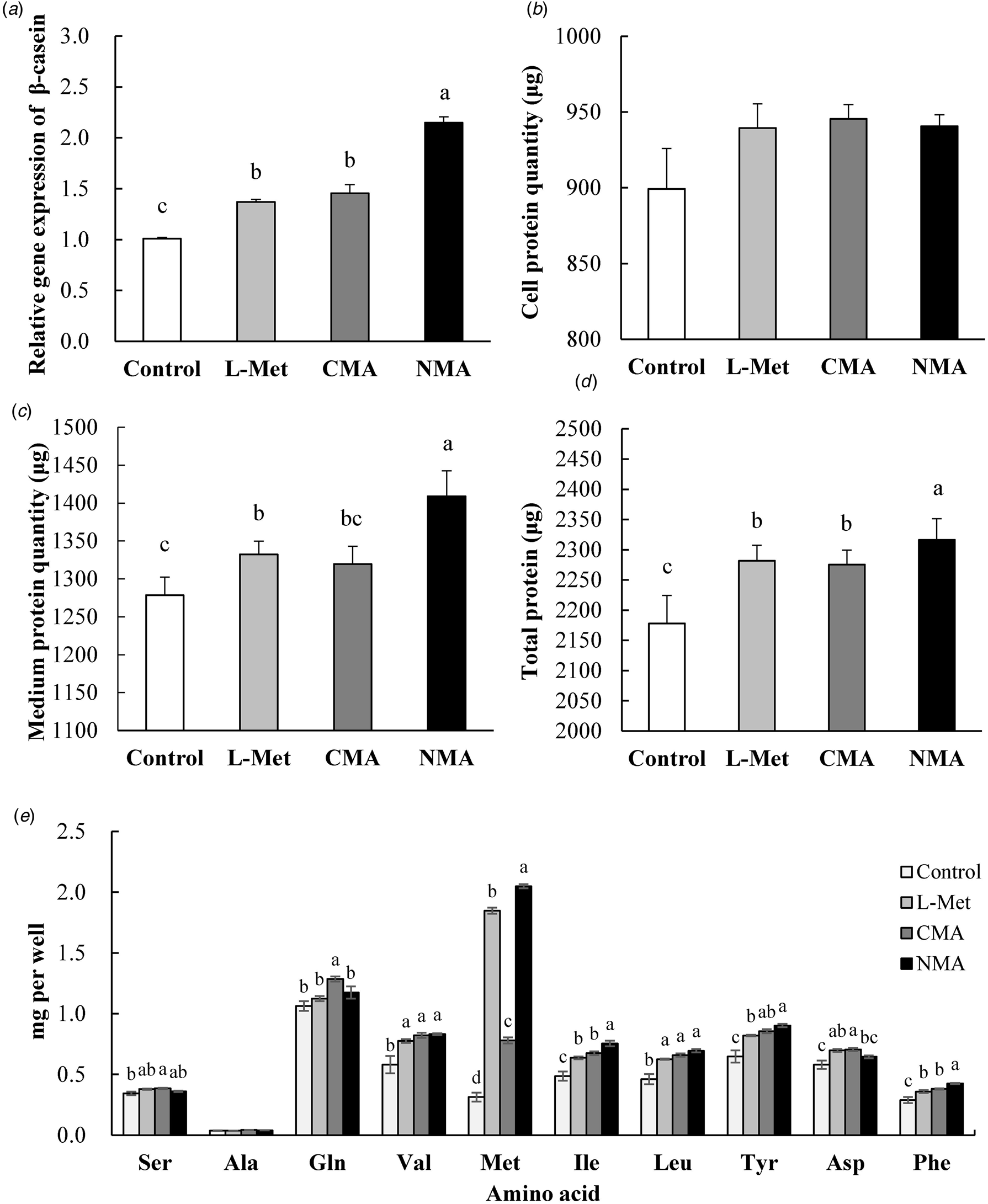
The following are the results obtained by using the optimal dosage and optimal time in MAC-T cells in the above-mentioned experiment. Upon addition of different forms of L-Met, NMA group showed the highest CSN2 mRNA relative expression followed by CMA and L-Met (Fig. 1a). This shows that NMA is the most efficient in stimulating CSN2 gene expression. Although there was no significant differences among treatments in cell protein content upon addition of different forms of L-Met in MAC-T cells, all the treatments increased protein synthesis numerically and has the tendency to increase (P < 0.10) (Fig. 1b).
CSN2 mRNA expression level (a), cell protein quantity (b), medium protein quantity (c), total protein quantity (cell and medium) (d) and amino acid quantity (e) in MAC-T cells incubated with control (0 mm), L-methionine (0.6 mm), CMA (0.6 mm) and NMA (0.6 mm). Values are expressed as means ± se (n = 6 per group) and a–c indicate significant differences at P < 0.05 by Duncan Multiple Range Test (DMRT).

Protein synthesis in culture medium with NMA treatment showed the highest protein synthesis rate (P < 0.05). L-Met group had the second highest level of protein synthesis rate compared to the control (P < 0.05) but CMA only tended to increase (P < 0.10) (Fig. 1c). On the other hand, total protein synthesis with NMA treatment showed the highest protein synthesis quantity (P < 0.05). L-Met and CMA had the second highest levels of protein synthesis quantity and control is the lowest (P < 0.05) (Fig. 1d). NMA had the highest protein synthesis quantity in overall total protein synthesis combining the protein synthesis in cells and in the culture medium.
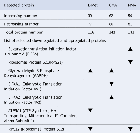
A total of 39 upregulated and 77 downregulated proteins, 62 upregulated and 80 downregulated proteins, 50 upregulated and 81 downregulated proteins were observed in L-Met, CMA, and NMA treated groups, respectively (Table 1) using semi-quantification relative ratio (≧2 or ≦0.5). In addition, several proteins involved in protein and energy metabolism were examined due to the effects of experimental treatments. Eukaryotic Translation Initiation Factor 3 Subunit A (EIF3A) was upregulated by NMA while EIF4A2 (Eukaryotic Translation Initiation Factor 4A2) was upregulated by L-Met. Glyceraldehyde-3-Phosphate Dehydrogenase (GAPDH) was upregulated by supplementing both L-Met and CMA. Upregulation of EIF3A by NMA and EIF4A2 by L-Met are important for stimulating protein synthesis because these proteins are important transcription factors for initiation of protein translation.
List of differently expressed proteins in MAC-T cells when supplemented with different forms of Methionine (Met) in comparison to control

L-Met, L-Methionine; CMA, conjugated L-Methionine and acetate; NMA, non-conjugated L-Methionine and acetate.
Selection Criteria:
▲ Upregulated: >2-fold in protein expression vs. Control
▼ Downregulated: <0.5-fold in protein expression vs. Control.
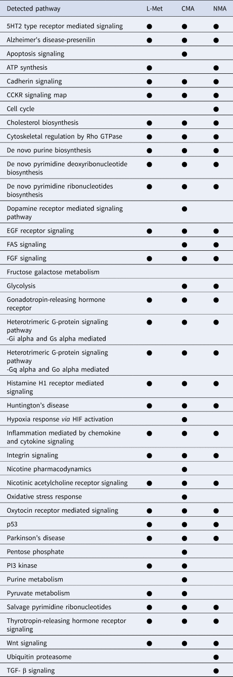
Protein pathway analysis revealed that NMA treatment, but not CMA treatment, stimulated ATP synthesis, cell cycle, ubiquitin proteasome, and TGF-β signaling pathways (Table 2). On the other hand, CMA and L-Met treatments stimulated PI3 kinase and pyruvate metabolism pathways. In case of CMA, there were increases in the proteins related to energy synthesis like glyceraldehyde-3-phosphate dehydrogenase (GAPDH) and H+ transporting, mitochondrial F1 complex, ATP synthase, alpha subunit 1 (ATP5A1) and also the stimulation of pentose phosphate pathway. As mentioned earlier, due to conjugation of acetate to L-Met, the acetate source cannot be utilized for energy source. However, it is important to note that the DMEM/F12 used (Cat# 11320033, Sigma-Aldrich Corp., St. Louis, MO, USA) contains 17.5 mm glucose which is higher than the normal glucose level in cows' blood which is around 3–5 mm glucose, implying that energy should be sufficient. In the case of NMA, it stimulated proteins related to protein synthesis like eukaryotic translation initiation factor 3 subunit A (EIF3A) and elongation factor 1-alpha 1 (EEF1A1) but, surprisingly, it decreased ribosomal protein S21 (RPS21) and ribosomal protein S12 (RPS21). There was also an increase in the glycolysis pathway in proportion to the increased protein synthesis. One interesting result was the increased cholesterol biosynthesis in NMA group due to availability of acetate from unconjugated form of acetate (Table 2).
List of all protein pathways affected by supplementation of different forms of Methionine (Met) in comparison to control

L-Met, L-methionine; CMA, conjugated L-methionine and acetate; NMA, non-conjugated L-methionine and acetate.
● Pathway analysis (Bos taurus). The analysis used increasing detected pathway at P < 0.05.
Intracellular amino acid levels in MAC-T cells were also analyzed (Fig. 1e). Supplementing NMA gave the highest increased in intracellular Met level. Aside from increasing Met intracellular level, supplementing L-Met, CMA and NMA treatments increased other amino acids such as Val, Ile, Leu, Tyr and Phe at the same time.
Based on metabolite pathway analysis, CMA supplementation stimulated Ala-Asp-Glu, aminoacyl-tRNA biosynthesis, and glyoxylate and dicarboxylate metabolism pathway. However, NMA, but not CMA, stimulated inositol phosphate metabolism and pentose phosphate metabolic pathway (online Supplemental Table S2).
Results of metabolite analysis also showed that CMA treatment caused increases of 15 metabolites (P < 0.05) (online Supplemental Table S3). On the other hand, NMA treatment caused increases of 15 metabolites and decreases of IMP and NAD+ (P < 0.05). Supplementation of L-Met stimulated the production of energy metabolites such as pyruvate, malate, and fumarate well-known as intermediates of Krebs cycle. On the other hand, CMA supplementation caused increases of energy metabolites such as malate, and xylulose-5-phosphate, an intermediate of pentose phosphate pathway. In the case of NMA supplementation, glucose intermediates (such as glucose-6-phosphate, fructose-6-phosphate, and glucose-1-phosphate) and pentose phosphate pathway intermediates (xylulose-5-phosphate and N-acetyl-L-glutamate) were increased.
Looking at the detected metabolites, there was a decrease in Met and acetate-glutamate level inside the cell of CMA compared to L-Met and NMA group which means that CMA might not be properly absorbed due to their larger conjugated size (online Supplemental Table S3). On the other hand, in response to NMA there was an increase in Met as well as other amino acids like valine, isoleucine, leucine, tyrosine and phenylalanine. The increase in Met and acetate-glutamate level in NMA but not in CMA may imply that Met as well as acetate-glutamate are readily available or properly absorbed inside the cell via the plasma membrane amino acid transport systems in their unconjugated form.
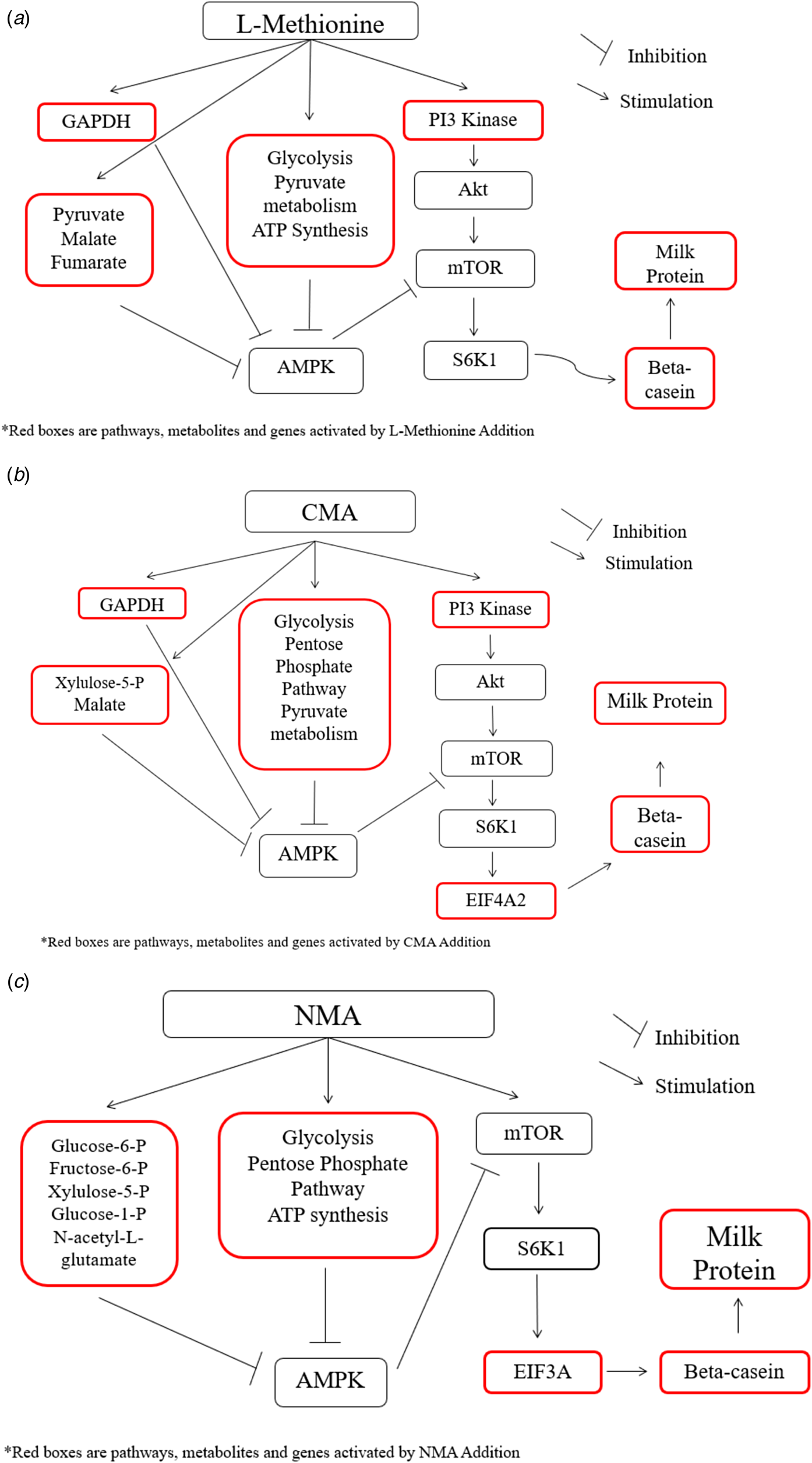
To summarize interactions of the addition of different forms of Met in MAC-T cells, diagrams were created to demonstrate different relationships among various components (Fig. 2). L-Met supplementation stimulated PI3 kinase and GAPDH as well as glycolysis, pyruvate metabolism, and ATP synthesis pathways. Metabolites such as pyruvate, malate, and fumarate were also stimulated. They are involved in energy production and metabolism (Fig. 1a). This stimulation may promote stimulation of mTOR activity that may elevate CSN2 expression that may eventually increase the protein synthesis.
Diagram showing the effects of L-Methionine (a), conjugated L-Met and acetate (CMA) (b) and non-conjugated L-Met and Acetate (NMA) (c) on protein synthesis pathway.

On the other hand, the addition of CMA stimulated PI3 kinase and GAPDH enzymes, EIF4A2 protein, glycolysis, pentose phosphate and pyruvate metabolism pathways, and xylulose-5-phosphate and malate metabolites (Fig. 2b), all of which may promote mTOR and CSN2 gene expression and, thus, increase protein synthesis.
Lastly, addition of NMA stimulated EIF3A protein, glycolysis, pentose phosphate, and ATP synthesis pathways as well glucose-6-phosphate, fructose-6-phosphate, xylulose-5-phosphate, glucose-1-phosphate, and N-acetyl-L-glutamate (Fig. 2c), all of which increased CSN2 gene expression resulting to increased protein synthesis. One difference that we can observe between the addition of conjugated form of CMA and non-conjugated form NMA is that pyruvate metabolism and PI3 kinase are activated in CMA, but not in NMA. When pyruvate metabolism and PI3 kinase are activated, cells are expected to produce energy from acetate (in case of pyruvate metabolism) and increase glucose utilization. This indicates that the conjugated form of L-Met and acetate (CMA) cannot utilize energy from acetate in vitro. However, the situation will be different in vivo because animals have enzymes (mainly in the intestine but also in the liver) that can cleave the conjugated form into its unconjugated form.
Discussion
L-Met dosage test showed that 0.6 mm level was the most effective concentration in increasing CSN2 mRNA expression indicating that 0.6 mm would be the optimal level. This level is in accordance with the result reported by Limin et al. (Reference Limin, Xuejun, Qingzhang, Jianguo, Rong and Huiming2012), showing that 0.6 mm L-Met is the best concentration to increase CSN2 protein expression. In addition, the best time when protein synthesis had the highest level coincided at the point where CSN2 mRNA expression was also at the highest level. This outcome suggests that 72 h should be selected to test different L-Met types because of its efficacy in increasing CSN2 mRNA expression and protein synthesis in MAC-T cells.
In general, protein synthesis is increased by the addition of different forms of L-Met especially in the culture medium where CSN2 protein was secreted outside the cell. The availability of amino acids for mammary epithelial cells is not only important for the regulation of translation, but also important for the transport rate of amino acids as one of the major limiting factors for protein synthesis (Baumrucker, Reference Baumrucker1985; Reynolds et al., Reference Reynolds, Harmon and Cecava1994; Shennan and Peaker, Reference Shennan and Peaker2000).
It is known that energy availability is also a crucial factor for the synthesis of other milk components, especially milk proteins (Reynolds et al., Reference Reynolds, Harmon and Cecava1994). Thus, acetate was also added as experimental treatment. NMA group showed higher protein synthesis than CMA conjugated group although both treatments contained the same components. The only difference was their conjugation form. This was validated by the analysis of metabolic pathway wherein based on the analysis, CMA treatment stimulated pyruvate metabolism pathway, indicating that acetate group conjugated in CMA could not be utilized as an energy source in MAC-T cells. Consequently, cells have to produce energy via the pyruvate pathway (Pietrocola et al., Reference Pietrocola, Galluzzi, Bravo-San Pedro, Madeo and Kroemer2015). Not cleaving L-Met and acetate into separate groups could cause some problem in the incorporation of L-Met into elongating peptides because ribosomes can discriminate amino acid structure within its peptidyl-transferase center (Englander et al., Reference Englander, Avinsb, Fleisherb, Liuc, Effraima, Wang, Schultenc, Leyhf, Gonzalez and Cornishb2015). That is, if the structure of an amino acid is different from its original form (in this case, it is conjugated with acetate), the ribosome cannot bind to the amino acid and will not be able to use the amino acid for protein elongation. Pyruvate is a precursor for the synthesis of acetate. In the case of NMA, enough or excess availability of acetate might cause feedback inhibition of pyruvate dehydrogenase which is the enzyme responsible for converting pyruvate into acetyl CoA and CO2 and eventually inhibiting the pyruvate pathway (Pietrocola et al., Reference Pietrocola, Galluzzi, Bravo-San Pedro, Madeo and Kroemer2015).
GAPDH enzyme translation was stimulated upon addition of L-Met and CMA but not in NMA. This enzyme stimulates the important energy-yielding process of carbohydrate metabolism during glycolysis and the reversible oxidative phosphorylation of the glyceraldehyde-3-phosphate in the availability of nicotinamide adenine dinucleotide and inorganic phosphate (NAD). The stimulation of GAPDH in L-Met or CMA group suggests that there is enough available energy supplied by acetate in NMA group so that there may be no need to produce more energy via the glycolysis pathway (Nicholls et al., Reference Nicholls, Li and Liu2012).
CMA and L-Met treatments stimulated pyruvate metabolism pathway indicating that acetate group conjugated in CMA could not be utilized as an energy source in MAC-T cells, consequently resulting for cells to produce energy via pyruvate pathway. Additional supply of acetate would cause feedback inhibition of pyruvate dehydrogenase enzyme which is responsible for converting pyruvate into acetyl CoA and CO2 which will eventually inhibit the pyruvate pathway. Upon addition of different forms of L-Met, the CMA group has a sharp decrease in terms of intracellular Met level compared to NMA group (P < 0.05). This could mean that the conjugated form of L-Met has low absorption into the cell probably due to its larger size.
Most pathway detection metabolites are involved in the metabolism of energy and amino acids. Such results are expected since the treatments are the addition of amino acid L-Met and acetate as energy source. Addition of different forms of methionine (L-Met, CMA and NMA) stimulated the production of intermediate metabolites for energy production (Fig. 2a–c). Previous researches have shown that energy intermediates (e.g. ATP, NADH) could stimulate protein synthesis indirectly by inhibiting AMPK signal (Sarbassov et al., Reference Sarbassov, Ali and Sabatini2005; Appuhamy et al., Reference Appuhamy, Knoebel, Nayananjalie, Escobar and Hanigan2012), a known direct inhibitor of mTOR pathway. The adenosine monophosphate kinase (AMPK) complex is a phylogenetically conserved fuel-sensing enzyme which is present in all mammalian cells (Richter and Ruderman, Reference Richter and Ruderman2009). AMPK may modulate the decrease in energy status, consequently inhibiting the very high energy consuming protein synthetic process (Proud, Reference Proud2007).
In conclusion, results of this study showed that NMA-treated group exhibited much better performance in stimulating the expression of CSN2 mRNA. NMA stimulated proteins and metabolites involved in energy and protein synthesis, and in addition protein and metabolic pathways involved in protein and energy synthesis were upregulated. As a result, NMA-treated group resulted in higher protein concentration in the culture medium. This outcome may suggest that CMA, which has lower bioavailability for synthesis of milk protein in mammary gland, could possibly be effectively utilized to increase protein synthesis in bovine mammary epithelial cells especially if they are separated into digested form of NMA.
Supplementary material
The supplementary material for this article can be found at https://doi.org/10.1017/S0022029919000979
Acknowledgements
This study was supported by CJ CheilJedang Co., Ltd. The authors thank CJ CheilJedang Co. Ltd. for provision of the material.