Introduction
Trypanosoma brucei is a unicellular eukaryote of the Excavata supergroup. It multiplies as the bloodstream form in man, rodents and ruminants, and as the procyclic form in the midgut of tsetse flies. Transcription of protein-coding genes is polycistronic, and individual mRNAs are co-transcriptionally excised by trans-splicing of a short-capped leader sequence at the 5′ end and polyadenylation at the 3′ end. Most regulation of gene expression is post-transcriptional, and numerous mRNA-binding proteins affect mRNA processing, translation, and decay (Clayton, Reference Clayton2019).
Pumilio domain proteins contain multiple ‘Puf’ repeats, each of which can bind to a single nucleotide in RNA. Classically, there are eight adjoined repeats, which together bind to a sequence with consensus of UGUA[U/C/A]AUU (Lu et al., Reference Lu, Dolgner and Tanaka Hall2009; Lu and Hall, Reference Lu and Hall2011; Miller and Olivas, Reference Miller and Olivas2011; Wang et al., Reference Wang, Oge, Perez-Garcia, Hamama and Sakr2018). Pumilio proteins are found in all eukaryotic supergroups, but their roles have been studied mainly in mammals and yeast. Investigations so far have revealed that some Pumilio-domain proteins have roles in rRNA processing, and are in the nucleolus, while the remainder are in the cytosol and bind mRNA. Studies of the latter have usually revealed that they suppress translation and/or stimulate mRNA decay; they are also involved in mRNA localization (see e.g. Abbasi et al., Reference Abbasi, Park and Choi2011; Miller and Olivas, Reference Miller and Olivas2011; Quenault et al., Reference Quenault, Lithgow and Traven2011; Goldstrohm et al., Reference Goldstrohm, Hall and McKenney2018). A Puf-domain protein has been shown to be important in regulating gene expression in Plasmodium (Gomes-Santos et al., Reference Gomes-Santos, Braks, Prudencio, Carret, Gomes, Pain, Feltwell, Khan, Waters, Janse, Mair and Mota2011).
The genome of T. brucei encodes 11 Pumilio-domain or ‘PUF’ proteins with different domain architectures (Fig. 1). Only PUF2, PUF3, PUF4 and PUF6 have the canonical eight Pumilio repeats, the remaining proteins having fewer. All except for PUF11 and PUF8 are conserved throughout Kinetoplastea, which includes not only parasitic kinetoplastida but also free-living bodonids (Fig. 2A). PUF7, PUF8 and PUF10 are found in the nucleolus (Dean et al., Reference Dean, Sunter and Wheeler2016) and both PUF7 and PUF10 are essential for rRNA maturation (Droll et al., Reference Droll, Archer, Fenn, Delhi, Matthews and Clayton2010; Schumann Burkard et al., Reference Schumann Burkard, Kaser, de Araujo, Schimanski, Naguleswaran, Knusel, Heller and Roditi2013). The remaining T. brucei PUF proteins are cytosolic (Dean et al., Reference Dean, Sunter and Wheeler2016) and all of them were found to be bound to mRNA in bloodstream forms (Lueong et al., Reference Lueong, Merce, Fischer, Hoheisel and Erben2016), implicating them in post-transcriptional control. PUF9 is required to stabilize some mRNAs in mid-to-late S phase (Archer et al., Reference Archer, van Luu, de Queiroz, Brems and Clayton2009). PUF2 is essential for normal parasite growth, but attempts to identify its mRNA targets were not successful (Jha et al., Reference Jha, Fadda, Merce, Mugo, Droll and Clayton2014). Trypanosoma brucei PUF5 appeared not to be essential (Jha et al., Reference Jha, Archer and Clayton2013). PUF1 binds preferentially to retroposon RNAs; it is dispensable in procyclic forms and depletion in bloodstream forms had no obvious effect (Luu et al., Reference Luu, Brems, Hoheisel, Burchmore, Guilbride and Clayton2006). The roles of T. brucei PUF4 and PUF6 have not yet been investigated in detail; preliminary RNA interference results suggested that they are not required in bloodstream forms (Luu et al., Reference Luu, Brems, Hoheisel, Burchmore, Guilbride and Clayton2006), but results from a genome-wide RNAi screen indicated that PUF4 is essential (Alsford et al., Reference Alsford, Turner, Obado, Sanchez-Flores, Glover, Berriman, Hertz-Fowler and Horn2011). Leishmania PUF6 binds to a retroposon-like sequence in the 3′-UTRs of some mRNAs (Azizi et al., Reference Azizi, Dumas and Papadopoulou2017).
Domains of T. brucei PUF proteins. Key to symbols and colours are given in the figure. The effects of tethering to a reporter mRNA are shown on the left, with upward arrows meaning an increase in reporter expression, downward a decrease and horizontal no significant effect. ‘na’ indicates that the open reading frame was not sufficiently represented. ‘Fragments’ refers to results from a high-throughput screen using random genomic fragments (Erben et al., Reference Erben, Fadda, Lueong, Hoheisel and Clayton2014) and ‘full’ refers to a screen using full-length open reading frames (Lueong et al., Reference Lueong, Merce, Fischer, Hoheisel and Erben2016). The colours of the protein labels refer to the predominant location, usually for GFP fusion proteins (Dean et al., Reference Dean, Sunter and Wheeler2016).

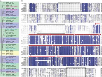
PUF3 sequence alignments. (A) Cladogram of PUF orthologues. (B) PUF3 protein sequences from T. brucei, T. vivax, T. congolense, T. theileri, T. cruzi, Leishmania major, Blechomonas ayalai, Endotrypanum monterogeii and B. saltans were aligned using Clustal Omega (EMBL-EBI). The dark shading highlights identical residues and paler shading shows similarities. Red bars indicate the position of the PUF repeats in T. brucei PUF3. The apparent gap in the seventh PUF repeat for T. vivax is caused by a gap in the sequence assembly. Boxes indicate inserts in the Bodo protein.

PUF3 (Tb927.10.310) is the only T. brucei Pumilio domain protein that lacks low complexity or coiled coil motifs (Fig. 1). Conservation between kinetoplastid PUF3 homologues is restricted to the eight repeats, a 17-residue region immediately C-terminal to the last repeat, and two short regions at the N-terminus (Fig. 2B). PUF3 was shown to bind to mRNA in T. brucei bloodstream forms (Lueong et al., Reference Lueong, Merce, Fischer, Hoheisel and Erben2016) and to repress expression when tethered, via a lambdaN peptide, to the 3′-untranslated region of a reporter mRNA (Erben et al., Reference Erben, Fadda, Lueong, Hoheisel and Clayton2014; Lueong et al., Reference Lueong, Merce, Fischer, Hoheisel and Erben2016). Moreover, results of a high-throughput RNAi screen suggested that reduced PUF3 expression caused a significant loss of fitness in all life-cycle stages tested (bloodstream forms, and early differentiation to the procyclic form) (Alsford et al., Reference Alsford, Turner, Obado, Sanchez-Flores, Glover, Berriman, Hertz-Fowler and Horn2011). We therefore investigated its function in more detail.
Materials and methods
Trypanosome culture and transfection
All the cells used in this study were based on stable cell lines constitutively expressing a tetracycline repressor (Alibu et al., Reference Alibu, Storm, Haile, Clayton and Horn2004), which for simplicity are referred to as wild-type. Lister 427 bloodstream forms (monomorphic) were cultured at 37°C and 5% CO2 in adapted HMI9 (Duszenko et al., Reference Duszenko, Mühlstädt and Broder1992): MEM including 3.24 g L−1 sodium bicarbonate, 23.2 mg L−1 bathocuprone sulphonate, 0.14 g L−1 hypoxanthine, 0.1 g L−1 sodium pyruvate, 39 mg L−1 thymidine, supplemented to give final concentrations of 1.5 mm l-cysteine and 0.2 mmβ-mercaptoethanol, 5% penicillin/streptomycin (Gibco), and 10% foetal bovine serum (heat inactivated at 55°C for 1 h). EATRO 1125 bloodstream forms (pleomorphic) were cultured in HMI-9 media containing 1.1% methyl cellulose (Sigma, M0512) to maintain their pleomorphism (Vassella et al., Reference Vassella, Kramer, Turner, Wankell, Modes, van den Bogaard and Boshart2001). Densities were kept below 5 × 10⁶ mL−1 for Lister 427 and below 1 × 10⁶ mL−1 for EATRO1125.
Procyclic forms were cultured at 27°C in MEM-Pros medium (Ziegelbauer et al., Reference Ziegelbauer, Quinten, Schwarz, Pearson and Overath1990) supplemented with 5% penicillin/streptomycin (Gibco), 7.5 mg L−1 haemin and 10% heat inactivated foetal bovine serum, at densities between 105 and 4 × 10⁶ cells mL−1.
Differentiation of bloodstream slender forms to stumpy forms was done by starting at a density of 5–7 × 10⁵ cells mL−1, and allowing the cell density to increase to 3–4 × 10⁶ cells mL−1 then decrease to 106 cells mL−1. This took 36–48 h. Differentiation to procyclic forms was induced by adding 6 mmcis-aconitate to slender or stumpy forms at 106 cells mL−1 and incubating at 27°C for 24 h before transferring the cells to MEM-Pros at 27°C. Growth was measured using media without selecting drugs, with 100 ng μL−1 of tetracycline when needed for transgene induction. Plasmids and oligonucleotides are listed in Table S1. Transfection of linearized plasmids, and drug selections, was performed as described in Alibu et al. (Reference Alibu, Storm, Haile, Clayton and Horn2004).
Immunofluorescence microscopy
Cells were incubated with tetracycline for 24 h to induce RNAi, then processed using immunofluorescence microscopy. Cells were fixed in 4% paraformaldehyde at room temperature for 20 min, washed and smeared on slides. The slides were either stored at 4°C or analysed immediately. All the following steps were performed at room temperature. The slides were placed in phosphate-buffered saline (PBS) for 5 min to rehydrate the cells then permeabilized using 0.2% Triton-X for 20 min. Cells were blocked with 0.5% gelatin for 1 h and incubated with a primary antibody (dissolved in 0.5% gelatin) for 1 h. The slides were washed three times with PBS and incubated in the dark with a secondary antibody containing a fluorescence dye for 1 h. Anti-V5 antibody (1:500, mouse) and secondary anti-mouse IgG antibody with Cy3 fluorophore (1:700, anti-mouse) were used. The slides were washed three times and incubated for 15 min with 1 μg mL−1 4′,6-diamidino-2-phenylindole solution to stain DNA. After three more washes, the slides were air-dried before mounting with 90% glycerol-PBS. The cover slips were sealed to the slides using nail polish. Images were acquired using an Olympus IX81 microscope and analysed with Olympus xCellence software or ImageJ.
RNA and DNA preparation and blotting
To obtain at least 10 μg of RNA, 3 × 10⁷ of log-phase bloodstream-form cells (1 × 10⁶ mL−1 for Lister 427, no more than 8 × 10⁵ mL−1 for EATRO1125) were collected at 900 × g for 10 min and RNA extracted using TriFast reagent according to the manufacturer's protocol. RNA concentrations were determined using a Nanodrop spectrophotometer and the RNA stored at −80°C or used immediately. rRNAs were depleted from whole RNA extracts before RNAseq using a cocktail of DNA oligonucleotides targeting rRNAs with RNaseH (Minia et al., Reference Minia, Merce, Terrao and Clayton2016). For blotting, RNA was electrophoresed on a formaldehyde agarose gel, blotted on a nylon membrane by downward capillary for at least 4 h followed by UV cross-linking of RNA to the membrane using Stratagene® UV cross-linker. Prior to probing, the blot was blocked using pre-hybridization buffer [5 × SSC (20 × SSC is 3 m NaCl, 0.3 m Na citrate), 0.5% sodium dodecyl sulphate (SDS), 5 × Denhardt's solution, 100 μg mL−1 Salmon sperm DNA]. For spliced leader probing, the pre-hybridization buffer was similar to the previous one except that 6 × SSC and 0.05% sodium pyrophosphate were included. Radioactive [32P] probes made from polymerase chain reaction (PCR) products were prepared using a PrimeIt kit (Stratagene), and those from oligonucleotides using polynucleotide kinase (NEB). The Northern blot was incubated overnight with at least 2 × 10⁶ cpm mL−1 of the probe at 42°C or 65°C respectively for oligonucleotide and PCR-product probes. The blot was washed with 2 × SSC, 0.1% SDS twice for 10 min each at room temperature and once for 15 min at 65°C then exposed to a phosphorimaging plate before developing the autoradiographs with a phosphorimager (Fujifilm FLA-3000).
For Southern blotting, at least 5 μg of genomic DNA was digested using restriction enzymes as shown in Figs 5C and 6C, and electrophoresed through an ethidium bromide-containing agarose gel. The gel was washed in hydrolysis buffer (0.25 m HCl) for 15 min, in denaturing buffer (1.5 m NaCl, 0.4 m NaOH) for 15 min to denature long DNA and in neutralizing buffer (1.5 m NaCl, 0.5 m Tris-Cl pH 7.5) for 15 min before blotting onto a nylon membrane and processed as described for Northern blotting.
Chloramphenicol acetyltransferase assay
A total of 2 × 10⁷ cells were collected at 900 × g for 10 min and washed three times with PBS. The pellet was re-suspended in 100 μL of CAT buffer (100 mm Tris-HCl pH 7.8) and freeze–thawed three times using liquid nitrogen and a 37°C heating block. The supernatants were collected by centrifugation at 10 000 × g for 5 min and kept in ice. The protein concentrations were determined by Bradford assay (BioRad) according to the manufacturer's protocol. For each reaction, 0.5 μg of protein in 50 μL of CAT buffer, 10 μL of [14C]-butyryl CoA (56 mCi mmol−1, 0.1 mCi ml−1), 2 μL of chloramphenicol (stock: 40 mg mL−1), 200 μL of CAT buffer and 4 mL of scintillation cocktail were mixed in a Wheaton scintillation tube HDPE (neoLab #9-0149) and the incorporation of the radioactive butyryl group into chloramphenicol was measured using a Beckman LS 6000IC scintillation counter.
Tandem affinity purification and mass spectrometry
A total of 6–7 × 10⁹ cells were collected at 900 × g at 4°C for 15 min in 500 mL centrifuge bottles. The cell pellet was washed twice in ice-cold PBS, snap frozen in liquid nitrogen and stored at −80°C or used immediately. The cells were lysed in IPP10 buffer [10 mm Tris-HCl (pH 7.8), 10 mm NaCl, 0.1% IGEPAL, 50 μg mL−1 leupeptin] by passing them through a 21-gauge needle 15–20 times. The lysate was cleared by centrifugation at 10 000 × g for 10 min and the concentration of NaCl was adjusted to 150 mm before loading onto a 0.8 × 4 cm Poly-Prep column (BioRad) containing IgG sepharose beads that had been prewashed in IPP150 buffer [10 mm Tris-HCl (pH 7.8), 150 mm NaCl, 0.1% octylphenoxypolyethoxyethanol (IGEPAL)]. This was incubated at 4°C for 2 h with gentle rocking. The beads were washed twice with 10 mL IPP150 buffer and once with 10 mL TEV cleavage buffer [IPP150 buffer, 1 mm dithiothreitol (DTT), 0.5 mm EDTA]. The beads were re-suspended in 800 μL TEV cleavage buffer and incubated with TEV protease at 15–20°C for 2 h with gentle rocking. The eluate was collected and transferred to a second column containing calmodulin beads in calmodulin binding buffer (IPP150 buffer, 10 mmβ-mercaptoethanol, 1 mm MgAc2, 1 mm imidazole, 2 mm CaCl2), and incubated for 2 h at 4°C. The beads were washed three times with calmodulin-binding buffer and the proteins were eluted using 2 mm EGTA. The proteins in the eluate were concentrated by trichloroacetic acid precipitation then dissolved in Laemmli buffer. This was electrophoresed for a short time so that the sample dye ran approximately 1.5 cm. The gel was then subjected to the sequencing facility at ZMBH (Heidelberg) for mass spectrometry.
RNA pull-down and RNASeq
This procedure was similar to the tandem affinity purification (TAP) with the exception that the cells were UV-irradiated before lysis and only the first purification step was performed. Approximately 3 × 10⁹ cells were collected at 900 × g at 4°C for 15 min, re-suspended in 30 mL of the supernatant on ice, transferred to a petri dish and UV-irradiated twice using a Stratagene UV crosslinker with an energy of 3 kJ. The cells were pelleted again in 4°C at 2000 × g for 5 min and washed twice with ice-cold PBS. Cells were lysed in IPP10 buffer consisting of KCl in the place of NaCl and containing RNase inhibitor (RNasin, Invitrogen), by passing them through a 21-gauge needle 15–20 times. The lysate was cleared by centrifugation at 10 000 × g for 10 min, the KCl concentration was adjusted to 150 mm. A small volume of the lysate equivalent to 7.5 × 10⁷ cells was collected as the input fraction and treated with 0.2 mg mL−1 proteinase K, 8 mm EDTA, 0.2% SDS at 45°C for 15 min before adding TriFast-FL reagent (Peqlab). The remainder of the lysate was loaded onto a column containing prewashed sepharose beads as for the TAP. The columns were incubated at 4°C for 2 h with gentle rocking and the unbound lysate collected. This was also subjected to proteinase K treatment as described for the input fraction. Subsequently, the beads on the column were washed twice with IPP150 buffer and once with TEV cleavage buffer, re-suspended in 500 μL of TEV cleavage buffer containing TEV protease and incubated at 15–20°C for 2 h with gentle rocking. The cleaved product (bound) was treated with proteinase K and mixed with TriFast-FL reagent. The three fractions in TriFast-FL were stored at −80°C and later used for RNA extraction according to the manufacturer's protocol. RNA concentrations were determined using a NanoDrop® or a Qubit® spectrophotometer and subjected to the next-generation sequencing facility in Bioquant (Heidelberg) for library preparation. Sequencing was performed at GeneCore EMBL (Heidelberg). Data were deposited as follows: RNAi sequence data: ENA, accession number ERP115825: https://identifiers.org/ena.embl:ERP115825. RIP-Seq sequence data: ENA, accession number ERP115855: https://identifiers.org/ena.embl:ERP115855.
Fastq files were used as input in TrypRNAseq v.4.3 – an in-house pipeline for RNAseq (https://github.com/klprint/TrypRNAseq; https://zenodo.org/record/158920#.Xpx03C97HYI) (Mulindwa et al., Reference Mulindwa, Leiss, Ibberson, Kamanyi Marucha, Helbig, Melo do Nascimento, Silvester, Matthews, Matovu, Enyaru and Clayton2018). Briefly, reads were taken through quality control, mapped on the T. brucei genome (TREU927) using Bowtie2, and the number of reads aligning to coding sequences (CDS) determined. Each sequence was allowed to align 20 times to allow for the repetitive nature of the T. brucei genome, but for statistical analyses, only one copy of repeated genes was considered (Siegel et al., Reference Siegel, Hekstra, Wang, Dewell and Cross2010). To analyse the RNAi data, fold changes were compared between RNAi-induced and un-induced samples using the DESeqUI adaptation (https://zenodo.org/record/290632#.Xpx1pi97HYI) of DESeq2 (Love et al., Reference Love, Huber and Anders2014).
To look for motifs, 3′-UTRs were downloaded from TritrypDB. Sequences between 10 and 5000 nucleotides were used: 66 out of 120 mRNAs in the ‘unbound’ set (more than 2-fold enriched in both purifications) and 1107 out of 1822 ‘unbound’ mRNAs (less than 1-fold enriched). The two sequence lists (sense strand only) were compared using DREME with standard settings (Bailey, Reference Bailey2011). Four selected 3′-UTRs were also examined manually using published transcriptome data.
Results
PUF3 is cytosolic and rapidly represses expression
To determine whether PUF3 was in the nucleus or cytoplasm, we used bloodstream-form trypanosomes expressing N-terminally V5-tagged PUF3 from the endogenous locus (V5-PUF3). By using immunofluorescence microscopy, V5-PUF3 was spread throughout the cytosol with no detectable signal from the nucleus (Fig. 3A); the localization was somewhat punctate, similar to that of T. cruzi PUF6 (Dallagiovanna et al., Reference Dallagiovanna, Perez, Sotelo-Silveira, Smircich, Duhagon and Garat2005). Since PUF3 has no canonical organellar localization signals the spots might be a fixation artefact, or might be small RNA-protein granules; results of a preliminary digitonin fractionation (not shown) indicated that PUF3 is not in glycosomes. Results obtained by others using high-throughput in-situ N-terminal GFP tagging (Dean et al., Reference Dean, Sunter and Wheeler2016), also indicated a cytosolic location.
PUF3 location and tethering. (A) Bloodstream-form trypanosomes with in situ tagged PUF3 and inducible RNAi targeting PUF3 were used. The upper image shows cells without tetracycline and the lower image after 24 h tetracycline treatment. Stains are for V5-PUF3 (red) and DNA (blue). DIC, differential interference contrast. (B) Results of the tethering screen (Erben et al., Reference Erben, Fadda, Lueong, Hoheisel and Clayton2014). The bloodstream-form trypanosomes used contained a tetracycline-inducible PGKB gene, with five copies of boxB between the open reading frame and the actin 3′-untranslated region (3′-UTR). They were transfected with a library designed for inducible expression of proteins encoded by ~1 kb genomic fragments, preceded by the lambdaN peptide. Tetracycline addition resulted in growth retardation, unless the expressed lambdaN-protein repressed PGKB expression. After induction, cells were grown, then the DNA inserts were amplified and sequenced. This selects for protein sequences capable of suppressing translation and/or inducing mRNA degradation. The graph shows the results for PUF3, with first codon positions on the x-axis, and the reads per million on the y-axis. Higher values with tetracycline than without tetracycline suggest suppressive activity. The cartoon below shows domains of PUF3, to scale: PUF domains are in cyan, and sequences conserved in Kinetoplastea (Fig. 1B) are in orange. A, B and C represent independent replicates. (C) Confirmation of tethering results for full-length lambdaN-PUF3-myc. The bloodstream-form trypanosomes used constitutively express a chloramphenicol acetyltransferase (CAT) mRNA with five B-boxes between the open reading frame and the actin 3′-UTR. Expression of lambdaN-PUF3-myc was induced by addition of tetracycline and CAT activity was measured. The graph shows results for three independent cultures. The Western blot below shows expression of lambdaN-PUF3-myc, with trypanothione reductase (TR) as control.

Cytosolic phosphoglycerol kinase (PGKB) is toxic to bloodstream-form trypanosomes. Tethering of full-length PUF3 to an mRNA encoding PGKB conferred a selective advantage (Lueong et al., Reference Lueong, Merce, Fischer, Hoheisel and Erben2016), suggesting that PUF3 suppresses expression. Using random fragments of the PUF3 gene (Erben et al., Reference Erben, Fadda, Lueong, Hoheisel and Clayton2014), most selected clones encoded tethered polypeptides that commenced within the first 100 amino acids (Fig. 3B). This result suggests that the suppressive sequence is N-terminal to the PUF repeats. We confirmed that tethering of lambdaN-PUF3-myc to a reporter mRNA encoding chloramphenicol acetyltransferase (CAT) was repressive: CAT activity decreased within 4 h (Fig. 3C). Consistent with this, PUF3 was not detectably associated with polysomal RNA (Klein et al., Reference Klein, Terrao, Inchaustegui Gil and Clayton2015). The tethering result also, incidentally, shows that addition of the C-terminal myc tag did not prevent the repressive activity.
A specific subset of mRNAs copurifies with tagged PUF3
To investigate mRNAs bound to PUF3, we made bloodstream-form trypanosomes in which one PUF3 open reading frame was deleted and the other was fused at the 5′-end with a sequence encoding a TAP tag. The cells grew normally. After UV cross-linking, cells were lysed, tagged PUF3 was selected, and bound and unbound RNAs were sequenced for two biological replicates. The unbound RNA fraction was subjected to rRNA depletion using RNase H and oligonucleotides complementary to rRNA (Minia et al., Reference Minia, Merce, Terrao and Clayton2016). To check the effect of the rRNA procedure on the transcriptome we also sequenced the input RNA from the experiment, either with or without rRNA depletion.
The transcriptome results showed that depletion of rRNA from the input RNA had no significant effect on the transcriptome (Fig. 4A and B; Table S2, sheet 3). By principal component analysis (Fig. 4A), the unbound RNA from the pull-down experiment clustered with the input samples, which is expected if only a few mRNAs are bound by the selected protein. Rather large numbers of cells were needed to obtain sufficient bound mRNA for standard sequencing library preparation, but these bound fractions were clearly different from the input and unbound RNAs (Fig. 4A). PUF3 mRNA was enriched in the bound fractions: this is expected because polysomal mRNA can be selected via the nascent polypeptide (Klein et al., Reference Klein, Terrao and Clayton2017; Mugo and Clayton, Reference Mugo and Clayton2017; Liu et al., Reference Liu, Marucha and Clayton2020) (Fig. 4C). 7SL RNA, which is a component of the signal recognition particle, was also, as expected, almost exclusively in the unbound fraction (Table S2, sheet 4). The 106 mRNAs that were reproducibly more than 2-fold enriched (‘bound’ mRNAs, Fig. 4C) were significantly shorter than mRNAs that were less than 1-fold enriched (unbound), suggesting that selection of bound mRNAs was not due to non-specific RNA binding to PUF3 or the matrix (Fig. 4D; Table S2, sheet 2). We do not know whether this short length has any significance. Very long-lived mRNAs were missing from the bound fraction: overall the half-lives of bound mRNAs were significantly shorter than those of the unbound mRNAs (Fig. 4E), which is consistent with destabilizing activity of PUF3. In contrast, there was hardly any difference in ribosome occupancy (Fig. 4F, Supplementary Table S2) (Antwi et al., Reference Antwi, Haanstra, Ramasamy, Jensen, Droll, Rojas, Minia, Terrao, Mercé, Matthews, Myler, Parsons and Clayton2016). The bound mRNAs mostly have similar abundances in procyclic forms and long-slender and stumpy bloodstream forms, and there was no significant enrichment or depletion of any particular functional protein class (not shown).
rRNA depletion and RNA pull-down. TAP-PUF3 was bound to IgG beads and input, bound and unbound RNAs were sequenced. Raw results are in Table S2 (see Underlying data). (A) Principal component analysis for all eight datasets. B, eluate or bound fraction; U, flow-through or unbound fraction. ‘R’ is input RNA after rRNA depletion, and ‘T’ is input RNA without rRNA depletion. (B). rRNA depletion using oligonucleotides and RNaseH does not affect the transcriptome. Average reads per million (RPM) for duplicate matched total RNA samples are shown, on a log 2 scale. Individual comparisons are in Table S1 (see Underlying data). (C) The ratios of bound/unbound reads per million were calculated for all unique genes. PUF3 mRNA was enriched 4.6-fold in replicate 1 and 1.88-fold in replicate 2 (orange dot). There was a weak positive correlation between the two sets of enrichment values (R = 0.35 for log-transformed results). The inset shows the enriched motif found by DREME. (D) Lengths of bound mRNAs relative to those of unbound mRNAs. mRNAs were classed as ‘bound’ if the reads per million in the bound fraction were more than twice those in the unbound fraction. The ‘unbound’ RNAs had ratios of less than 1. The boxes include values from the 25th to 75th percentiles, with a horizontal line at the median; notches are 1.58 × the interquartile range divided by the square root of the number of data points. Dotted lines (whiskers) extend to maximum and minimum values that are no further than 1.5× the inter-quartile distance, and dots are outliers. The bound mRNAs are significantly shorter than the unbound mRNAs (P = 1.2 × 10⁻¹⁶, two-tailed Student's t-test). (E) Half-lives of bound mRNAs and unbound mRNAs. Data are from (Fadda et al., Reference Fadda, Ryten, Droll, Rojas, Färber, Haanstra, Bakker, Matthews and Clayton2014). The PUF3-bound mRNAs are significantly (P = 7 × 10⁻¹², two-tailed Student's t-test) less stable than the unbound mRNAs: the ratio of medians is 0.67. (F) Ribosome densities on bound mRNAs and unbound mRNAs. Data are from Antwi et al. (Reference Antwi, Haanstra, Ramasamy, Jensen, Droll, Rojas, Minia, Terrao, Mercé, Matthews, Myler, Parsons and Clayton2016). The bound mRNAs have very slightly higher ribosome densities than the unbound ones: the ratio of medians is 1.2, P = 0.003.

To obtain further information about PUF3 binding specificity, we compared the annotated 3′-UTRs of bound and unbound mRNAs. Only one motif was significantly enriched. Of the 66 annotated 3′-UTRs from the bound fraction, 23 included the motif ‘UGUA[U/C]AUU’ (inset, Fig. 4C; E-value: 8.7 × 10⁻⁹). The same motif is present in the 3′-UTRs of only 15 of the 1107 unbound control mRNAs, even though those sequences were, on average, 1.6 times longer. The proportion of PUF3-bound mRNAs that contain the motif is definitely an under-estimate, because many annotated 3′-UTRs are truncated. (Of course, this annotation error affects the control set as well.) We individually examined three bound mRNAs whose annotated 3′-UTRs lacked the sequence, and found that the corrected 3′-UTR of one of them, Tb927.3.2380, contains two copies of the motif; it was also present in the (non-annotated) 3′-UTR of Tb927.9.11760. The lack of the motif in remaining mRNAs could mean that PUF3 binds a slightly more relaxed motif; alternatively some of the mRNAs in the ‘bound’ list may have been enriched by chance.
We also subjected TAP-PUF3 to TAP and identified co-purifying proteins by mass spectrometry (Table S3). The only proteins that appeared to co-purify, relative to TAP-GFP controls, were abundant proteins that had previously been identified in various other purifications. The results therefore gave no hint of a mechanism by which PUF3 might inhibit mRNA translation or promote mRNA degradation.
PUF3 is not required in growing bloodstream forms
To assess the function of PUF3 we first reduced its expression by RNAi. We used two trypanosome cell lines expressing the tet repressor. One, of the EATRO1125 strain, is capable of full differentiation from the bloodstream to the procyclic form. When they reach a density of 2.5–3.0 × 10⁶ mL−1, they arrest in G0, and become ‘stumpy’ – that is, they are shorter and fatter, and express the stumpy-form marker PAD1 (Dean et al., Reference Dean, Marchetti, Kirk and Matthews2009) and various mitochondrial proteins required for survival in tsetse. When the growth temperature is reduced from 37 to 27°C and cis-aconitate is added, they differentiate to procyclic forms and start to grow in appropriate medium within 1–2 days. Our Lister 427 strain bloodstream forms, in contrast, can grow to densities of 6–7 × 10⁶ mL−1 and do not make stumpy forms or PAD1. If these cells are treated at 27°C with cis-aconitate, they are able to express some procyclic-specific mRNAs and proteins, including the procyclic surface protein EP procyclin, but they are unable to grow as procyclic forms, so are unsuitable for studying full differentiation.
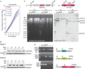
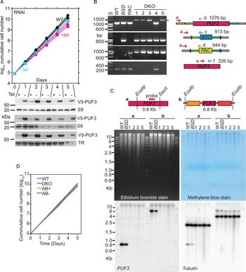
Depletion of PUF3 in Lister 427 strain bloodstream-form trypanosomes had only a marginal effect on growth (Fig. 5A and B) and gene deletion was also possible (Fig. 5C and D); the growth rates of these cells were not measured carefully but were not noticeably different from wild-type. Induction of RNAi in pleomorphic EATRO1125 bloodstream-form trypanosomes (EATRO1125 strain) also resulted in extremely mild growth retardation (Fig. 6A), but once again, clones lacking both genes were readily obtained (Fig. 6B–D). Three independent EATRO1125 clones lacking PUF3 had the same growth rates as cells containing both, or only one PUF3 gene, all having doubling times between 6.9 and 7.5 h. This suggested that if PUF3 has any role in cultured bloodstream forms, its loss can be compensated during clonal selection.
PUF3 is not essential for growth and survival of monomorphic bloodstream forms. (A) Growth of bloodstream-form Lister 427 trypanosomes with in situ N-terminally V5-tagged PUF3 and tetracycline-inducible PUF3 RNAi. Results are shown for wild-type cells and for two independent clones grown with (+) and without (−) tetracycline. (B) Western blots showing the expression of a V5-tagged PUF3 in the cell lines in (A). Antibody to aldolase was used as a loading control. (C) The upper panel represents the design of a Southern blot. Coding sequences of genes are represented by rectangles and vertical double-strokes show restriction sites. Genomic DNA of cells selected for homozygous knockout (transfected with plasmids pHD2851 and pHD2852) was digested with Eco RI (experiment a) or Eco NI and Sac II (experiment b). The digested DNAs for wild-type (WT), single knockouts (BSD or PAC) and three candidate homozygous knockout clones were analysed by Southern blotting using a probe targeting PUF3 (as indicated in the upper panel), with TUT3 as a loading control. L: marker ladder. (D) DNA from (C) was used in a polymerase chain reaction using the primers shown. Oligonucleotides were: (a) CZ6220; (b) CZ3542; (c) CZ5153; (d) CZ5155 (Table S4, see Underlying data). Double stroke lines represent the ends of the cloned fragments used for transfections.

PUF3 is not essential for growth and survival of pleomorphic bloodstream forms. (A) Bloodstream-form EATRO1125 trypanosomes with inducible RNAi targeting PUF3 were grown with or without tetracycline and diluted as necessary. Three replicates are shown. ‘WT’ is the parental line without RNAi. A western blot showing depletion of V5-PUF3 from each replicate is shown below, with ribosomal protein S9 or trypanothione reductase (TR) as loading controls. (B) PCR test for PUF3 knockout. Genomic DNA from wild-type, single knockouts (BSD and PAC) and candidate homozygous knockout (DKO) clones were used in a polymerase chain reaction. Double stroke lines represent the ends of the cloning fragment used for transfections. Oligonucleotides were: (a) CZ6220; (b) CZ3542; (c) CZ5153; (d) CZ5155; (e) CZ6328; (f) CZ6329 (Table S4, see Underlying data). PCR fragment sizes are on the right. Clone 4 did not have a homozygous deletion. For primers ‘a’ and ‘b’, the identity of the slowly-migrating band (asterisk) is unknown, but gene deletion was confirmed by the absence of bands for primers ‘e’ and ‘f’. (C) Southern blot analysis. The upper panel represents the design of the digests. Coding sequences of genes are represented by rectangles and vertical double-strokes show restriction sites. Genomic DNA of cells selected for homozygous knockout (transfected with plasmid pHD2851 and pHD2852) was digested with Eco NI and Sac II (experiment a) or Eco RI (experiment b). The digested DNAs for wild-type (WT), single knockouts (BSD) and four candidate homozygous knockout clones were analysed by Southern blotting first using a probe targeting PUF3 (as indicated in the upper panel) and second with a probe for beta-tubulin. (D) Cumulative growth of the bloodstream forms, mean values. Variation (not shown) was so low that all of the values overlapped. AB: Add-back of tetracycline-inducible PUF3-myc, +tet or −tet.

To investigate why EATRO1125 cells with PUF3 RNAi had slightly slower growth (Fig. 6A), we examined their transcriptomes. RNA was extracted 1 day after tetracycline addition. Depletion of V5-PUF3 protein was confirmed, but unexpectedly, the number of reads for PUF3 was about 1.5-fold increased (Fig. 7A; Table S4). In this case, therefore, the RNAi appeared to have stopped translation without complete mRNA destruction. Only a single additional mRNA (Tb927.9.10170, encoding anaphase-promoting complex subunit 11) was significantly increased but the difference was only 1.5-fold and is probably just random variation (Fig. 7A; Table S4). mRNAs that were at least 2-fold enriched in the PUF3-bound fraction did not increase after RNAi (Fig. 7B). We concluded that depletion of PUF3 for 24 h has no significant effect on the transcriptome.
Effects of PUF3 RNAi on the transcriptome. RNA was prepared from trypanosomes with tetracycline-inducible RNAi targeting PUF3. RNA was prepared 24 h after tetracycline addition, with cells without tetracycline serving as controls. (A) RNA reads that aligned to coding sequences were analysed by DEseq2 and fold changes (± tetracycline) represented on a scatter plot relative to the total number of normalized counts. The red arrow points to the only significantly increased mRNA; the cyan arrow is PUF3. The panel below is the Western blot demonstrating PUF depletion. (B) Average binding ratios for each mRNA (see Fig. 3) are plotted against the fold change after RNAi. None of the RNAi effects was statistically significant. Most mRNAs with higher bound/unbound ratios showed no RNAi effect at all. Orange dots represent mRNAs for which bound/unbound was >2 in both experiments. (C) Distribution of reads mapping on the region of the genome encoding PUF3. Bam files generated from PUF3 RNAi reads were loaded in Integrative Genomics Viewer (IGV version 2.4.14) using default settings. The coverages were viewed using the same data range for all the bam files and screenshots taken. The cartoons in blue are the gene models of PUF3 (with untranslated regions adjusted to correspond to the RNA read coverage) and flanking genes while the magenta bar represents the PUF3 region targeted by RNAi. In the alignments, grey represents reads that aligned perfectly, red are reads with insertions or deletions, and the other colours represent sequences that are also present elsewhere in the genome.

To investigate the peculiar increase in PUF3 reads after RNAi, we examined the read distribution over the PUF3 gene. Without RNAi, the sequence reads were fairly evenly distributed (Fig. 7C), as expected for non-poly(A)-selected transcriptomes. In contrast, after RNAi there was an accumulation not only over the region covered by the dsRNA, but also towards the 5′-end of the transcript (Fig. 7C). Comparison with the neighbouring gene (Tb927.10.300) shows that the read density in the 5′ part of PUF3 was at least 2-fold higher than in uninduced cells; it also clearly exceeded that seen 3′ to the dsRNA fragment. We have no explanation for this, but a similar discrepancy between mRNA and protein levels was seen after RNAi targeting the CPSF30 mRNA: the CPSF30 mRNA migrated slightly slower than without RNAi and the amount slightly increased, although the protein amount was strongly reduced (Hendriks et al., Reference Hendriks, Abdul-Razak and Matthews2003).
PUF3 may assist bloodstream-to-procyclic form differentiation
We next tested whether PUF3 has a role in differentiation of bloodstream forms to procyclic forms. Using Lister 427 trypanosomes, RNAi was induced 24 h before cells attained a density of 1.8–2.0 × 10⁶ cells mL−1. Then, to induce differentiation, cis-aconitate was added and the temperature was reduced from 37 to 27°C. Depletion of PUF3 caused a slight delay in appearance of EP procyclin (Fig. 8A) and the defect was greatly accentuated in Lister 427 trypanosomes completely lacking PUF3 (Fig. 8B). To check that the defect was due to the lack of PUF3, we inducibly re-expressed the (untagged) protein. In one cloned line, re-expression of PUF3 mRNA was tetracycline-dependent, and in the other expression was constitutive (Fig. 8C). Restoration of EP procyclin expression correlated with PUF3 expression (Fig. 8D), although it was incomplete, perhaps because the amount of PUF3 expressed was too low or too high. C-terminally myc-tagged PUF3 could not complement the defect (Fig. 8E). N-terminally myc-tagged PUF3 was extremely poorly inducibly expressed (Fig. 8F) so was not tested.
PUF3 is required for procyclin expression by differentiation-stimulated Lister 427 bloodstream forms. (A) RNAi targeting PUF3 in Lister 427 bloodstream forms gave a transient differentiation defect. RNAi was induced 24 h before cells attained a density of 1.8–2.0 × 10⁶ cells mL−1. cis-Aconitate (CA) was added to the culture (final concentration 6 mm) and the temperature was reduced from 37 to 27°C. The time in hours after CA treatment is indicated. The Western blot shows expression of EP procyclin and V5-tagged PUF3, with ribosomal protein S9 as a control. Protein size markers are in kilodaltons. (B) Monomorphic bloodstream forms were grown to 1.8–2.0 × 10⁶ cells mL−1, then cis-aconitate (CA) was added to the culture (final concentration 6 mm) and the temperature was reduced from 37 to 27°C. After 24 h the medium was changed to standard procyclic medium (very-low-glucose, high-proline minimal essential medium, MEM). The time in hours after CA treatment is indicated. EP procyclin, and ribosomal protein S9, were detected by Western blotting. WT: cells with wild-type complement of PUF3 genes; KO: cells lacking PUF3. (C) Northern blot showing inducible expression of PUF3 mRNA in the add-back clones 3 and 4. The 7SL signal recognition particle RNA (SRP) and rRNA were detected as loading controls. (D) As in (A), but cells with inducible expression of PUF3 (‘add-back’, AB) were also included, either with (+) or without (−) tetracycline addition (final concentration 100 ng mL−1) 24 h before the start of the experiment. The add-back results are for two different clones, clone 3 (with inducible expression) and clone 4 (with constitutive expression). GPEET, EP and S9 were detected by western blotting. (E) Expression of PUF3-myc did not complement the defect in differentiation, as judged by EP procyclin expression. The experiment is similar to that in (C) and (D) except that PUF3-myc was expressed. (F) Inducible expression of N-terminally myc-tagged PUF3 in five Lister 427 bloodstream-form clones. The protein (arrow) was barely detectable. The upper band just above 75 kDa is background from the anti-myc antibody.

Next, we followed differentiation in pleomorphic EATRO1125 cells with PUF3 depletion or knock-out (Fig. 9). These were first grown to high density in methyl cellulose-containing media to promote stumpy-form differentiation, with or without tetracycline, and then switched to differentiation conditions (Fig. 9A). First, we examined cells after PUF3 RNAi. In these cells, we could also see that PUF3 RNAi caused reduced expression of the stumpy-form marker PAD1 (Fig. 9B and C). Expression of the bloodstream-form marker RBP10 is reduced in stumpy forms, and this was not prevented by PUF3 RNAi (Fig. 9C). These results corroborated and extended those previously obtained with the monomorphic Lister 427 line.
Differentiation of PUF3-depleted EATRO1125 bloodstream forms. (A) Expression of EP procyclin during differentiation of cells with PUF3 RNAi. RNAi was induced in EATRO1125 bloodstream forms expressing V5-PUF3, at a density of 5 × 10⁵, in methyl cellulose-containing medium. 48 h later, cis-aconitate (CA) was added and the temperature changed to 27°C. LS: long slender cells, WT; wild-type, PC: established procyclic forms. Ribosomal protein S9 and the Ponceau protein stain serve as controls. (B) Replica of (A) with detection of PAD1. (C) Expression of PAD1 and RBP10 after PUF3 RNAi in EATRO1125 bloodstream forms. The experiment was as shown in Fig. 5B. (D) The experiment is as in (A) but here PUF3 single knockout cells expressing V5-tagged Drosophila adenosine deaminase-PUF3 (AD) or TAP-PUF3 (T) from the PUF3 locus were used; ‘W’ indicates cells with two intact copies of PUF3. Expression of the differentiation markers PAD1 (Dean et al., Reference Dean, Marchetti, Kirk and Matthews2009) and the phosphatase PIP39 (Szoor et al., Reference Szoor, Wilson, McElhinney, Tabernero and Matthews2006) are also illustrated. (E) The experiment is as in (A) but here PUF3 single knockout cells expressing V5-tagged Drosophila adenosine deaminase-PUF3 (AD) or double knockout cells expressing V5-AD-GFP (G) from the PUF3 locus were used. (F) Cells adapted to the absence of PUF3 are not defective in EP expression in response to differentiation stimuli. The experiment is as in (A), but here we used cells completely lacking the original PUF3 genes (KO). ‘AB’ indicates cells with a tetracycline-inducible copy of PUF3-myc, which were grown with (+) or without (−) tetracycline.

We also checked the differentiation capability of EATRO1125 trypanosomes that lacked wild-type PUF3, and constitutively expressed either TAP-tagged PUF3, or PUF3 that was N-terminally fused with V5-tagged Drosophila adenosine deaminase (AD) (Fig. 9D and E). In addition, we tested cells that lacked PUF3, but instead expressed V5-AD-GFP from the PUF3 locus (Fig. 9E). Cells without PUF3, or expressing only V5-AD-PUF3, did not express any EP procyclin after 24 h (Fig. 9D and E). Cells with TAP-PUF3 did express EP procyclin, PAD1 (Dean et al., Reference Dean, Marchetti, Kirk and Matthews2009) and the phosphatase PIP39 (Szoor et al., Reference Szoor, Wilson, McElhinney, Tabernero and Matthews2006), but induction was slower than wild-type (Fig. 9D). These results are difficult to interpret. We do not know how much PUF3 was expressed relative to the wild-type level so the delay in expression of differentiation markers in the tagged-PUF3 lines might be due to the presence of abnormal amounts of PUF3. However since the various tags impaired differentiation to different extents, it is likely that PUF3 activity is impaired by both N-terminal and C-terminal tags. The abundances of tagged PUF3s did not reproducibly change during differentiation (Fig. 9A–D). It is however possible that its activity changes, since it has several phosphorylation sites (Urbaniak et al., Reference Urbaniak, Martin and Ferguson2013), and three of them are modified predominantly in the S-phase (Benz and Urbaniak, Reference Benz and Urbaniak2019).
We next examined the differentiation of the pleomorphic EATRO1125 cells in which both PUF3 genes had been replaced by selectable markers (Fig. 6B and C). To our immense surprise, EP procyclin expression was induced normally during differentiation (Fig. 9F). Not unexpectedly, expression of PUF3-myc in those cells made no difference (Fig. 9F). Preliminary results (not shown) confirmed PAD1 expression in the cells without PUF3. These results suggest that during selection of the pleomorphic knock-out clones, loss of PUF3 had been compensated for by some other unknown changes. This is not unprecedented: for example, T. brucei lacking UMP synthase were initially avirulent for mice, but recovered their virulence after prolonged in vitro culture (Ong et al., Reference Ong, Sienkiewicz, Wyllie, Patterson and Fairlamb2013).
Correct expression of PUF3 in procyclic forms is required for optimal growth
Pleomorphic bloodstream-form trypanosomes lacking PUF3 could differentiate, and grew as procyclic forms when we transferred them to appropriate medium (examples are shown in Fig. 10A and B). Cells with inducible PUF3, but without tetracycline, grew similarly. However, if expression of PUF3 was induced during and after differentiation, growth was repressed (PUF3+ in Fig. 10A and B). Nevertheless, despite an adaptation time of several weeks, both cells lacking PUF3 and the uninduced line grew slower than wild-type (Fig. 10C). Unfortunately induction of PUF3 transgene expression did not correct the defect: instead, growth was repressed (Fig. 10C). This suggests that the correct level of PUF3 is required for optimal growth of procyclic forms and that an excess is toxic. Since we have no antibody to PUF3, however, we could not measure the protein levels.
Correct expression of PUF3 is required for normal growth of procyclic forms. Three different EATRO1125 cell lines were used: wild-type (WT); lacking endogenous PUF3 genes (DKO), and DKO cells with inducible expression of PUF3, grown either with (PUF3+) or without (PUF3−) tetracycline. (A) Cells were grown to 106 mL−1, then cis-aconitate was added, with or without tetracycline, and the temperature was reduced to 27°C (27°C/CA). After 24 h the cells were transferred to procyclic-form medium (MEM). (B) As (A) except that cells were kept at high density to induce differentiation to stumpy forms before differentiation to procyclic forms was induced. (C) Differentiated cells were grown as procyclic forms for 4 weeks, then tetracycline was added to induce PUF3 over-expression. Results are for three independent cultures, indicated by the different symbols.

Finally, we investigated whether PUF3 is required for differentiation of procyclic forms to bloodstream forms. This can be tested in vitro only by ectopic expression of the RNA-binding proteins RBP6 (Kolev et al., Reference Kolev, Ramey-Butler, Cross, Ullu and Tschudi2012) or RBP10 (Mugo and Clayton, Reference Mugo and Clayton2017). RBP10 expression in procyclic forms forces them to differentiate to metacyclic-like forms without intervening stages and very small proportion of these metacyclic forms can replicate when placed in bloodstream-form culture conditions (Mugo and Clayton, Reference Mugo and Clayton2017). We transfected bloodstream-form EATRO1125 cells lacking PUF3 genes (Fig. 6B) with a construct for inducible expression of RBP10-myc (Mugo and Clayton, Reference Mugo and Clayton2017). Regulated expression was tested (not shown), then the cells were differentiated to procyclic forms. One clone survived. The cells were cultured for 2 weeks, then RBP10-myc expression was induced with tetracycline. Two days later the cells were placed under bloodstream-form culture conditions. After 2 weeks, replicating bloodstream-form trypanosomes were readily detectable. Procyclic forms lacking PUF3 are therefore capable of RBP10-induced differentiation to the bloodstream form.
Discussion
PUF3 bound to a specific subset of mRNAs, and could suppress expression when tethered. Transient depletion of PUF3 caused a minor growth defect in bloodstream forms, but it was not essential for growth of either bloodstream forms or procyclic forms. Transient depletion also led to defects in differentiation, but upon prolonged cultivation those defects were lost. Artificial tethering of PUF3 to a reporter suppressed its expression, and PUF3 target mRNAs were on average less stable than non-targets, but RNAi in bloodstream forms had no significant effect on the transcriptome. Taking these results together, we suggest that PUF3 slightly destabilizes the mRNAs to which it binds, but that a different protein can take over this function when PUF3 is absent. In other organisms, also, Pumilio domain proteins tend to be needed in order to fine-tune gene expression, rather than to effect big changes. Indeed, laboratory yeast that lacks all five cytosolic mRNA-binding Pumilio domain proteins is still viable (Fischer and Olivas, Reference Fischer and Olivas2018).
PUF3 suppressed expression of an mRNA when tethered. This is unlikely to be an artefact of the tethering, since the mRNAs bound to PUF3 had significantly shorter half-lives than the remainder (Fig. 4E). The region of PUF3 that is responsible for suppression is probably towards the N-terminus (Fig. 3B). Two short N-terminal sequences are unique to PUF3 and conserved throughout Kinetoplastea: they are WTV[QH][QE]D[DE][TYC] at residues 3–10, and YT[KQ][SC]SQVF at residues 38–45 (Fig. 2B). One or both sequences might be implicated in suppression. The mechanism, however, remains unknown since no interacting protein partners were identified. The N-terminally tagged version that was used for the purifications clearly was capable of specific RNA binding, but only partially complemented the differentiation defect in cells lacking wild-type PUF3, so it is possible that the bulky N-terminal affinity purification tag (unlike the lambdaN peptide) interfered with protein–protein interactions.
Depletion of PUF3 by RNA interference had only a marginal effect on growth of bloodstream forms, with no effect on the transcriptome, and the genes could be deleted in both bloodstream and procyclic forms. In contrast, RNA interference caused a clear defect in differentiation from the bloodstream to the procyclic form, as measured by procyclin expression and, in pleomorphic cells, failure to express the stumpy-form marker PAD1. These results suggest that the main role of PUF3 is in modulating gene expression during differentiation – presumably in order to suppress the expression of particular proteins. Differentiation to the stumpy form is accompanied by a general decrease in translation, and even a minor delay in this can impair subsequent differentiation (Terrao et al., Reference Terrao, Kamanyi Marucha, Mugo, Droll, Minia, Egler, Braun and Clayton2018), so it is quite feasible that abnormal translation or degradation of a particular subset of mRNAs right at the beginning of the process could prevent subsequent steps, even in monomorphic trypanosomes. Which of the PUF3 targets might be relevant is not obvious. If a protein needs to be removed during differentiation, then RNAi targeting it might enhance differentiation. RNAi targeting 19 of the bound mRNAs (17%) promoted survival of differentiating trypanosomes (Supplementary Table S2) (Alsford et al., Reference Alsford, Turner, Obado, Sanchez-Flores, Glover, Berriman, Hertz-Fowler and Horn2011), but this was true for 14% of mRNAs overall – not a significant difference. None of the PUF3-bound mRNAs is significantly decreased in stumpy forms (Silvester et al., Reference Silvester, Ivens and Matthews2018) and their ribosome occupancies and mRNA levels mostly do not differ significantly between cultured long slender bloodstream forms and procyclic forms. There is no enrichment of any particular functional protein class, very few have been specifically studied, and none has (to our knowledge) previously been implicated specifically in trypanosome differentiation.
The sequence UGUA[U/C]AUU, which was significantly enriched in the 3′-UTRs of PUF3-bound mRNAs (Fig. 4C), is an exact match with the consensus Pumilio binding motif (Lu and Hall, Reference Lu and Hall2011; Miller and Olivas, Reference Miller and Olivas2011; Wang et al., Reference Wang, Oge, Perez-Garcia, Hamama and Sakr2018). It contains the same number of nucleotides as the number of repeats (Figs 1 and 2), and resembles the UGUA[A/C/U]AUA motif that is recognized by metazoan Pumilio proteins and yeast Puf5 (Miller and Olivas, Reference Miller and Olivas2011). This convincingly confirms the specificity of association of mRNAs in the PUF3-bound fraction. As expected, the motif differs from the trypanosome PUF9 recognition motif, UUGUACC (Archer et al., Reference Archer, van Luu, de Queiroz, Brems and Clayton2009). A caveat is that two-thirds of the investigated PUF3-bound sequences lacked the motif. In some cases, this could be because the annotated 3′-UTRs are truncated; alternatively additional motif variants may also be recognized by PUF3, but not detected as significant. Apart from PUF9, there are five other cytosolic PUF proteins, which are all likely to bind to related motifs. Some may even compete with PUF3 for binding to the same motif (Valley et al., Reference Valley, Porter, Qiu, Campbell, Hall and Wickens2012). As a consequence, the unbound fraction is certain to contain mRNAs bearing Pumilio recognition motifs.
After the prolonged culture that was required to select pleomorphic bloodstream-form trypanosomes completely lacking PUF3 genes, the cells behaved normally. We suggest, therefore, that the functions of PUF3 can be taken over by changes in the levels of, or altered modification of, another protein – most likely, another member of the PUF family. This would not be unusual: the functions of Puf proteins also overlap in Saccharomyces cerevisiae (Fischer and Olivas, Reference Fischer and Olivas2018). In T. brucei, the most closely related proteins to PUF3 are PUF1 and PUF4 (Figs 1 and 2A). PUF1 also suppresses expression when tethered (Lueong et al., Reference Lueong, Merce, Fischer, Hoheisel and Erben2016), but it probably binds to retroposon sequences (Luu et al., Reference Luu, Brems, Hoheisel, Burchmore, Guilbride and Clayton2006); PUF4 is probably essential; fragments did not suppress expression when tethered (Erben et al., Reference Erben, Fadda, Lueong, Hoheisel and Clayton2014), but no data are available for the full-length protein (Lueong et al., Reference Lueong, Merce, Fischer, Hoheisel and Erben2016) (Fig. 1). The only other cytoplasmic PUF protein that is known to have suppressive function is PUF2, which is essential in bloodstream forms (Jha et al., Reference Jha, Fadda, Merce, Mugo, Droll and Clayton2014). An alternative explanation is that PUF3 antagonizes the action of an activating RNA-binding protein. If so, a decrease in the level or activity of that protein might compensate for PUF3 loss.
Ectopic expression of PUF3 was deleterious in procyclic forms that lacked constitutively-expressed wild-type PUF3. This is difficult to interpret, especially as the loss of PUF3 genes during generation of the knockout line might have been accompanied by compensatory changes in other genes. Over-expression of PUF3 could cause excessive repression of genuine targets, but might also result in binding to mRNAs that have sub-optimal recognition sequences, displacing other PUF proteins from their target mRNAs and thus disrupting regulation.
PUF3 is conserved throughout Kinetoplastea, including the free-living Bodo saltans, and in parasites restricted to arthropods. We therefore suggest that in addition to assisting differentiation to the procyclic form, PUF3 may confer a selective advantage within invertebrate hosts.
Supplementary material
The supplementary material for this article can be found at https://doi.org/10.1017/S003118202000092X.
Acknowledgements
We thank Ute Leibfried and Claudia Helbig for technical assistance, and Keith Matthews (University of Edinburgh) for the antibodies to PAD1 and PIP39. We thank the ZMBH mass spectrometry facility for protein analysis and David Ibberson (BioQuant, Heidelberg) for RNASeq library construction. An earlier version of this article can be found at bioRxiv (https://doi.org/10.1101/682542).
Financial support
Kevin Marucha was supported by a fellowship from the Deutsches Akademisches Austauschdienst (DAAD). The work was also supported by the Deutsche Forschungsgemeinshaft (CL112/24).
Conflict of interest
None.
Ethical standards
Not applicable.