Introduction
The interaction between the archenteron tip and the roof of the blastocoel has been of interest to developmental biologists for over a century (Herbst, Reference Herbst1900; Ernst, Reference Ernst1997). It is a cellular interaction of fundamental importance to the development of the archenteron and to basic morphogenesis during sea urchin gastrulation. It is a classic cellular interaction in an embryo that has been designated by the National Institutes of Health as a model organism to study fundamental principles important in human health and disease (Davidson & Cameron, Reference Davidson and Cameron2002; Davidson, Reference Davidson2006). An understanding of the molecular mechanisms that control this cellular interaction will contribute to a better understanding of the nature of adhesive interactions in many other systems and suggest what might go wrong when adhesive interactions become deranged in human diseases such as cancer (Davidson & Cameron, Reference Davidson and Cameron2002; Davidson, Reference Davidson2006).
This laboratory has been studying this cellular interaction for many years (Latham et al., Reference Latham, Tully and Oppenheimer1999; Khurrum et al., Reference Khurrum, Hernandez, Eskalaei, Badali, Coyle-Thompson and Oppenheimer2004; Razinia et al., Reference Razinia, Carroll and Oppenheimer2007; Carroll Jr et al., Reference Carroll, Hutchins-Carroll, Coyle-Thompson and Oppenheimer2008; Ghazarian et al., Reference Ghazarian, Coyle-Thompson, Dalrymple, Hutchins-Carroll, Metzenberg, Carroll and Oppenheimer2009; Idoni et al., Reference Idoni, Ghazarian, Metzenberg, Hutchins-Carroll, Oppenheimer and Carroll2010). Recent work from our laboratory has shown that specific glycosidases added to whole developing embryos inhibit archenteron organization and development (Idoni et al., Reference Idoni, Ghazarian, Metzenberg, Hutchins-Carroll, Oppenheimer and Carroll2010). This previous work utilized a microplate assay that could not be used to identify the site of action of glycosidases because of the complexities associated with work in whole, living embryos (Idoni et al., Reference Idoni, Ghazarian, Metzenberg, Hutchins-Carroll, Oppenheimer and Carroll2010). In the study reported here the archenteron and blastocoel roof were microdissected out of intact, formaldehyde-fixed embryos (Coyle-Thompson & Oppenheimer, Reference Coyle-Thompson and Oppenheimer2005). These fixed isolated components display surface interactions similar to those in the living structures (Coyle-Thompson & Oppenheimer, Reference Coyle-Thompson and Oppenheimer2005).
We show here that specific glycosidases inhibit the cellular interaction, and that they are active on fixed and live embryo material. We demonstrate that these glycosidases are not detectibly contaminated with protease activities, and that they maintain their expected enzymatic activities in the presence of embryos. To our knowledge, this is the first study that utilizes specific glycosidases on the actual isolated components of an important sea urchin cellular interaction, providing direct evidence for a role of specific glycans in mediating this model adhesive interaction.
Materials and methods
Enzyme assays
Unless otherwise noted all reagents were purchased from Sigma-Aldrich (Sigma-Aldrich Corp., St. Louis, MO, USA). Solutions of each of the enzymes were prepared following the manufacturer's instructions. One unit/ml solutions of α-amylase (from Bacillus sp.; Sigma-Aldrich product #A6814-1MU; lot 018K0759V) and β-amylase (from sweet potato; Sigma-Aldrich product #A7005-25KU; lot #030M7032V) were prepared in cold deionized water (Bernfeld, Reference Bernfeld1955). Next, 1% (w/v) starch in 20 mM sodium phosphate buffer (pH 6.9) was used as a defined 2× substrate solution for α-amylase and the same 2× substrate solution prepared in 16 mM sodium acetate buffer (pH 4.8) was used for β-amylase. Amylase activity was measured by the amount of maltose released during a 15-min incubation at 20°C, using a colorimetric solution of 5.3 M sodium potassium tartrate and 96 mM 3,5-dinitrosalicylic acid (DNS) and measuring absorbance at 540 nm. A solution containing 0.15–0.30 units/ml of α-glucosidase (from Saccharomyces cerevisiae product #G0660-750UN; lot #129k1426) was freshly prepared in cold deionized water for each assay (Bergmeyer, Reference Bergmeyer1984; Carrea et al., Reference Carrea, Riva, Secundo and Danielli1989), and diluted 1:30 for a reaction conducted in 57 mM potassium acetate (pH 6.8), 0.1 mM reduced glutathione, and 0.85 mM p-nitrophenyl α-d-glucoside to allow for colorimetric measure of p-nitrophenyl release at 400 nm. A solution containing 1.8 units/ml β-glucosidase (from almonds; Sigma-Aldrich product #G4511-KU; lot #020M4071) was prepared in cold deionized water, and 1.0% (w/v) salicin in 100 mM sodium acetate buffer (pH 5.0) was used as a 1.2× substrate solution. Activity was measured by release of d-glucose by the Somogyi method (Nelson, Reference Nelson1944; Somogyi, Reference Somogyi1945, Reference Somogyi1952) at 540 nm. A 0.6 unit/ml solution of β-galactosidase (from E. coli; Sigma-Aldrich product #G4155-1KU; lot #059K86001) was prepared in 100 mM cold sodium phosphate buffer (pH 7.3) (Craven et al., Reference Craven, Steers and Anfinsen1965), and diluted 1:30 for a reaction conducted in 90 mM sodium phosphate (pH 7.3), 1 mM MgCl2, 110 mM 2-mercaptoethanol, and 2 mM O-nitrophenyl β-d-galactopyranoside (ONPG) to allow for a colorimetric assay of o-nitrophenol release at 410 nm. Enzyme assays for α-glucosidase, β-glucosidase and β-galactosidase were performed at 37°C. Samples were analyzed using a ThermoScientific NanoDrop 2000 micro-volume spectrophotometer (Wilmington, DE, USA).
Denaturation assays
In order to establish that the enzymatic activities could be abrogated by denaturation, a measured number of units of each enzyme were boiled for 7 h. Twenty-five ml of enzyme solution representing the same number of units used in non-denatured enzyme assays were prepared and these were capped tightly to decrease evaporative loss. The heat-treated enzymes were then assayed using the same procedures described above.
SDS-PAGE
Sodium dodecyl sulphate polyacrylamide gel electrophoresis (SDS-PAGE) was performed by the method of Laemmli, using a 10–12% separating gel and an 8% stacking gel (Laemmli, Reference Laemmli1970) using a Bio-Rad (Hercules, CA, USA) Mini Protean III gel apparatus (0.75 mm spacers) and an electrophoretic kit (Sigma-Aldrich product #08091; lot #BCBC3644). Protein samples prepared for loading were mixed with loading buffer and were boiled for 5 min. A sample of 10 μl of each enzyme solution was analyzed by gel electrophoresis alongside known molecular weight markers (ThermoScientific PageRuler™ Prestained Protein Ladder). The gels were run for 1 h at 170 V, and stained with Coomassie Blue dye. The images of the gels were captured using a Canon PowerShot SD1400 IS 14.1 mega-pixel camera (Lake Success, NY, USA).
Assay for contaminating glycolytic activities in enzymes
Solutions of β-glucosidase (1.8 units/ml) and β-galactosidase (0.6 units/ml) were prepared in deionized water and tested against a substrate solution of 1% (w/v) starch in artificial seawater, under the same conditions used for incubation of fixed embryos. The products were tested using the DNS colorimetric assay described previously to determine if these enzymes have contaminating activities that release measurable amounts of reducing sugars (e.g. maltose or glucose) over the 12-h period of the experiment.
Assay for protease contamination of enzymes
A 0.5% (w/v) solution of Azocoll™ (Sigma-Aldrich product #A4341–1G; lot #080M5305V) (Jiang et al., Reference Jiang, Tan, Ho and Ding2007) was prepared in 100 mM potassium phosphate buffer with pH adjusted to 7.0 at 37°C using NaOH. The colorimetric assays in the Azocoll™ tests were calibrated using a standard curve of known amounts of proteolytic activity (Chavira et al., Reference Chavira, Burnett and Hageman1984), with serial dilutions prepared from a stock solution of 13.83 units/ml protease (19.1 units/mg, from Bacillus licheniformis, Sigma-Aldrich product #P5380; lot #090M1310V) in 1 M cold potassium phosphate buffer (pH 7.8). For the colorimetric tests 100 μl glycosidase enzyme (30 μg of protein) or a known quantity of Bacillus licheniformis protease was mixed with 25 μl of Azocoll™ solution and incubated for 12 h at 37°C. The solution was clarified by centrifugation at 6000 rpm (2817 g) for 15 min and spectrophotometric absorbance measured at 520 nm at 1, 2, 3 and 12 h.
Preparation of live and fixed embryos
Artificial seawater (ASW) was prepared according to a recipe from Marine Biological Laboratory (Woods Hole, MA, USA; Bidwell & Spotte, Reference Bidwell and Spotte1985; 423 mM NaCl, 9 mM KCl, 9.3 mM CaCl2.2H2O, 22.9 mM MgCl2.6H2O, 25.5 mM MgSO4.7H2O, 2.1 mM NaHCO3, pH 8.0). In order to collect gametes for fertilization 1 ml of 0.55 M KCl was injected intracoelomically into Lytechinus pictus urchins (Marinus Scientific, Garden Grove, CA, USA). Males that released sperm were kept on Petri dishes over ice immediately, and sperm were observed for motility with a light microscope. Eggs were collected in beakers and were washed three times with ASW. Here, 500 μl of suspended sperm were used to fertilize eggs in excess ASW in glass trays (9.5×5.5 inches). These trays were covered with aluminum foil and placed in an incubator at 15–17°C for 48 h, by which point embryos reached the late gastrula stage. Embryos for fixation were collected in 50-ml plastic centrifuge tubes and treated with 37% formaldehyde (1.7 M final).
Glycosidic enzyme action on live and fixed embryos
Live and fixed sea urchin embryos were treated separately with α-amylase or β-amylase to determine the amount of maltose released through action of these enzymes. Live or fixed embryos at the late gastrula stage (1 ml packed volume, in ASW) were disrupted physically by centrifugation at 8000 rpm (9302 g) for 20 min, and introduced into the enzyme assays (described above). The assay products were clarified by centrifugation 8000 rpm (9302 g) for 5 min before spectrophotometry at their respective wavelengths. Background levels of absorbing substances released from fixed embryos by the action of α-glucosidase (at 400 nm) and β-galactosidase (at 410 nm) were established in the absence of any added colorimetric substrate.
Enzyme assays were also performed using mixed samples of fixed sea urchin embryos and the respective enzyme substrates (starch for α- and β-amylase, p-nitrophenyl α-d-glucoside for α-glucosidase, and ONPG for β-galactosidase). This procedure was done to determine if these enzymes were active and not inhibited by the presence of sea urchin material.
Dissections of sea urchin embryos for adhesion assays
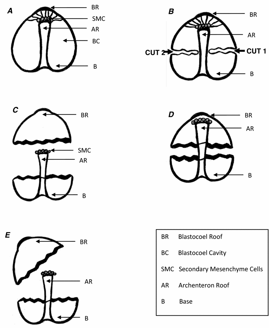
Embryos fixed in formaldehyde were dissected on Sigmacote® coated glass slides using fine Black Enameled insect pins, gauge size 00 (Australia, BioQuip, Gardena, CA, USA) according to Coyle-Thompson & Oppenheimer (Reference Coyle-Thompson and Oppenheimer2005). Before the microdissection step (Fig. 1) the embryos were washed five times in ASW by transferring them from one Petri dish to another. Washed embryos were dissected using a Swift SM90 Series Stereo dissecting microscope (Fisher Scientific, Pittsburgh, PA, USA) in such a way that archenterons and blastocoel roofs were separated. Glycosidases were then tested for their ability to prevent re-adherence of the separated archenteron and blastocoel roof (incubating the structures at 22°C with the enzymes under the previously specified conditions for 1–2 min or 12 h). The scoring of archenteron and blastocoel roof pairings as ‘attached’ (adhering) or ‘non-attached’ (non-adhering) was based on their adhesive stability while attempting to physically separate them (Coyle-Thompson & Oppenheimer, Reference Coyle-Thompson and Oppenheimer2005). Attempted pairings were recorded using a Canon PowerShot SD1400 IS 14.1 mega-pixel camera with a Zeiss (Oberkochen, Germany) Axiolab compound light microscope at ×100 magnification.
Diagrammatic representation of sea urchin embryo dissection using microdissecting needles. Intact embryo (a) was dissected by introducing two cuts in the blastocoel area (b) removal of archenteron from the roof of blastocoel (c). Treatment with (or without) enzyme and testing for adhesion (d); or non-adhesion (e). AR, archenteron; B, base; BC, blastocoel cavity; BR, blastocoel roof; SMC, secondary mesenchymal cells.

Statistical analysis
Statistical analyses were carried out using the IBM SPSS statistics V19 program (McCullagh & Nelder, Reference McCullagh and Nelder1989). The method of the analysis was a generalized linear model; more specifically a log–linear model, which was based on factorial experiments. The log–linear model was used because the distribution of the data was binary and the response variable was either yes or no (attached or not attached).
Results
Characterization of glycosidase activity
The commercially available glycosidases used in this study were first characterized for activity on defined substrates and fixed and live sea urchin material, and for purity and contaminating protease activity. The activities of native and denatured enzymes against defined substrates are shown in Table 1, with the native enzymes having activities close to their expected values (from supplier data sheets). Denatured enzymes showed little or no remaining glycosidase activity.
Comparison of the activity of five glycosidases (unit activities specified by Sigma-Aldrich) with that determined by our independent enzyme assays with active and denatured glycosidases in units/mg

The data reported above for each enzyme were used to calculate the amount of glycosidases for treating sea urchin embryo components.
Assessment of glycosidases purity and condition
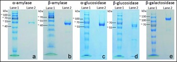
In order to check the purity of the glycosidases and to determine if the enzymes showed signs of physical degradation, each glycosidase was tested by SDS-PAGE under denaturing conditions. The amounts of enzyme tested ranged from 1.1 to 18 μg, as shown in Fig. 2. While there was no detectable smearing or degradation of the principal bands (the subunit components of the enzymes) trace bands of lower molecular weight were detected in the cases of β-amylase (Fig. 2 b), β-glucosidase (Fig. 2 d) and β-galactosidase (Fig. 2 e).
Sodium dodecyl sulphate-polyacrylamide (SDS-PAGE) gel electrophoresis of glycosidases. (a) α-Amylase: 3.75 μg; MW = 49 kDa (each subunit). (b) β-Amylase: 2.9 μg; MW = 50 kDa. (c) α-Glucosidase: 1.1 μg; MW = 68.5 kDa. (d) β-Glucosidase: 18 μg; MW = 65 kDa (each subunit). (e) β-Galactosidase: 10 μg = 116 kDa (each subunit). A PageRuler™ Prestained Protein Ladder is included in lane 1 of each gel (a–e).

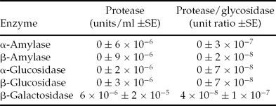
The glycosidases were tested for cryptic proteolytic activity against the substrate Azocoll™, an Azo dye-impregnated collagen, used as a chromogenic non-specific substrate for proteases, with released soluble proteolytic products detected by absorbance at 520 nm (Jiang et al., Reference Jiang, Tan, Ho and Ding2007). By comparison with a standard curve of known concentrations of Bacillus licheniformis protease as a control, the equivalent units of contaminating protease activity in each glycosidase was determined (Table 2). These data indicated the presence of low to undetectable levels of protease, with only β-galactosidase showing a measurable amount (6 × 10⁻⁶ equivalent units protease per assay) that was still not significantly different from zero (being within the standard error). We conclude that our enzymes are essentially free of protease for the purposes of these studies.
Protease contamination of glycolytic enzyme represented in unit equivalents of B. licheniformis protease per unit glycosidic enzyme

SE, standard error.
We also measured whether the β-glucosidase and β-galactosidase enzymes might be contaminated with an amylase-like or α-glucosidase-like activity by testing these enzymes against a defined substrate (starch) that should have yielded no product. Released reducing sugars would be detectable with the same colorimetric DNS assay used for α- and β-amylase. We determined that the levels of contamination were low, but might be significant over long time periods of incubation (6 × 10⁻³ ± 2 × 10⁻³ standard error (SE) contaminating amylase unit equivalents per unit β-glucosidase and 0.00 ± 5 × 10⁻³ SE contaminating amylase unit equivalents per unit β-galactosidase). In the case of β-galactosidase the levels of contaminating enzyme were undetectable but the standard error uncertainty was sufficiently high that we cannot conclude that either enzyme is essentially free of amylase-like or α-glucosidase contaminants. It was not possible for us to assess the levels of contaminating amylase enzyme in α-glucosidase, because the glucose released from starch by that enzyme is readily detected by the colorimetric DNS assay.
Glycosidase activity on fixed and live urchin embryos
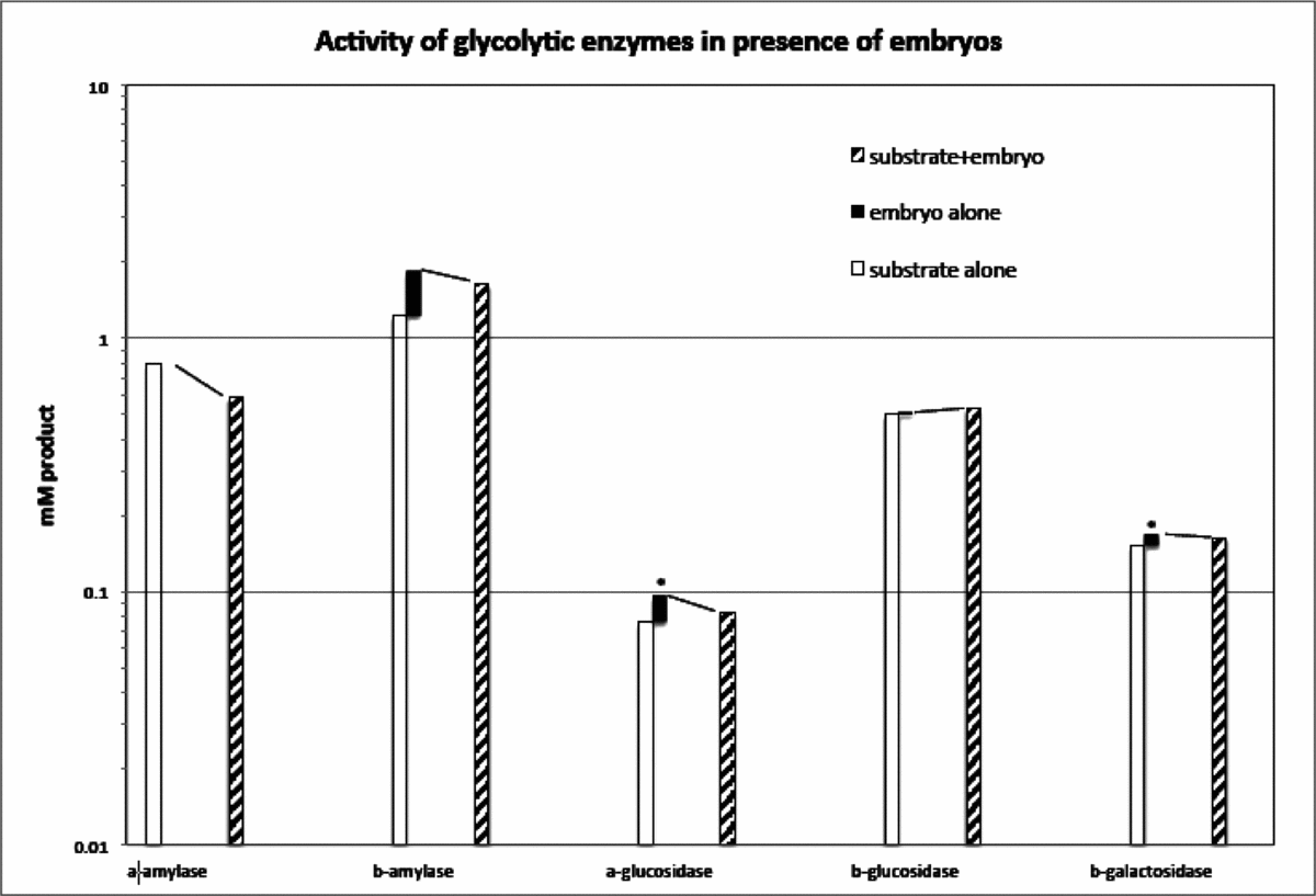
In order to understand the action of enzymes on fixed embryos, we assessed whether measurable amounts of maltose can be released from fixed sea urchin embryos through the actions of α- or β-amylase, and more generally whether the embryos release inhibitors of these and other enzymes that we used for this study. Fixed embryos were used as substrates for α- or β-amylase, and maltose release was detected by a colorimetric assay with 3,5-dinitrosalicylic acid. These results were compared with parallel assays with a defined substrate (starch) and a mixed substrate (both fixed embryo and starch). As shown in Fig. 3, between the two amylase enzymes only the β-amylase released measurable amounts of product from embryos alone (black bar) and α-amylase did not. However, there was no evidence of significant inhibition by the embryos of either the α- or β-amylase enzymes, as the sum of the assays using defined substrate (white bar) and mixed substrates (hatched bars) were similar (dashed connecting lines). β-Amylase released a substantial amount of maltose from fixed embryos (0.210 mg/ml), indicating that glycans with terminal amylose-like structures must exist in the embryo and be accessible to the enzyme even after formaldehyde fixation. The α-amylase and β-amylase enzymes were both capable of releasing maltose from disrupted live embryos at the gastrulation stage (Table 3), suggesting that the α-amylase enzyme has specific sites of cleavage that may become inaccessible during formaldehyde fixation.
Product obtained for glycosidases tested on live/fixed embryo material

Activity of glycosidases in the presence and absence of embryos. Product yield (log10 mM product) is indicated for each enzyme, with defined substrate (open bar), fixed embryo as substrate (closed bar), and combined defined substrate plus embryo (hatched bar). The sum of the open and filled bars is compared with that of the hatched bar (dashed lines). Background absorbance at the detection wavelength, in the absence of colorimetric substrate (*).

The α- and β-glucosidases and β-galactosidase may release monosaccharides from sea urchin glycans, but a direct test for these products is difficult to interpret in the context of the entire embryo being present because of high levels of background. However, the α-glucosidase, β-glucosidase, and β-galactosidase enzymes were tested in mixed reactions with their defined colorimetric substrates and fixed sea urchin embryos, and there was no evidence of inhibition of these enzymes (Fig. 3). There was also no evidence of natural pigments that could generate an artifact signal at the wavelengths used in colorimetry (410 and 540 nm).
Effects of glycosidases on adhesion of archenteron and blastocoel roof
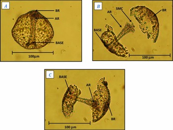
In previous studies we have shown that the blastocoel roof and archenteron show adherence in vitro after their dissection from fixed late gastrula stage embryos (Coyle-Thompson & Oppenheimer, Reference Coyle-Thompson and Oppenheimer2005). The strength of this interaction can be verified by attempting to separate physically the microscopic embryonic components (Fig. 4). In this study we measure the effect of each glycosidase on adhesion following enzymatic reactions of 1–2 min and 12 h incubation. The number of enzyme units used in these experiments for α-amylase, β-amylase, α-glucosidase and β-glucosidase was initially based on dose–response curves (Idoni et al., Reference Idoni, Ghazarian, Metzenberg, Hutchins-Carroll, Oppenheimer and Carroll2010) and subsequently increased if low enzyme concentration showed no effect on adherence.
Photographs of example of embryo dissection results. (A) Represents intact embryo with blastocoel roof (BR), archenteron (AR) and base. (B) Shows the dissected embryo with archenteron removed from the BR. At the tip of archenteron some secondary mesenchymal cells (SMC) are seen, which play an important role in cellular adhesion. (C) Shows the attachment of the dissected BR and the archenteron when they are brought back together in cases in which they did attach.

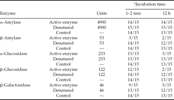
As shown in Table 4, β-amylase strongly inhibited the adherence of blastocoel roof and archenteron after only 1–2 min exposure (only three out of 15 trials showed adherence), compared with no enzyme or denatured enzyme controls (14 out of 15 trials showed adherence, the difference having a 99% statistical confidence P-value < 0.01). These results indicate that adherence is mediated in part by terminal amylose-like glycans, the specific substrate of β-amylase. Exposure to α-amylase did not generate any statistically significant effect on adhesion (with 14 out of 15 trials continuing to show adherence of blastocoel roof and archenteron after 12 h incubation). These results suggest a fundamental difference between the ‘exo’ β-amylases and ‘endo’ α-amylases in their ability to recognize or gain access to cleavage sites on the embryonic material.
Numbers of attached archenterons and roofs following treatment with glycosidases

a E.g. 14/15 means 14 microdissected embryos showed adhesion, out of 15 trials.
Number of enzyme units was based on demonstrated activity on the known substrates.
The α- and β-glucosidases both showed only slight effects on adherence after 1–2 min, but show highly significant effects after 12 h exposure (p < 0.01) whereas only two or three trials out of 15 showed retention of adherence (Table 4). The α-glucosidase enzyme hydrolyzes terminal d-glucose in an α-1,4 linkage and would be expected to degrade amylose-like glycans, albeit one monomer at a time. Almond β-glucosidase hydrolyzes terminal β-d-glucosides, β-d-galactosides, and β-d-fucosides, and so the observed reduction in adherence after 12 h may result from interference of recognition by C-type lectins specific for galactose (Kolatkar & Weis, Reference Kolatkar and Weis1996) and/or fucose (Lee et al. Reference Lee, Ichikawa, Fay, Drickamer, Shao and Lee2011). β-Galactosidase similarly had a small effect after 1–2 min (nine out of 15 trials showing adherence) and a significant effect (P < 0.01) on adhesion after 12 h exposure (three out of 15 trials showing adherence), and this could be explained as interference of an adhesion mediated by a C-type lectin specific for galactose.
An issue that complicates the interpretation of effects of α- and β-glucosidases and β-galactosidase on fixed embryos is the extended time required to detect interference. In all three cases it is possible that a contaminating protease was responsible for the gradual inhibition of adhesion, or that a contaminating glycosidase (e.g. a β-amylase activity) generated a gradual reduction in adhesion. For example, when tested against a starch substrate over a 12-h period, the β-glucosidase released an amount of reducing sugar equivalent to 1.4 mg maltose per ml, comparable in amount with that released in our β-amylase assay within minutes. It is conceivable that the starch substrate has a minor contaminant of cellulose, which could account for the released sugar, but we cannot exclude the possibility that the β-glucosidase has a minor contaminant that is only problematic in very long (e.g. 12 h) experimental incubations with embryos.
Discussion
We are not aware of any past studies that have investigated a model sea urchin embryo cellular interaction using all of the approaches included here: (1) the components of the cellular interaction were dissected out of the embryo proper allowing direct analysis of the adhesive interaction without the factors present in intact embryos that could confuse results on the sites of action of specific glycosidases; (2) the five glycosidases used here were independently characterized for activity on known substrates as well as on live and fixed sea urchin materials; (3) the glycosidases were examined electrophoretically for purity; (4) the glycosidases were examined for protease contamination using Azocoll™; (5) the glycosidases were tested on the components of the cellular interaction at 1–2 min and at 12 h incubation; (6) both untreated and heat denatured enzymes were tested for activity on known substrates and on live and fixed sea urchin material; and (7) both α- and β-glycosidases were used to suggest possible structural conformations of the glycans that may be involved in the cellular interaction.
The results indicate that only β-amylase was immediately active in inhibiting the cellular interaction and that it was essentially pure and active on both known substrate and sea urchin gastrula substrate. As the β-amylase inhibited the adhesive interaction within 1–2 min of incubation (rather than 12 h), we conclude that the enzyme was acting in accordance with its known specificities. It is well known that long term incubation might allow enzyme contaminants to influence the results (Chang, Reference Chang1983), and only β-amylase fulfilled the criterion of very rapid effect at relatively low numbers of units.
In contrast to these unambiguous results with β-amylase, the enzyme α-amylase had no measurable effect on adhesion even at a unit concentration nearly 100 times that used for β-amylase. We have shown that the α-amylase is not inhibited by embryonic materials (nor were the other four glycosidases in this study), so it is possible that the different results with β- and α-amylase are attributable to their respective ‘exo’ and ‘endo’ specificities on glycans. β-Amylase cleaves α-1,4 polyglycans but it only does so from exposed 1,4 terminal ends (Pili et al., Reference Pili, Chang, Partis, Mueller, Chrest and Passaniti1995), and α-amylase also cleaves α-1,4 glucose polymers (Elodi et al., Reference Elodi, Mora and Krysteva1972). Our results suggest that polyglucose residues are in a conformation with exposed α-1,4 terminal ends on the archenteron and/or blastocoel roof that can interact with β-amylase but not α-amylase.
Cleavage of internal α-1,4 linkages by α-amylase may reduce the overall size of a heavily branched glycan without having a qualitative effect on binding activity, whereas cleavage of terminal maltose residues by β-amylase may lead to the equivalent of a terminal dextran that has lost its adhesive properties. α-Amylase has been shown to be active on structures in sea urchin eggs (Daily et al., Reference Daily, Latham, Garcia, Hockman, Chun, Oppenheimer, West, Rostamiany, Chao, Pollock and Oppenheimer1994), but did not show any effect in the gastrula embryos studied here. It is possible that the different effects of α- and β-amylase are due to differences in enzyme penetration into formaldehyde-fixed embryos, or that the interior of a large branched glycan may be inaccessible after formaldehyde fixation while distal residues remain susceptible at least to β-amylase. The β-amylase results presented here are consistent with those found using intact embryos where β-amylase blocked archenteron organization and development and α-amylase did not (Idoni et al., Reference Idoni, Ghazarian, Metzenberg, Hutchins-Carroll, Oppenheimer and Carroll2010). However, in that study it was not possible to know where the enzyme acted in or on the intact embryos (Idoni et al., Reference Idoni, Ghazarian, Metzenberg, Hutchins-Carroll, Oppenheimer and Carroll2010) and in this work we demonstrate the site of action was clearly on adhesion between the surfaces of the fixed blastocoel roof and archenteron.
It is known that glycosyl transferases are present in sea urchin embryos (Evans & Bosmann, Reference Evans, Bosmann1977). There are concanavalin A-binding ligands and Lens culinaris-binding ligands (both have glucose- and mannose-binding specificity) on the outside and inside of sea urchin embryo surfaces (Latham et al., Reference Latham, Tully and Oppenheimer1999). In addition, the glucose-containing sea urchin embryo glycoprotein hyalin (Callebout et al., Reference Callebout, Gilges, Vignon and Mornon2000) blocked archenteron development and archenteron–blastocoel roof interaction (Razinia et al., Reference Razinia, Carroll and Oppenheimer2007; Ghazarian et al., Reference Ghazarian, Coyle-Thompson, Dalrymple, Hutchins-Carroll, Metzenberg, Carroll and Oppenheimer2009) and periodate-treated hyalin blocked hyalin's inhibitory activity (Ghazarian et al., Reference Ghazarian, Coyle-Thompson, Dalrymple, Hutchins-Carroll, Metzenberg, Carroll and Oppenheimer2009). Hyalin is a large glycoprotein (Carroll, Jr. et al., Reference Carroll, Hutchins-Carroll, Coyle-Thompson and Oppenheimer2008) that may contain the polyglucans involved in archenteron–blastocoel roof interaction. Taken together there is evidence that glucose-containing molecules are present on sea urchin embryo components and that they may be involved in the interaction between the archenteron and blastocoel roof (Latham et al., Reference Latham, Tully and Oppenheimer1999; Khurrum et al., Reference Khurrum, Hernandez, Eskalaei, Badali, Coyle-Thompson and Oppenheimer2004; Ghazarian et al., Reference Ghazarian, Coyle-Thompson, Dalrymple, Hutchins-Carroll, Metzenberg, Carroll and Oppenheimer2009; Idoni et al., Reference Idoni, Ghazarian, Metzenberg, Hutchins-Carroll, Oppenheimer and Carroll2010). The results presented here, however, are the best characterized and most direct evidence in implicating polyglucans with exposed α-1,4 terminal ends in the specific isolated cellular interaction.
The glycan target of β-amylase may be a branched structure similar to other adhesive glycans that range in size from 2 kDa to 580 kDa in sea urchin embryos (Papakonstantinou & Misevic, Reference Papakonstantinou and Misevic1993) and in marine sponges (Misevic & Burger, Reference Misevic and Burger1993; Garcia-Manyes, Reference Garcia-Manyes2006). Branched glycans can allow for a degree of adhesive specificity that could not be easily achieved by linear structures (Garcia-Manyes, Reference Garcia-Manyes2006; Taniguchi & Korekane, Reference Taniguchi and Korekane2011). While our results are consistent with a branched glycan structure, it is also possible that the target of β-amylase on the embryo is a much simpler molecule with as few as two d-glucose residues (Genghof et al., Reference Genghof, Brewer and Hehre1978). A growing body of recent evidence suggests that glycan structures are important in many types of cellular interactions (llyas et al., Reference Ilyas, Wallis, Soilleux, Townsend, Zehnder, Tan, Sim, Lehnert, Randeva and Mitchell2011; Vokhmyanina et al., Reference Vokhmyanina, Rapoport, Ryzhov, Korchagina, Pazynina, Severov, Kaltner, Andre, Gabius and Bovin2011; Handa & Hakomori, Reference Handa and Hakomori2012; Rachmaninov et al., Reference Rachmaninov, Zinger-Yosovich and Gilboa-Garber2012; Schultz et al., Reference Schultz, Swindall and Bellis2012; Yang et al., Reference Yang, Yu and Yang2012; Zheng et al., Reference Zheng, Jia, Zhao, Wei and Liu2012) and we believe that this is the first study that presents thoroughly characterized direct evidence for a role of polyglucans with exposed α-1,4 terminal ends in mediating a cellular interaction that has been of interest to developmental biologists for over a century (Herbst, Reference Herbst1900; Ernst, Reference Ernst1997). As the NIH designated model adhesive system described in this report is isolated from the embryo, making it one of the most accessible systems available anywhere for cell adhesion studies, continued work on this cellular interaction will provide important information on the role of specific glycans in mediating adhesive interactions of importance in many systems including humans (Davidson & Cameron, Reference Davidson and Cameron2002; Davidson, Reference Davidson2006).
Acknowledgements
This work was supported by grants from NIH NIGMS SCORE (S064680), NIH NIGMS RISE, NIH NIGMS MARC, the Joseph Drown Foundation, the Sidney Stern Memorial Trust; NSF US Presidential Award 0731633 to SBO, the CSUN office of Graduate Studies and Julie Gorchynski, MD Research Scholarships for Graduate Student Research. We would like to thank Dr Mark Steele and Arash Afsar for help with statistical analysis.