INTRODUCTION
Avian coccidiosis is caused by infection by species of Eimeria and is a major parasitic disease of poultry. It inflicts severe economic loss on the poultry industry, with an estimated worldwide annual loss of more than $3 billion (Shirley et al. Reference Shirley, Ivens, Gruber, Madeira, Wan, Dear and Tomley2004). Current conventional disease control strategies mainly rely on prophylactic medication. However, with the emergence of drug-resistant parasites and close attention to food safety, a novel control approach is needed as a substitute for prophylactic medication. Live oocyst vaccines are a useful alternative to prophylactic medication, but have disadvantages such as difficulty of conservation, diffusion of species, and danger from potential coccidiosis, and are not widely used on poultry. The development of subunit or recombinant vaccines are also limited. Therefore, novel control approaches are urgently need to study to control coccidiosis effectively (Dalloul and Lillehoj, Reference Dalloul and Lillehoj2005; Shirley et al. Reference Shirley, Smith and Tomley2005, Reference Shirley, Smith and Blake2007).
Eimeria belongs to the phylum Apicomplexa, which contains obligate intracellular parasites including other pathogens of medical and veterinary importance such as Toxoplasma, Plasmodium, Cryptosporidium, Sarcocystis and Neospora. These protozoan parasites are characterized by a peculiar organelle complex at their apical end (Blackman and Bannister, Reference Blackman and Bannister2001). The life cycle of Eimeria is complex, and consists of many different developmental stages. As some developmental stages have different morphological characteristics and living environments, different life-cycle stages are likely to have different gene expression profiles (Ng et al. 2002; Miska et al. Reference Miska, Fetterer and Barfield2004). Sporozoites are the first developmental stage to invade the epithelial cells lining the intestine. The differentially expressed genes of sporozoites might be involved in Eimeria invasion (Kinnaird et al. Reference Kinnaird, Bumstead, Mann, Ryan, Shirley, Shiels and Tomley2004; Wiersma et al. Reference Wiersma, Galuska, Tomley, Sibley, Liberator and Donald2004; Peroval et al. Reference Peroval, Pery and Labbe2006). Systematic isolation and identification of differentially expressed genes from the stages of Eimeria could provide important information about invasion processes and identify targets for new coccidiosis vaccines and drugs. Eimeria tenella is one of the most important species causing avian coccidiosis and is frequently used as a model to study species of Eimeria. It also is the only one for which the genome has been sequenced (http://www.genedb.org/Homepage/Etenella). In our previous report, differentially expressed genes of E. tenella sporozoites were analysed using suppression subtractive hybridization (SSH) and cDNA microarrays. Some expressed sequence tags (ESTs) with differential expression were found (Han et al. Reference Han, Lin, Zhao, Dong, Jiang, Xu, Zhu and Huang2010). BLAST searches showed that a protein encoded by one of the ESTs (clone number ZB1-H07, GenBank number: ES351365·1) shared significant identity with a calcium-dependent protein kinase (CDPK) of Toxoplasma gondii and other species.
The family of CDPKs contains unique enzymes found only in plants, green algae, ciliates and apicomplexan parasites, but not in bacteria, fungi, yeasts, worms and animals (Ishino et al. Reference Ishino, Orito, Chinzei and Yuda2006). CDPKs are special calcium-binding serine/threonine protein kinases which consist of several canonical characteristic domains including a N-terminal variable region, a kinase domain containing a serine/threonine protein kinase active site responsible for phosphorylating a target protein, a calmodulin-like domain containing 4 EF-hands responsible for calcium binding, a junction domain that connects the kinase domain and a calmodulin-like domain. The kinase activity of CDPKs is directly activated by calcium binding to the EF hands in the absence of calmodulin or phospholipids (Harper and Harmon, Reference Harper and Harmon2005). They are different from calmodulin-dependent protein kinase and protein kinase C, which play central roles in calcium signalling in mammals (Ishino et al. Reference Ishino, Orito, Chinzei and Yuda2006). Cytoplasmic calcium is widely used as a tightly regulated secondary signal, and calcium-binding proteins must be highly efficient in transmitting this signal to other enzymes (Reddy and Reddy, Reference Reddy and Reddy2004). CDPKs receive the calcium signal and enact a critical step in a series of intracellular cascades (Harper and Harmon, Reference Harper and Harmon2005). Recent research has revealed the function of several CDPKs in key parasitic life cycles, including T. gondii and Plasmodium (Ishino et al. Reference Ishino, Orito, Chinzei and Yuda2006; Lourido et al. Reference Lourido, Shuman, Zhang, Shokat, Hui and Sibley2010). Since no members of the CDPK family have been found in the animal hosts of apicomplexan parasites, these enzymes could be suitable targets for the development of novel anti-parasitic chemotherapeutics (Kieschnick et al. Reference Kieschnick, Wakefied, Narducci and Beckers2001). In this study, we applied the rapid amplification of cDNA ends (RACE) technique to clone the full-length cDNA sequence, based on the EST sequence described above, encoding a novel CDPK of E. tenella, designated EtCDPK3. The expression of EtCDPK3 was examined in different developmental stages. The involvement of EtCDPK3 in parasitic invasion and development was also investigated through inhibition tests and immunofluorescence analysis.
MATERIALS AND METHODS
Parasites and in vitro culture of E. tenella
Eimeria tenella was provided by the Shanghai Veterinary Research Institute, Chinese Academy of Agricultural Sciences and maintained and propagated by passage through coccidia-free 2-week-old chickens as previously described (Tomley, Reference Tomley1997). Unsporulated oocysts and sporulated oocysts were obtained and purified using standard procedures (Shirley, Reference Shirley, Coudert, Eckert, Braun and Shirley1995; Han et al. Reference Han, Lin, Zhao, Dong, Jiang, Xu, Zhu and Huang2010). Sporozoites were purified from cleaned sporulated oocysts by in vitro excystation and second-generation merozoites were collected from the caecal mucosa of chickens at 112 h post inoculation (Shirley, Reference Shirley, Coudert, Eckert, Braun and Shirley1995).
The chicken embryo fibroblast cell line DF-1, derived from East Lansing Line (ELL-0) chicken embryos, was used for infection, inhibition assays and immunofluorescence experiments (Jiang et al. Reference Jiang, Lin, Han, Zhao, Dong, Zhu and Huang2012a).
Cloning of EtCDPK3 entire cDNA
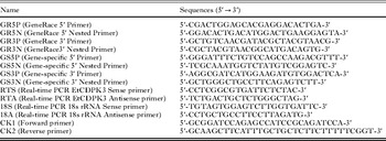
A 696 bp EST (GenBank number: ES351365·1) sequence for CDPK, a gene differentially expressed in E. tenella sporozoites, was obtained using SSH and cDNA microarrays (Han et al. Reference Han, Lin, Zhao, Dong, Jiang, Xu, Zhu and Huang2010). BLAST searches showed that the protein encoded by this EST sequence shared significant identity with the putative CDPKs of T. gondii, Perkinsus marinus and Plasmodium falciparum. The full-length 5′- and 3′- ends of the cDNAs were obtained by RNA ligase-mediated rapid amplification of cDNA ends (RACE) using the GeneRacer kit (Invitrogen, USA). All procedures were performed according to the manual instructions. Primers supplied with the kit were used (GR5P, GR5N, GR3P and GR3N) and gene-specific primers (GS5P, GS5N, GS3P and GS3N) are in Table 1. All the fragments were cloned into the pCR2·1-TOPO cloning vector (Invitrogen, USA) and sequenced. After aligning and assembling of sequences of the original EST, the 5′- and 3′-ends of cDNAs, the full-length cDNA sequence of EtCDPK3 gene was obtained and submitted to NCBI GenBank (Accession number: JQ679091).
Primer sequence and name

Sequence analysis, multiple sequence alignment, and phylogenetic analysis
The full-length cDNA sequence of EtCDPK3 gene was analysed for similarity using the BLAST programs at the National Center for Biotechnology Information (http://www.ncbi.nlm.nih.gov/BLAST/) and the genome sequence of E. tenella (http://www.genedb.org/Homepage/Etenella). The deduced amino-acid sequence, molecular mass and theoretical isoelectric point were obtained using translating tool software at the ExPASy server of the Swiss Institute of Bioinformatics (http://www.expasy.org/tools/protparam.html). The signal peptide, transmembrane (TM) regions and protein motifs were predicted using SignalP (http://www.cbs.dtu.dk/services/SignalP/), TMHMM (http://www.cbs.dtu.dk/services/TMHMM-2·0/) and Motifscan (http://hits.isb-sib.ch/cgi-bin/motif_scan), respectively. Multiple sequence alignment was performed by using the program Clustal W (http://www.ebi.ac.uk/Tools/msa/clustalw2/) and phylogenetic analysis was used by Clustal X and Mega 4·0 software using the neighbour-joining algorithm. Tree topology was evaluated by 1000-replication bootstrap.
Real-time quantitative PCR of E. tenella CDPK3 gene transcripts
The mRNA expression profiles of EtCDPK3 in different developmental stages (unsporulated oocysts, sporulated oocysts, sporozoites, merozoites and intracellular parasites) of E. tenella were examined using real-time quantitative PCR (qPCR). The qPCR method was performed using the SYBR1 Green I dye method. Total RNA was isolated with TRIzol reagent (Invitrogen, USA) and treated with DNase I (Invitrogen, USA). First-strand cDNA templates were synthesized from 2 μg of total RNA by SuperScript II reverse transcriptase (Invitrogen, USA) using random primers. A fragment from E. tenella 18S r RNA gene was used as a control. Primers for the EtCDPK3 gene (RTS, RTA) and 18S r RNA gene (18S, 18A) (Table 1) were designed manually using the Beacon Designer program (www.premierbiosoft.com). Each reaction was carried out in triplicate and each experiment was performed twice.
Expression and purification of recombinant EtCDPK3 protein
The entire EtCDPK3 open reading frame (ORF) was amplified by RT-PCR. Sequence-specific primers were designed to contain sites for restriction enzyme Bam HI in the forward primer (CK1) and Hind III in the reverse primer (CK2) (Table 1). Amplified fragments were cloned into pET28a (+) expression vectors (Novagen, USA) and transformed into E. coli BL21 (DE3) competent cell. Expression of recombinant protein was induced by isopropylthio-α-D-galactoside (IPTG) (Sigma, USA) and purified by His Bind Resin (Merck, Germany) under denaturing conditions (8 M urea), then urea was removed by gradient dialysis according to standard protocols (Wang et al. Reference Wang, Xu, Wu, Xu and Wang2005). Purified protein was stored in aliquots at −20 °C until further use.
Polyclonal sera against recombinant EtCDPK3
Recombinant EtCDPK3 (rEtCDPK3) protein was used to immunize a 2-month-old male rabbit by intraperitoneal injection of 0·2 mg purified protein emulsified in Freund's complete adjuvant (Sigma, USA). The rabbit was boosted 2 weeks later using the same conditions. This was repeated 3 times at intervals of 2 weeks with purified proteins emulsified in Freund's incomplete adjuvant (Sigma, USA). One week after the final immunization, rabbit antiserum against rEtCDPK3 was collected. The study was approved by the Institutional Animal Experiment Commission in accordance with the Chinese regulations of animal experimentation.
SDS-PAGE and Western blot analysis
Protein lysate from 4 different life-cycle stages of E. tenella (unsporulated oocysts, sporulated oocysts, sporozoites and merozoites) were prepared using cell lysis buffer for Western and IP (Beyotime, China), respectively. Protein concentrations were determined using a BCA protein assay kit (Beyotime, China). Then 100 μg protein lysate and 20 μg rEtCDPK3 were resolved by 10% SDS-PAGE and transferred to polyvinylidene difluoride (PVDF) transfer membranes (Millipore, USA). Western blot assays were performed according to standard procedures (Peroval et al. Reference Peroval, Pery and Labbe2006). Sporozite-immunized rabbit sera or rEtCDPK3-immunized rabbit sera were used at 1:100 dilution. Anti-His6 monoclonal antibody or mouse monoclonal anti-α-tubulin (at 1:2000 dilution) (Beyotime, China) or naive rabbit sera (1:100 dilution) were used as negative controls. Horseradish peroxidase-conjugated goat anti-rabbit IgG or goat anti-mouse IgG (1:2000 dilution, Sigma, USA) was added as the secondary antibody and detection used diaminobenzidine (DAB) (Sigma, USA) or the Super Signal® West pico chemiluminescence substrate (Thermo Scientific, USA).
Immunofluorescence of EtCDPK3 during first schizogony
Sporozoites were purified and incubated for 1 h at 41 °C in PBS or complete DMEM (Invitrogen, USA) and air-dried on a glass slide before fixation (Labbe et al. Reference Labbe, Peroval, Bourdieu, Girard-Misquich and Pery2006; Peroval et al. Reference Peroval, Pery and Labbe2006). Sporozoites incubated in complete DMEM were inoculated into DF-1 cells. At various times post-inoculation, DF-1 cells were collected and washed. Sporozoites and cells were fixed with 2% paraformaldehyde in PBS and permeabilized with 1% Triton X-100 in PBS for 15 min, then blocked with PBS containing 2% (w/v) bovine serum albumin (BSA) for 20 min. Antibody against recombinant EtCDPK3 diluted 1:100 was added and incubated for 1 h at 37 °C. Goat anti-rabbit IgG fluorescein isothiocyanate (FITC)-conjugated antibody (1:500 dilution; Sigma, USA) was added to the slides for 1 h at 37 °C. Cell nuclei were labelled with 10 μg mL−1 of DAPI (4,6-diamidino-2-phenylindole, Beyotime, China) for 10 min. After every step, the slides were washed 3 times in PBS with 0·05% Tween 20-PBS. Slides were mounted with 60 μL of Fluoromount Aqueous Mounting Medium (Sigma, USA).
Invasion inhibition assay
The invasion inhibition assay was based on the observation that E. tenella sporozoites invade DF-1 cells (Jahn et al. Reference Jahn, Matros, Bakulina, Tiedemann, Schubert, Giersberg, Haehnel, Zoufal, Mock and Kipriyanov2009; Jiang et al. Reference Jiang, Lin, Han, Zhao, Dong, Zhu and Huang2012a). All antibodies were purified by using Protein A + G agarose (Beyotime, China). Freshly isolated sporozoites were irreversibly labelled with the dye carboxyfluorescein diacetate succinimidyl ester (CFSE-Molecular Probes, Beyotime, China) according to the manufacturer's instructions. The labelled sporozoites were pre-incubated at 37 °C with 50 μg mL−1, 100 μg mL−1, 200 μg mL−1, 300 μg mL−1 or 400 μg mL−1 purified IgG against rEtCDPK3 for 2 h. The same quantity of purified IgG from naive rabbit sera, or non-treated sporozoites were used as controls. After culturing for 16 h at 41 °C, infected cells were trypsinized, washed and fixed in 2% paraformaldehyde in PBS for 10 min. Cells were washed with PBS and analysed by flow cytometry using a Cytomics FC 500 (Beckman Coulter, USA). As a control, non-infected DF-1 cells were also analysed. Infected cells, non-infected cells and free sporozoites were gated using RXP software for delineation and counting of infected (containing labelled sporozoites) and non-infected (fluorescence-free) cells. All assays were performed in triplicate. Deduced percentages of infected cells in the presence or absence of inhibitory antibody were used to calculate inhibition rates as previously described; the inhibition rates =100 × (1 − [%infected cellsAb/%infected cellsneg.control]) (Jahn et al. Reference Jahn, Matros, Bakulina, Tiedemann, Schubert, Giersberg, Haehnel, Zoufal, Mock and Kipriyanov2009).
RESULTS
Cloning and sequence analysis of EtCDPK3 entire cDNA
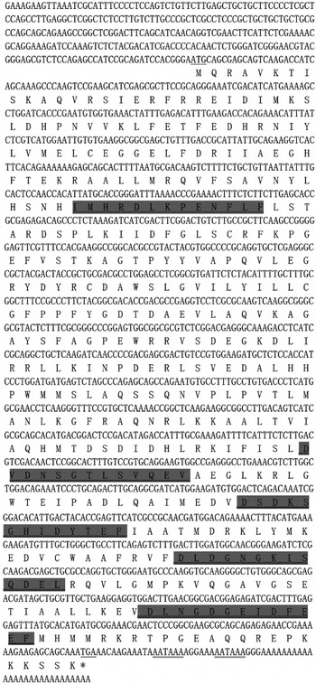
The 1637 nucleotides of the full-length cDNA were cloned and confirmed by RACE and sequencing based on the EST sequence. The full-length cDNA included a 5′-untranslated region (UTR) of 276 bp, a 3′-UTR of 59 bp with 2 canonical polyadenylation signal sequences AATAAA and a ploy(A) tail, and an ORF of 1302 bp (positions 277–1578) encoding 433 amino acids with a predicted molecular mass of 49·26 kDa and a theoretical isoelectric point of 6·00 (Fig. 1). The deduced amino-acid sequence analysis showed the protein had no signal peptide or TM regions, but had a N-myristoylation site. Motifscan predicted that the protein contained distinct characteristic domains of the calcium-dependent protein kinase, including an amino-terminal kinase domain containing a typical serine/threonine kinase active site and a carboxy-terminal calmodulin-like domain with 4 EF-hand motifs for calcium-binding (Fig. 1). By using Blast to search the E. tenella genome database (http://www.genedb.org/Homepage/Etenella), the ORF sequence of this gene had very high identity of about 99% (1301/1302 nucleotides) and the deduced amino acid sequence, 100% identity with 1 gene (ETH_00025085 on supercontig_156) encoding a putative CAM kinase, belonging to CDPK family. This gene was designated EtCDPK3 (GenBank Accession no. JQ679091).
Full cDNA and deduced amino-acid sequence of EtCDPK3. Underlined, start and stop codons; double underlined, classical polyadenylation signal in the 3′UTR; underlined and shaded, serine/threonine kinase active site; double underlined and shaded, 4 calcium-binding EF-hand motifs.

Database searches revealed that the amino acid sequence of EtCDPK3 was highly homologous to other apicomplexan CDPKs, including CDPKs from T. gondii (Genbank number: EEB00700, 76% identity and 87% similarity), P. marinus (Genbank number: EER18260, 60% identity and 79% similarity), P. falciparum (Genbank number: CAA68090, 57% identity and 77% similarity) and Neospora caninum (Genbank number: CBZ51964, 52% identity and 68% similarity).
Multiple sequence and phylogenetic analysis
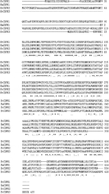
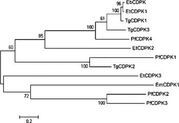
Multiple sequence analysis of the deduced amino acid sequence of EtCDPK3 and other known Eimeria CDPKs revealed that EtCDPK3 had a lower sequence identity to other Eimeria CDPKs, with only about 40% identity (Fig. 2). A phylogenetic tree was constructed using deduced amino-acid sequences from cloned apicomplexan CDPKs. The phylogenetic tree based on the amino-acid sequence of the protein kinase domains suggested that EtCDPK3 was related to E. maxima CDPK1 (EmCDPK1), P. falciparum CDPK2 (PfCDPK2) and P. falciparum CDPK3 (PfCDPK3) (Fig. 3).
Multiple alignment analysis of EtCDPK3 with other Eimeria CDPKs. Deduced protein sequences were used in analysis using the Clustal W sequences alignment program. Asterisk indicates identical amino acids. GenBank/EMBL database Accession numbers are: EtCDPK1 (E. tenella, ABA60894·1), EtCDPK2 (E. tenella, ABA60893·1), EmCDPK1 (Eimeria maxima, CAA96438·1), EbCDPK1 (Eimeria bovis, ACP74204·1).

Phylogenetic analysis of EtCDPK3 relative to CDPKs of other apicomplexan parasites based on amino-acid sequences of the protein kinase domains. Tree topology was evaluated by 1000 replications bootstraps. Numbers on branches are bootstrap support values. Sequences of other CDPKs were from Genbank; EtCDPK1 (Eimeria tenella, ABA60894·1), EtCDPK2 (E. tenella, ABA60893·1), EmCDPK1 (E. maxima, CAA96438·1), EbCDPK1 (E. bovis, ACP74204·1), PfCDPK1 (Plasmodium falciparum, CAA47704·1), PfCDPK2 (P. falciparum, CAA68090·1), PfCDPK3 (P. falciparum, AAF63154·1), PfCDPK4 (P. falciparum, CAD50923·1), TgCDPK1 (Toxoplasma gondii, AAG53993·1), TgCDPK2 (T. gondii, AAG53994·1) and TgCDPK3 (T. gondii, ABA60892·1).

EtCDPK3 transcript expression in different developmental stages of E. tenella
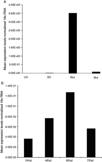
To determine the expression profile of EtCDPK3 in different development stages (unsporulated oocysts, sporulated oocysts, sporozoites, merozoites and intracellular parasites) of E. tenella, total RNA was extracted and subjected to qPCR analysis. Among 4 development stages (unsporulated oocysts, sporulated oocysts, sporozoites and merozoites), the mRNA level of EtCDPK3 was much higher in sporozoites than the other 3 stages, and the transcripts were undetectable in unsporulated oocysts (Fig. 4A). The EtCDPK3 mRNA levels in intracellular parasites at various times after sporozoites were incubated with DF-1 cells in vitro were also determined. The results showed that the expression level of EtCDPK3 in intracellular parasites increased from 24 h p.i. to 60 h p.i., reached the highest at 60 h p.i., then decreased at 72 h p.i. (Fig. 4B). So the expression level of this gene in E. tenella had stage specificity.
Quantitative real-time RT-PCR analysis of EtCDPK3 expression in different Eimeria tenella development stages. (A) UO, unsporulated oocysts; SO, sporulated oocysts; Spz, sporozoites; Mrz, merozoites. (B) Infected DF-1 cells were collected at different times (h) post-infection (p.i.).

Western blot analysis for recombinant EtCDPK3 and native protein of EtCDPK3
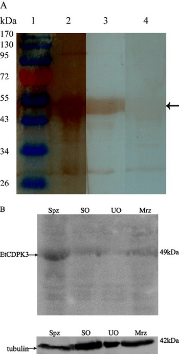
Western blot analysis showed the purified rEtCDPK3 was recognized by rabbit sera against sporozoite protein or anti-His6 monoclonal antibody. Naive rabbit sera failed to stain any protein band corresponding to the expected size of rEtCDPK3 (Fig. 5A). These results indicated that rEtCDPK3 reacted with antiserum generated against sporozoites. The presence of native EtCDPK3 in different stages of E. tenella was determined by immunoblotting using rabbit antiserum against rEtCDPK3. Mouse monoclonal anti-α-tubulin was used as control. Western blot analysis also showed that antibody against rEtCDPK3 could recognize a native protein of approximately 49 kDa in the lysate from different stages of E. tenella. The expression level of this protein in sporozoites was higher than in the other 3 stages, and was very weak in the unsporulated oocysts (Fig. 5B).
Western blot analysis. (A) Purified recombinant EtCDPK3(rEtCDPK3) protein (diaminobenzidine as substrate). Rabbit sera against sporozoites of Eimeria tenella or anti-His6 monoclonal antibody was used as primary antibody. Lane1, protein marker. Lane 2, anti-sporozoite serum. Lane 3, anti-his6 monoclonal antibody. Lane 4, naive rabbit serum. (B) Protein lysates from 4 different life-cycle stages of E. tenella (Super Signal® West pico-chemiluminescence as substrate). Rabbit serum against rEtCDPK3 was used as primary antibody. Spz, sporozoites; SO, sporulated oocysts; UO, unsporulated oocysts; Mrz, merozoites.

Immunofluorescence localization of EtCDPK3 in E. tenella-infected DF-1 cells
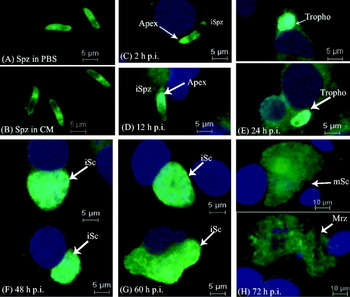
Using an antibody against rEtCDPK3, the localization of EtCDPK3 in sporozoites and during first schizogony in vitro was examined by immunofluorescence. EtCDPK3 had a dispersed distribution in the cytoplasm, especially near the nucleus and on the surface of whole sporozoites after incubation in PBS or complete medium (Fig. 6A and 6B). After sporozoites were added to DF-1 cells for 2 h and 12 h, EtCDPK3 was concentrated at the apical end of the intracellular sporozoites and the specific staining intensified (Fig. 6C and D). When sporozoites developed into trophozoites in DF-1 cells, the specific staining was more intense (Fig. 6E). At later times post-infection, the specific staining had a uniform distribution in immature and mature schizonts. Moreover, the staining intensified in immature schizonts (Fig. 6F and G) and decreased in mature schizonts (Fig. 6H). After the release of merozoites from mature schizonts, the labelling became very weak (Fig. 6H).
Localization of EtCDPK3 during in vitro DF-1 cell infection by indirect immunofluorescence using antibody against rEtCDPK3 protein. (A) Sporozoites (Spz) were incubated in PBS. (B) Spz incubated in complete medium (CM). Infected DF-1 cells were collected at different hours post-infection (p.i.). (C) 2 h p.i., intracellular sporozoites (iSpz). (D) 12 h p.i., intracellular sporozoites (iSpz). (E) 24 h p.i., trophozoites (Tropho). (F) 48 h p.i., immature schizonts (iSc). (G) 60 h p.i., immature schizonts (iSc). (H) 72 h p.i., mature schizonts (mSc) (up) and mature merozoites (Mrz) (down).

Inhibition of E. tenella invasion by antibodies against rEtCDPK3
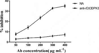
To evaluate EtCDPK3 in the invasion of DF-1 cells by E. tenella sporozoites, an invasion inhibition assay was performed by blocking protein function by pre-incubation of sporozoites with antibody against rEtCDPK3 (AbEtCDPK3) before DF-1 cell infection. The data showed that AbEtCDPK3 inhibited the invasion to 35% of the level seen with infection compared with non-treated sporozoites. The observed inhibition effect was dose dependent. Under the experimental conditions, an inhibition plateau of 33–35% was reached at an antibody concentration of 300 μg mL−1. By comparative analysis, the naive rabbit sera IgG did not have a significant effect on invasion (Fig. 7).
Inhibition of sporozoite invasion in vitro by antibody against rEtCDPK3. Anti-rEtCDPK3, IgG purified from rabbit antiserum against recombinant EtCDPK3 protein; NA, IgG from naive rabbit serum. All assays were performed in triplicate.

DISCUSSION
In this work, a new calcium-dependent protein kinase (EtCDPK3) of E. tenella was cloned and identified (GeneBank Accession number JQ679091). The entire cDNA of EtCDPK3 contains 1637 bp, with a 1302 bp ORF encoding 433 amino acids. Alignment of multiple sequences revealed that EtCDPK3 is highly homologous to putative CDPKs of T. gondii, P. marinus and other species. Sequence analysis indicated that the protein contained typical CDPK characteristic domains: a catalytic domain at the N-terminal protein kinase domain, a C-terminal calmodulin-like domain of 4 calcium-binding EF-hand motifs, a junction domain and a very short N-terminal variable region. By blasting the E. tenella genome database (http://www.genedb.org/blast/submitblast/GeneDB_tenella), the deduced amino-acid sequence had 100% identity with 1 gene (ETH_00025085 on supercontig_156) encoding a putative CDPK. All of the above results suggested that EtCDPK3 was a member of the CDPKs family.
CDPKs, encoded by multigene families, are widespread in plants and apicomplexans. By analysis of the apicomplexan genome database, P. falciparum contains 8 CDPK-like genes, C. parvum contains 7 (Billker et al. Reference Billker, Lourido and Sibley2009) and T. gondii contains over 20 CDPKs or CDPK-related kinases (Nagamune and Sibley, Reference Nagamune and Sibley2006). However, only 4 genes encoding for putative CDPKs of E. tenella were found in the E. tenella genome database (http://www.genedb.org/Homepage/Etenella), including EtCDPK3. In apicomplexans, CDPKs have been found to be involved in a number of critical events in the complex life cycles, including cell attachment and migration, cell invasion, egress, gametogenesis (Kieschnick et al. Reference Kieschnick, Wakefied, Narducci and Beckers2001; Billker et al. Reference Billker, Tewari, Franke-Fayard and Brinkman2004; Ishino et al. Reference Ishino, Orito, Chinzei and Yuda2006; Dvorin et al. Reference Dvorin, Martyn, Patel, Grimley, Collins, Hopp, Bright, Westenberger, Winzeler, Blackman, Baker, Wandless and Duraisingh2010) and also to participate in development (Choi et al. Reference Choi, Kim, Moon, Lee, Sattabongkot, Na, Kim, Suh, Kim, Cho, Lee, Rhie and Kim2010; Lourido et al. Reference Lourido, Shuman, Zhang, Shokat, Hui and Sibley2010). Moreover, some apicomplexans CDPKs have canonical domain structures, while others are more diverse. According to the number of EF hands, apicomplexan CDPKs were classified into 4 major classes (Billker et al. Reference Billker, Lourido and Sibley2009). The first class contains canonical CDPKs containing 4 C-terminal EF hands. The second class contains 3 C-terminal EF hands. The other 2 classes of CDPKs have several N-terminal EF hands. In addition, the first class of canonical apicomplexan CDPKs is divided into 2 groups. The first group contains relatively short N-termini that are predicted to be acylated and includes PfCDPK1, PfCDPK4, TgCDPK3, and TgCDPK1. The second group has somewhat longer N-termini of unknown function and is typified by PfCDPK3, PfCDPK5 and TgCDPK2 (Billker et al. Reference Billker, Lourido and Sibley2009). In this report, according to the motif scan results, EtCDPK3 belongs to the first group of canonical apicomplexan CDPKs.
The mRNA levels and protein expression of EtCDPK3 were examined in the 4 different developmental stages. Results from both of the qPCR and Western blot showed that the mRNA and protein levels of EtCDPK3 were found to the highest in sporozoites, and the lowest was found in unsporulated oocysts. These results showed that the EtCDPK3 gene is expressed predominantly at a distinct phase of the parasite life cycle, which was consistent with the previous reports. The EtCDPK3 gene was cloned by RACE based on a single EST, which was a differentially expressed gene obtained from sporozoites using SSH and cDNA microarrays. Furthermore, qPCR showed that the EST was highly expressed in sporozoites (Han et al. Reference Han, Lin, Zhao, Dong, Jiang, Xu, Zhu and Huang2010). Previous studies of apicomplexans found that each CDPK gene is expressed predominantly at a distinct phase of the parasite life cycle. For example, another CDPK isoform of E. tenella, EtCDPK1, was found to be expressed in sporulated oocysts, sporozoites and merozoites, but not present in unsporulated oocysts by Western blot (Dunn et al. Reference Dunn, Bumstead and Tomley1996). In P. falciparum, PfCDPK1 was first identified and found to be expressed in the asexual blood stages of the parasite, particularly in late-stage schizonts (Zhao et al. Reference Zhao, Franklin and Kappes1994a; Möskes et al. Reference Möskes, Burghaus, Wernli, Sauder, Durrenberger and Kappes2004). In contrast, PfCDPK3 is expressed specifically in the sexual erythrocytic stage (Li et al. 2000). PfCDPK4 was also only detected in P. falciparum gametocytes (Ranjan et al. Reference Ranjan, Ahmed, Gourinath and Sharma2009). In another apicomplexan, T. gondii, TgCDPK1 and TgCDPK3 are produced in the tachyzoite stage, but TgCDPK2 protein is not detectable in this stage (Kieschnick et al. Reference Kieschnick, Wakefied, Narducci and Beckers2001; Donald et al. Reference Donald, Zhong, Wiersma, Nare, Yao, Lee, Allocco and Liberator2006).
Using an antibody raised against the rEtCDPK3, we have shown by indirect immunofluorescence that the kinase is located on the surface and in the cytoplasm of E. tenella sporozoites. CDPKs are often found in several subcellular localizations (membranes, cytoplasm) and some isoforms are located in more than one compartment (Moskes et al. Reference Möskes, Burghaus, Wernli, Sauder, Durrenberger and Kappes2004). Indirect immunofluorescence also showed that the EtCDPK1 was situated around the apical end and generally on the surface of E. tenella sporozoites (Dunn et al. Reference Dunn, Bumstead and Tomley1996). PfCDPK1 has been shown to be present in the membrane and organelle fraction of the blood-stage parasites and in the membrane fraction of ring-stage infected erythrocytes (Zhao et al. Reference Zhao, Franklin and Kappes1994a, Reference Zhao, Franklin and Kappes1994b). Although these proteins have no transmembrane regions, they had a N-myristoylation site (Moskes et al. Reference Möskes, Burghaus, Wernli, Sauder, Durrenberger and Kappes2004). Myristoylation plays a vital role in membrane targeting and signal transduction in plant responses to environmental stress (Podell and Gribskov, Reference Podell and Gribskov2004).
EtCDPK3 localization in intracellular parasites was also investigated and it appeared to be mobilized to the end of the sporozoites to DF-1 cells. With the parasite developing in DF-1 cells, the specific staining becomes more intense in trophozoites and immature schizonts, but decreases in mature schizonts and merozoites. These results were confirmed by the qPCR analysis, which showed that the expression of EtCDPK3 increased significantly in immature schizonts of intracellular parasites (60 h p.i.), then decreased in mature schizonts (72 h p.i.). This has similarity to EtCDPK1 that was also located at the apical end of the sporozoites after addition to Mardin–Darby bovine kidney (MDBK) cells. However, the expression of EtCDPK1 was higher in mature schizonts than in immature schizonts. EtCDPK1 appeared to be specifically involved in sporozoite invasion of host cells and in the release of merozoites from mature E. tenella schizonts (Dunn et al. Reference Dunn, Bumstead and Tomley1996). These results suggest that EtCDPK3 is possibly related to the invasion and survival of the parasitic intracellular stages.
Western blot analysis showed that anti-rEtCDPK3 antibody could strongly recognize a main polypeptide of approximately 49 kDa, a similar molecular size to that of the rEtCDPK3 protein in different stages of E. tenella, and especially in sporozoites. Although the antibody faintly stained some other bands in the sporozoites and other stages, these bands were very weak and hardly visible. In addition, EtCDPK3 shares very low identities with other EtCDPKs isoforms. So we can assume that anti-EtCDPK3 antibody is specific to EtCDPK3 and the cross-reactivity with other EtCDPKs isoforms seems unlikely. Previous studies showed that invasion inhibition assays in vitro clearly reduced sporozoite invasion in the presence of monoclonal antibody or specific polyclonal antibody (Peroval et al. Reference Peroval, Pery and Labbe2006; Jahn et al. Reference Jahn, Matros, Bakulina, Tiedemann, Schubert, Giersberg, Haehnel, Zoufal, Mock and Kipriyanov2009; Jiang et al. Reference Jiang, Lin, Han, Zhao, Dong, Zhu and Huang2012a, b). In this study, invasion inhibition assays using antibody against rEtCDPK3 showed that this specific antibody blocked the invasion of sporozoites into cells. While further studies are needed to determine the exact function of EtCDPK3, because this kinase family is absent from hosts, it represents a valid target that might be exploitable for chemotherapy against apicomplexans.
ACKNOWLEDGEMENTS
This work was supported by grants from the National Natural Science Foundation of China (Grant No. 31201699), the Shanghai Science Foundation of China (Grant No. 09ZR1438700) and Central Public Welfare Research Institutions and Basic Scientific Research Business Expenses (Grant No. 2011JB01). We thank Zhixin Zhao, Beiming Zhang and Dingtao Liu for their excellent technical assistance.