Introduction
At least 20 phytophagous Cecidomyiidae (Diptera) species are known to occur on Vitis Linnaeus (Vitaceae) worldwide (Gagné and Jaschhof Reference Gagné and Jaschhof2017), including one species of Neolasioptera Felt (Cecidomyiinae: Alycaulini), one species of Rhizomyia Kieffer (Cecidomyi-inae: Brachineurini), one species of Ampelosucta De Stefani (Cecidomyiinae: Cecidomyiini), two species of Contarinia Rondani (Cecidomyiinae: Cecidomyiini), two species of Clinodiplosis Kieffer (Cecidomyiinae: Clinodiplosini), one species of Dasineura Rondani (Cecidomyiinae: Dasineurini), three species of Vitisiella Fedotova and Kovalev (Cecidomyiinae: Dasineurini), two species of Lasioptera Meigen (Cecidomyiinae: Lasiopterini), three species of Asphondylia Loew (Cecidomyiinae: Asphondyliini: Asphondyliina), and four species of Schizomyia Kieffer (Cecidomyiinae: Asphondyliini: Schizomyiina). Recently, an additional species of Asphondylia was found infesting grape berries in the southwestern United States of America and northern Mexico (Gagné et al. Reference Gagné, Kim, Uechi and Yukawa2018).
In Japan, Vitisiella oenephila (Haimhoffen) (previously in the genus Janetiella Kieffer) was listed by Matsumura (Reference Matsumura1898) with reference to grape pests in Europe, but this gall midge has been neglected in Japan because no distributional records existed from the country (Yukawa Reference Yukawa2014). In 1937, Asphondylia baca Monzen, a cecidomyiid species that induces fruit galls on Ampelopsis glandulosa (Wallich) Momiyama var. heterophylla (Thunberg) Momiyama (Vitaceae), was described from Japan and Korea (Monzen Reference Monzen1937). Later, Yukawa and Masuda (Reference Yukawa and Masuda1996) recorded cultivated Vitis vinifera Linnaeus as a host plant used by this species, and Sunose (Reference Sunose1992) and Uechi et al. (Reference Uechi, Yukawa and Yamaguchi2004) found that A. baca uses leaf buds of Weigela Thunberg (Caprifoliaceae) as overwintering hosts.
Conical leaf galls on wild Vitis coignetiae Pulliat ex Planchon, V. flexuosa Thunberg, and Vitis ficifolia Bunge and cultivated Vitis vinifera have been reported from Japan (e.g., Monzen Reference Monzen1929; Yukawa and Masuda Reference Yukawa and Masuda1996) and those on Vitis amurensis Ruprecht from Korea (Saito Reference Saito1932). Shinji (Reference Shinji1944) mentioned that a cecidomyiid species responsible for these conical leaf galls was very similar to “Dasineura vitis O.S.” though this Nearctic species (Gagné Reference Gagné2009) has not reported in these areas (Gagné and Jaschhof Reference Gagné and Jaschhof2017). In addition, this is probably a misidentification or misspelling of D. vitis Felt, although the shape of the galls is not similar to the Japanese species, or Schizomyia viticola (Osten Sacken), a Nearctic species associated with Vitis (Yukawa Reference Yukawa2014; Gagné and Jaschhof Reference Gagné and Jaschhof2017). Unfortunately, Shinji’s (Reference Shinji1944) work only illustrated the shape of the galls, not the adults or immature stages (Yukawa Reference Yukawa2014).
Such leaf galls were found again on cultivated V. coignetiae cultivar Suzumi-murasaki in Fukushima Prefecture, northern Honshu, and adults were successfully reared from the leaf galls on V. coignetiae in 2017, and later determined that this species belongs to the subtribe Schizomyiina (Asphondyliini) based on the distinctively longer female sternite VII than preceding sternites, the dorsally situated gonostyli of male terminalia, unfused gonostylus tooth, and ventrally situated larval anus (Tokuda Reference Tokuda2012). For these reasons, this species does not belong to the genus Dasineura of Dasineurini, which is a quite distinct group from Asphondyliini and is easily separated each other by many morphological features (e.g., Gagné Reference Gagné1989, Reference Gagné1994). Following further morphological examination of these gall midges and by comparison with the Nearctic Vitis-associated species of Schizomyiina, namely Schizomyia petiolicola Felt, S. viticola, S. vitiscoryloides (Packard), and S. vitispomum (Osten Sacken), we concluded that the Japanese species is new to science. Furthermore, the Vitis-associated species of Schizomyia, including the new Japanese species and three of the Nearctic species (i.e., S. viticola, S. vitiscoryloides, and S. vitispomum), should be removed from that genus and placed in the new genus, which is described here. We conducted a molecular phylogenetic analysis to establish that the new species is oligotrophic.
Materials and methods
Collecting gall midges and rearing methods
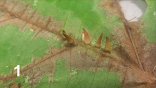
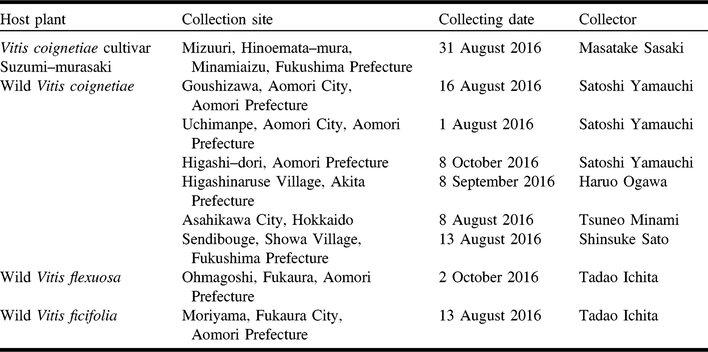
Approximately 800 conical leaf galls (Fig. 1) were collected in 2016 from wild V. flexuosa, V. ficifolia, V. coignetiae, and cultivated V. coignetiae cultivar Suzumi-murasaki in various localities of northern Japan (Table 1). Galls induced on V. coignetiae were collected in October 2016 from Higashi-dori, Aomori Prefecture were put in plastic cups (120 mm in diameter, 110 mm in depth) containing a mixture of peat moss and sand (1:1). Each cup was covered with a plastic lid, in which the central circle portion (35 mm in diameter) was punched 60–70 small holes. These cups were half buried in the soil and maintained from October 2016 to April 2017 at the Hikosan Biological Laboratory, Kyushu University, Fukuoka Prefecture (about 700 m in elevation) to allow mature larvae of the gall midge to overwinter under cool temperate conditions. In April 2017, the cups were brought back to the laboratory and maintained at room temperature to rear adults. Adults emerged in May 2017.
Conical leaf galls induced by Ampelomyia conicocoricis on Vitis coignetiae.

Collecting data of conical leaf galls induced by Ampelomyia conicocoricis on Vitis (Vitaceae) in Japan.

A subsampled of galls collected from separate localities were dissected to obtain larval specimens after measurements of gall length and diameter, while the remaining galls were used to rear adults following the methods mentioned above. The cups were kept in the Laboratory of Systems Ecology, Saga University, Saga Prefecture (about 5.5 m in elevation). Adults emerged in May and early June 2017. Specimens of mature larvae, adults, and pupal exuviae were preserved in 75% ethanol for morphological examination or in 99.5% ethanol for molecular phylogenetic study.
Morphological examination of gall midges
Specimens were mounted on slides in Canada balsam using the technique outlined in Gagné (Reference Gagné1994), except for the clearing step following Elsayed et al. (Reference Elsayed, Shimizu-Kaya, Itioka, Meleng, Yukawa and Tokuda2018). The slide-mounted specimens were examined under bright-field and phase-contrast microscopy (H550L, Nikon, Tokyo, Japan) and illustrations were made with the aid of a drawing tube. A semi-motorised fluorescence microscope (BX53, Olympus, Tokyo, Japan) was used to photograph some of the characters from mounted specimens with the aid of the microscope-attached colour camera (DP22, Olympus, Tokyo, Japan).
The morphological terminology follows McAlpine et al. (Reference McAlpine, Peterson, Shewell, Teskey, Vockeroth and Wood1981) for adults, but the thoracic sclerite “mesanepisternum” follows Gagné (Reference Gagné1968) and the wing venation follows Yukawa (Reference Yukawa1971). The larval and pupal terminology follows Gagné (Reference Gagné1994). The type materials including the holotype of the newly described species are deposited in the collection of the Entomological Laboratory, Faculty of Agriculture, Kyushu University, Japan (KUEC).
Specimens of three Vitis-associated species of Schizomyiina, that is, S. viticola, S. vitiscoryloides, and S. vitispomum, were examined in September 2016 at the National Museum of Natural History, Smithsonian Institution, Washington, District of Columbia, United States of America (USNM). Because only larval specimens of these Nearctic species were mounted on slides in good conditions and available for comparison, we distinguished species based on the larval characters. Adults of S. petiolicola Felt (Felt Reference Felt1908), purportedly from grape, do not match other Schizomyiina known from grape. Instead, the larvae and galls originally attributed to this species fit Neolasioptera vitinea Felt (Alycaulini). So, we consider that S. petiolicola is not associated with Vitis and the adults were a contamination in the rearing container. Moreover, S. petiolicola lacks the ventroapical extension on tarsomere I, which is characteristic of the new genus erected in this paper (R.J. Gagné, personal communication, Reference Gagné, Kim, Uechi and Yukawa2018). Thus, this species was excluded from the present study.
Genetic analysis of gall midge
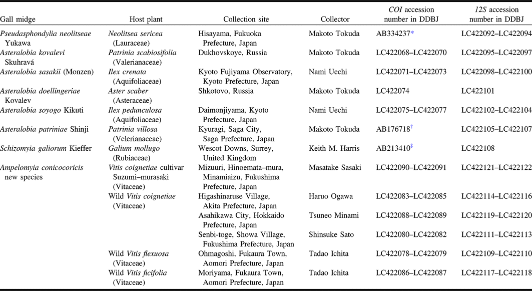
The total DNA was extracted from larval specimens collected from three host plant species in the four localities in Japan. Fragments of the mitochondrial genes, cytochrome oxidase subunit I (COI) and 12S small ribosomal subunit, were sequenced and aligned following Elsayed et al. (Reference Elsayed, Ogata, Kaburagi, Yukawa and Tokuda2017). The following sets were used for cytochrome oxidase subunit I (COI) gene by the primers: J–1718 (5′–GGA GGA TTT GGA AAT TGA TTA GTT CC–3′) (Simon et al. Reference Simon, Frati, Beckenbach, Crespi, Liu and Flook1994) and COIA (5’–CCC GGT AAA ATT AAA ATA TAA ACT TC–3′), COIS (5′–GGA TCA CCT GAT ATA GCA TTC CCA TAT TGG–3′) and COIA (Funk et al. Reference Funk, Futuyma, Orti and Meyer1995), or LCO1490 (5′–GGT CAA CAA ATC ATA AAG ATA TTG G–3′) and HCO2198 (5′–TAA ACT TCA GGG TGA CCA AA AAT CA–3′) (Folmer et al. Reference Folmer, Black, Hoew, Lutz and Vrijenhoek1994). The following primer set was used for 12S small ribosomal subunit gene: SR-J-14199 (5′–TAC TAT GTT ACG ACT TAT–3′) and SR-N-14594 (5′–AAA CTA GGA TTA GAT ACC C–3′) (Kambhampati and Smith Reference Kambhampati and Smith1995). The obtained sequences were deposited in the DNA Data Bank of Japan (DDBJ), European Molecular Biology Laboratory (EMBL), and GenBank (www.ncbi.nlm.nih.gov/genbank) under accession numbers shown in Table 2. The sequence data were analysed with the maximum likelihood method using MEGA (version 6.0) (Tamura et al. Reference Tamura, Stecher, Peterson, Filipski and Kumar2013). The software jModelTest 2 (Guindon and Gascuel Reference Guindon and Gascuel2003; Darriba et al. Reference Darriba, Taboada, Doallo and Posada2012) was used to determine the best model, which was general time reversible model and rates among sites gamma distributed rate variation among sites (GTR+G) on the basis of hierarchical likelihood ratio tests (hLRT). Sequences of Asphondylia gennadii Marchal (AB198012 and AB115580) were used as outgroup species. Asteralobia kovalevi Skuhravá, Asteralobia sasakii (Monzen), Asteralobia doellingeriae Kovalev, Asteralobia soyogo Kikuti, Asteralobia patriniae Shinji, Schizomyia galiorum Kieffer, and Pseudasphondylia neolitseae Yukawa were used as ingroup species beside the gall midges associated with Vitis from different localities in Japan: Hokkaido, Akita, Fukushima, and Aomori Prefectures.
Gall midge species used for genetic analysis.

DDBJ, DNA Data Bank of Japan.
*Tokuda et al. (Reference Tokuda, Yukawa and Suasa-ard2008); †Tokuda et al. (Reference Tokuda, Tabuchi, Yukawa and Amano2004); ‡Tokuda et al. (Reference Tokuda, Harris and Yukawa2005).
Ampelomyia Elsayed and Tokuda, new genus
http://zoobank.org/urn:lsid:zoobank.org:act:57BCB00A-B21C-44B5-83B6-BA13F82BE08F
Type species. A. conicocoricis Elsayed and x here designated.
Diagnosis. This genus belongs to the tribe Asphondyliini because of the following synapomorphies: the female sternite VII is distinctively much longer than preceding sternites, and the gonostyli are dorsally situated, short, and compact (Tokuda Reference Tokuda2012). The tribe Asphondyliini is divided into two subtribes, Asphondyliina and Schizomyiina (Gagné and Jaschhof Reference Gagné and Jaschhof2017). The new genus, Ampelomyia, belongs to the subtribe Schizomyiina because the larval anus is situated ventrally and the gonostylus tooth is unfused (Tokuda Reference Tokuda2012). Ampelomyia can be distinguished from other genera of Schizomyiina, such as Schizomyia and Asteralobia, by the following combination of characters: tarsomere I of all legs with ventroapical extension, which is common in Asphondyliina (Gagné Reference Gagné1994) and does not exist elsewhere in Schizomyiina, except Illiciomyia Tokuda (Tokuda Reference Tokuda2004) and two species of Schizomyia (Sousa and Maia Reference Sousa and Maia2007; Kolesik and Butterill Reference Kolesik and Butterill2015); the shifted dorsalmost pair of pleural papillae of the larval abdominal segment VIII to a dorsal position between the spiracles, which is unique for Schizomyiina; the shifted pair of dorsal papillae to the posteromedial lobe on the larval abdominal segment VIII. For these reasons, Ampelomyia was erected here as a new genus to science.
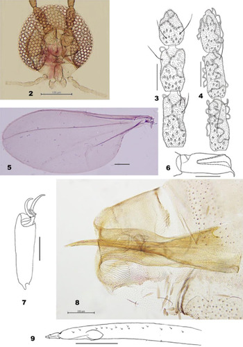
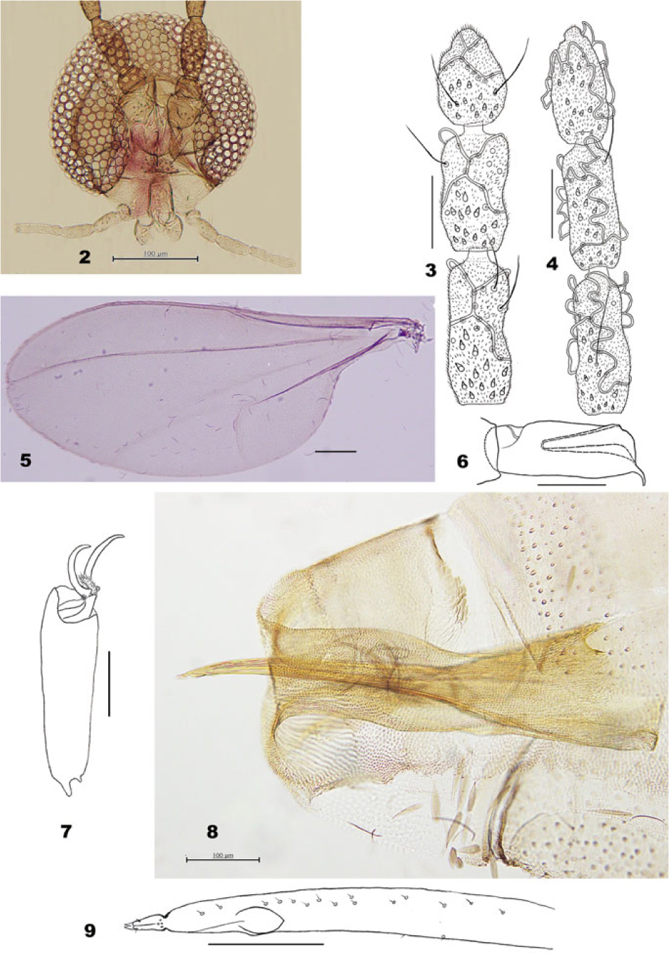
Description. Adult. Head. Compound eyes with rounded facets (Fig. 2). Mouthparts: palpus four-segmented, labrum triangular, labella well-developed. Antenna: flagellomeres I and II not fused; female flagellomeres with two connected rings of circumfila (Fig. 3); male flagellomeres with sinuous circumfila (Fig. 4).
Thorax. Wing (Fig. 5): R1 joining C before wing midlength, R5 joining C just before wing apex, C broken after the conjunction with R5; wing fold present; M3+4 forked with CuA. Legs with ventroapical extension on tarsomere I (Fig. 6); tarsal claws curved, untoothed; empodia less than half the length of claws, covered with long setulae; pulvilli short (Fig. 7).
Female abdomen. Ovipositor (Figs. 8–9): protrusible; with needle-like portion, broadly notched at base, with scattered fine setae posteriorly; ending with fused cerci.
Ampelomyia conicocoricis. 2, Head; 3, female terminal antennal flagellomeres; 4, male terminal antennal flagellomeres; 5, wing; 6, tarsomere I; 7, terminal tarsomere; 8, female post-abdomen showing the ovipositor; 9, the terminal part of the needle-like portion of the ovipositor. Scale bars = 50 µm but 100 µm for Figs. 2, 5, and 8.
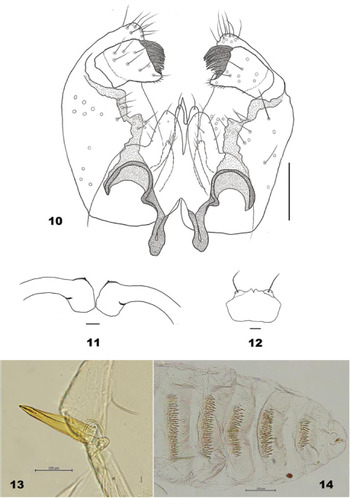
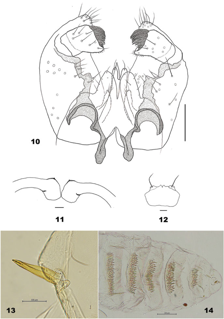
Male abdomen. Terminalia (Fig. 10): Gonocoxite extending ventrally as convex lobe beyond insertion of gonostylus. Gonostylus with unfused denticles. Cerci emarginate. Hypoproct bilobed. Aedeagus tapered apically.
Pupa. Exuviae not pigmented except the prothoracic spiracle and the abdominal dorsal spines. Four cephalic papillae present, two setose and two asetose (Fig. 12). Prothoracic spiracle short, pointed apically, with trachea extending to the tip (Fig. 13). Abdominal terga I–VIII each with anterior pair of trichoid sensilla and two pleural papillae. Four setose and two asetose dorsal papillae present on abdominal terga I–VII; only two setose dorsal papillae present on tergum VIII. Abdominal terga II–VIII with 4–5 rows of dorsal spines (Fig. 14).
Mature larva. Spatula bidentate or quadridentate, usually with anterior free portion more sclerotised than the posterior portion. Lateral papillae in two groups, each group with two setose and one asetose papillae. Two asetose ventral papillae present on all thoracic segments and abdominal segments I–VII. Anus located ventrally, with simple opening. Four setose pleural papillae present on all thoracic segments and abdominal segments I–VIII, dorsalmost pair of abdominal segment VIII shifted to dorsal position. A pair of dorsal papillae of abdominal segment VIII shifted posteriorly on a large mediodorsal lobe. Terminal segment with 8–12 terminal papillae, 2 corniform and at least 6 setiform.
Etymology. The generic name is composed of two Greek words, ampelos, meaning grapevine, and myia, meaning midge or fly. This generic name is feminine in gender.
Composition. There are four species in this new genus: A. conicocoricis Elsayed and Tokuda new species; Ampelomyia viticola (Osten Sacken) new combination, A. vitiscoryloides (Packard) new combination, and A. vitispomum (Osten Sacken) new combination.
Ampelomyia conicocoricis Elsayed and Tokuda, new species
http://zoobank.org/urn:lsid:zoobank.org:act:B744EA8E-530C-45F5-A8CB-4A4565A6FF16
Dasineura vitis: Shinji Reference Shinji1944 (misidentification).
Description. Head (Fig. 2). Compound eyes separated on vertex by diameter of 0.25–1.5 facets, eye bridge 6–7 facets long. Frontoclypus with 25–37 setae (n = 13). Mouthparts: palpus four-segmented, first approximately 27.4 μm (24–33 μm, n = 11), second approximately 62 μm (57–70 μm, n = 11), third approximately 68.1 μm (51–82 μm, n = 11), fourth approximately 95 μm (80–110 μm, n = 11); labrum with microtrichous edges; labella setose. Antenna: the terminal flagellomere with apical constriction in both sexes (Figs. 3 and 4).
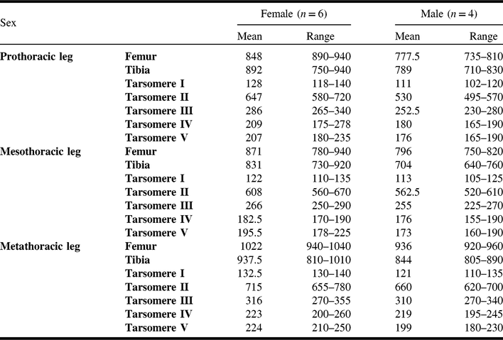
Thorax. Wing (Fig. 5): CuA and M3+4 weak. Legs almost equal in length (Table 3). Anepimeral setae 22–32 (n = 12); mesanepisternum scales 23–43 (n = 8); and lateral scutal setae 101–128 (n = 11).
Measurements of leg length (µm) in Ampelomyia conicocoricis.

Female abdomen. Tergites with anterior pair of trichoid sensilla; tergites I–VII rectangular; tergites I–VI with one posterior row of setae and some lateral median setae; tergite VII with 2–3 posterior rows of setae, scattered setae on the posterior half and fewer scales anteriorly than the preceding; tergite VIII sclerotised only anteriorly, without dorsal lobes on the posterior margin. Sternites II–VII with pair of median anterior trichoid sensilla; sternites II–VI bare and less pigmented medially, with several scattered setae and scales on the anterior half, and 1–2 rows of setae posteriorly; sternite VII about two times as long as preceding; posterior two-thirds covered with setae. Ovipositor (Figs. 8–9): protrusible portion about twice as long as sternite VII.
Male abdomen. Tergites I–VII as in female. Tergite VIII weakly sclerotised medially, about 0.25 times as wide as tergite VII and with anterior pair of trichoid sensilla. Sternites II–VII as sternites II–VI in female; sternite VIII setose. Terminalia (Fig. 10): gonocoxite about two times as long as width. Gonostylus about 1.5 times longer than wide, pointed anteroapically. Cerci without blunt strong setae. Hypoproct with one strong seta on each lobe. Parameres about half as long as aedeagus.
Pupa (Figs. 11–14). Antennal horns slightly developed. Four frontal papillae present, two setose and two asetose; three lateral facial papillae present, one with short seta and two without setae. Abdominal segments covered with spinules, except posterior third of each segment.
Ampelomyia conicocoricis. 10, Male terminalia; 11, pupal antennal horns; 12, pupal cephalic plate; 13, pupal prothoracic spiracle; 14, dorsal view of the pupal posterior abdominal segment. Scale bars = 50 µm but 100 µm for Figs. 13–14.
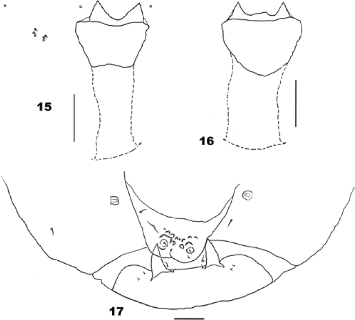
Mature larva. Cylindrical, yellow in colour, body length about 2 mm. Spatula bidentate (Figs. 15–16). Anus with one setose anal papilla on each side. Two setose sternal papillae present on all thoracic segments and abdominal segments I–VIII. Mediodorsal lobe of abdominal segment VIII notched apically, with pointed verrucae. Terminal segment with two corniform, eight setose, and two usually asetose terminal papillae (Fig. 17).
Ampelomyia conicocoricis larval characters. 15–16, Spatula; 17, dorsal view of the abdominal segment VIII and the terminal segment. Scale bars = 50 µm.

Etymology. The species name is composed of two Greek words, konikos meaning conical and korykis meaning leaf galls.
Type material. Holotype: 1♂ (KUEC): reared from conical leaf galls on the wild Vitis coignetiae (Vitaceae) by A.K. Elsayed, collected by Satoshi Yamauchi from Uchimanpe, Aomori City, Aomori Prefecture, Japan, on 1.viii.2016, emerged on 23.v.2017. Paratypes: all paratypes were reared from conical leaf galls on the wild V. coignetiae in Japan. Five larvae: collected from Higashinaruse Village, Akita Prefecture, on 8.ix.2016, Haruo Ogawa, dissected on 14.ix.2016 by A.K. Elsayed; one larva: collected from Goushizawa, Aomori City, Aomori Prefecture, on 16.viii.2016, Satoshi Yamauchi, dissected on 21.viii.2016 by A.K. Elsayed; one adult ♂: collected from Higashinaruse Village, Akita Prefecture, on 8.ix.2016, Haruo Ogawa, emerged on 18.v.2017; four pupal exuviae and one adult ♂: collected from Higashinaruse Village, Akita Prefecture, on 8.ix.2016, Haruo Ogawa, emerged on 19.v.2017; one adult ♂: collected from Higashinaruse Village, Akita Prefecture, on 8.ix.2016, Haruo Ogawa, emerged on 23.v.2017; two pupal exuviae and three adult ♂: collected from Higashinaruse Village, Akita Prefecture, on 8.ix.2016, Haruo Ogawa, emerged on 22.v.2017; one adult ♀: collected from Uchimanpe, Aomori City, Aomori Prefecture, Japan, on 10.viii.2016, Satoshi Yamauchi, emerged on 23.v.2017; four adult ♀: collected from Uchimanpe, Aomori City, Aomori Prefecture, Japan, on 10.viii.2016, Satoshi Yamauchi, emerged on 24.v.2017.
Distribution. Japan: Hokkaido, Aomori, Fukushima, and Akita Prefectures. In addition, Saito (Reference Saito1932) reported similar cecidomyiid galls on V. amurensis from Korea, but further studies are needed to confirm the taxonomic position of this gall midge.
Natural history. Ampelomyia conicocoricis is univoltine and induces conical leaf galls on Vitis coignetiae, V. flexuosa, and V. ficifolia in Japan. Mature galls can be collected from August to October, with diameter 2.19–5.15 mm and length 6.64–10.84 mm. Larvae overwinter in the galls on the fallen leaves and pupate in the galls. Adults emerge from galls in May and early June.
Remarks. Three Nearctic species associated with Vitis species, namely S. viticola, S. vitiscoryloides, and S. vitispomum, also have a ventroapical spur on tarsomeres I. In addition, on abdominal segment VIII of their larvae, the dorsalmost pair of pleural papillae shifted to dorsal position and a pair of dorsal papillae to the mediodorsal lobe. Based on these characters, we also place these species within Ampelomyia.
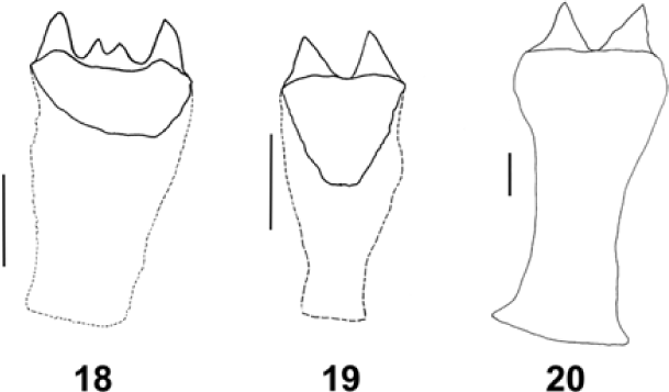
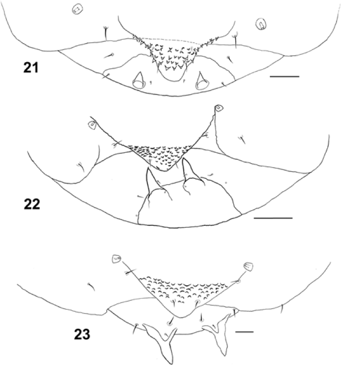
The newly described species A. conicocoricis can be separated easily from the other three Nearctic species based on the larval characters as follows: A. conicocoricis have a notched apically mediodorsal lobe on abdominal segment VIII and only two setose sternal papillae on the abdominal segments I–VIII, while the other three Nearctic species have an entire mediodorsal lobe on the abdominal segment VIII and four asetose sternal papillae on abdominal segments I–VIII; A. conicocoricis, A. vitiscoryloides, and A. vitispomum have bidentate spatulae, but A. conicocoricis has a wider gap between the anterior lobes than the other two species, while A. viticola has a quadridentate spatula (Figs. 18–20); A. conicocoricis has setose anal papillae and 12 terminal papillae, but the three Nearctic species possess asetose anal papillae and at most 10 terminal papillae (2 corniform and 8 setiform papillae in A. viticola; 2 corniform, 6 setiform, and 2 asetose papillae in A. vitiscoryloides; 2 corniform and 6 setiform in A. vitispomum (Figs. 21–23)); A. conicocoricis induces conical leaf galls, similar to those of A. viticola, but A. vitispomum and A. vitiscoryloides induce apple-like and hazelnut-shaped stem galls, respectively (Gagné Reference Gagné1989). For these reasons, we consider A. conicocoricis a new species.
The larval spatula of the Nearctic Ampelomyia. 18, Ampelomyia viticola; 19, A. vitiscoryloides; 20, A. vitispomum. Scale bars = 50 µm.

Dorsal view of the larval abdominal segment VIII and the terminal segment of the Nearctic Ampelomyia. 21, Ampelomyia viticola; 22, A. vitiscoryloides; 23, A. vitispomum. Scale bars = 50 µm.

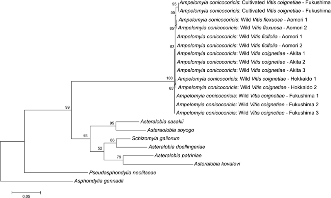
A phylogenetic reconstruction based on partial sequences of cytochrome oxidase subunit I (COI) and 12S small ribosomal subunit genes. The topology and branch length were produced by the maximum likelihood method (note the scale bar). Bootstrap values are indicated at branches gaining more than 50% support (103 replications). The sequences in clade of Ampelomyia conicocoricis includes the host plant name and locality.

Molecular phylogenetic analysis. The clade of A. conicocoricis was distinctly separated from the clade including species of Asteralobia and Schizomyia, being supported with 100% bootstrap value (Fig. 24). Genetic divergence among individuals of A. conicocoricis on cultivated V. coignetiae and wild Vitis species was very small (ranging from 0.00 to 0.69%), indicating that they are identical species.
Discussion
In this study, we place three Nearctic species, previously in the genus Schizomyia, within Ampelomyia based on larval characters and tarsomere I. Because we studied in detail only larval material, adults of those species may shed more light on our observations and collecting new specimens is required to analyse the phylogenetic relationships of the genus.
Although pupation inside galls in Asphondyliini usually results in size diminishment of the larval terminal abdominal segment and degeneration of its papillae (Möhn Reference Möhn1961), the larval terminal segment of Ampelomyia is normal in size and the number of papillae is not degenerated. This may imply that Ampelomyia has evolved its pupation habit inside galls relative recently. The pointed verrucae of the mediodorsal lobe on the abdominal segment VIII, the sclerotisation of the dorsomedially part of the terminal segment, and the unusual number of terminal papillae suggest that they all together play an important role in the larval movement inside galls.
Although the number of the larval dorsal terminal papillae in Cecidomyiidae is eight or less (R.J. Gagné, personal communication, Reference Gagné, Kim, Uechi and Yukawa2018), at least three Ampelomyia species, A. conicocoricis, A. viticola, and A. vitiscoryloides, are found to have more than eight terminal papillae in the present study, and they represent the first examples in Cecidomyiinae of apparent papillae rising de novo. These extra papillae are without setae so one might wonder whether they are anal papillae shifted from the venter. This does not appear likely as two or four pairs of anal papillae are still present in the genus. Four anal papillae are the most reported for Asphondyliini (Möhn Reference Möhn1961) so they could not be the origin of the extra papillae found in A. conicocoricis.
Molecular phylogenetic analysis supported the results of our morphological examination and showed that A. conicocoricis specimens collected from three Vitis species in Japan had a genetically very close pairwise divergence distances of less than 1%. This indicates that A. conicocoricis is an oligophagous species, and further molecular phylogenetic study using more samples collected from different hosts in Japan is needed to clarify the host range of A. conicocoricis on Japanese Vitis.
Acknowledgements
We thank Raymond J. Gagné (Systematic Entomology Laboratory, United States Department of Agriculture, Washington, District of Columbia, United States of America) for his valuable comments on an early draft, information about Schizomyia petiolicola, and fruitful discussions about larval characters. We also thank Yukio Nagano (Analytical Research Center for Experimental Sciences, Saga University) for his careful assistance in molecular studies. We are grateful to Tadao Ichita, Tsuneo Minami, Haruo Ogawa, Masatake Sasaki, Shinsuke Sato, and Satoshi Yamauchi for collecting galls, and Daisuke Yamaguchi (Kyushu University) for rearing larvae during the winter.