Introduction
Classical swine fever virus (CSFV) is an enveloped single positive stranded RNA virus of the Genus Pestivirus within the Flaviviridae family (Lindenbach et al., Reference Lindenbach, Murray, Thiel, Rice, Knipe and Howley2013). It causes the eponymous notifiable disease of domestic and feral pigs and can cause serious economic losses.
The genome comprises one large open reading frame (ORF) flanked by two non-translated regions (NTRs) (Rümenapf et al., Reference Rümenapf, Meyers, Stark and Thiel1991; Collett, Reference Collett1992). The ORF codes for a polyprotein of about 3900 amino acids which is co- and post-translationally processed by viral and cellular proteases into eleven viral proteins (Thiel et al., Reference Thiel, Stark, Weiland, Rümenapf and Meyers1991; Rümenapf et al., Reference Rümenapf, Unger, Strauss and Thiel1993; Tautz et al., Reference Tautz, Elbers, Stoll, Meyers and Thiel1997; Xu et al., Reference Xu, Mendez, Caron, Lin, Murcko, Collett and Rice1997; Bintintan and Meyers, Reference Bintintan and Meyers2010). The core (C) protein and the three envelope glycoproteins Erns, E1, and E2 constitute the viral structure. Furthermore, seven non-structural (NS) proteins are encoded, namely Npro, p7, NS2-3, NS4A, NS4B, NS5A, and NS5B (Thiel et al., Reference Thiel, Stark, Weiland, Rümenapf and Meyers1991; Elbers et al., Reference Elbers, Tautz, Becher, Stoll, Rumenapf and Thiel1996; Meyers et al., Reference Meyers, Thiel and Rumenapf1996; Lattwein et al., Reference Lattwein, Klemens, Schwindt, Becher and Tautz2012).
Over the past decades, different genomic regions were routinely sequenced for virus characterization and molecular epidemiology: (i) a 150 nucleotide (nt) fragment of the 5′-NTR; (ii) a 190 nt fragment of the E2 encoding region (E2 is the main immunogen of CSFV and indispensable for virus entry); and (iii) a 409 nt fragment of the polymerase gene NS5B (Paton et al., Reference Paton, McGoldrick, Greiser-Wilke, Parchariyanon, Song, Liou, Stadejek, Lowings, Bjorklund and Belak2000). A vast collection of these sequences is held at the European Union and OIE Reference Laboratory for Classical swine fever (CSF) in Hannover (Greiser-Wilke et al., Reference Greiser-Wilke, Depner, Fritzemeier, Haas and Moennig1998; Dreier et al., Reference Dreier, Zimmermann, Moennig and Greiser-Wilke2007). However, approaches using longer fragments or even whole-genomes have now become feasible due to the technological advances and thus economic solutions. Recently, full-length E2 encoding sequences were recommended for reliable in detail phylogenetic analyses (Postel et al., Reference Postel, Schmeiser, Bernau, Meindl-Boehmer, Pridotkas, Dirbakova, Mojzis and Becher2012), and next-generation sequencing has been employed to provide full-length sequences that allow studying, e.g. the role of quasispecies and virulence determinants (Töpfer et al., Reference Töpfer, Höper, Blome, Beer, Beerenwinkel, Ruggli and Leifer2013; Fahnoe et al., Reference Fahnoe, Pedersen, Risager, Nielsen, Belsham, Hoper, Beer and Rasmussen2014).
Accessible data on the current diversity of CSFV strains and their geographical distribution on a global scale are missing. For this reason, published reports on phylogenetic analyses of CSFV strains over the past two decades were compiled. Apart from molecular epidemiology, sequence data can give an insight in virulence determinants. In this review, a brief overview is given on current knowledge with regard to genetic virulence markers and the impact of quasispecies variability.
Diversity and distribution
CSFV can be assigned to three genotypes with three to four sub-genotypes (Lowings et al., Reference Lowings, Ibata, Needham and Paton1996; Paton et al., Reference Paton, McGoldrick, Greiser-Wilke, Parchariyanon, Song, Liou, Stadejek, Lowings, Bjorklund and Belak2000). Only recently, a new sub-genotype was added to genotype 1 (Postel et al., Reference Postel, Schmeiser, Perera, Rodriguez, Frias-Lepoureau and Becher2013b). The newly formed sub-genotype 1.4 comprises Cuban isolates that were so far placed into sub-genotype 1.2 (de Arce et al., Reference de Arce, Ganges, Barrera, Naranjo, Sobrino, Frias and Nunez2005; Pereda et al., Reference Pereda, Greiser-Wilke, Schmitt, Rincon, Mogollon, Sabogal, Lora, Sanguinetti and Piccone2005). Regrouping was done after phylogenetic analyses of full-length E2 sequences revealed that these isolates formed a separate sub-genotype and were quite divergent from other genotype 1 strains. An additional differentiation and sub-grouping was suggested for sub-genotypes 2.1 (Cha et al., Reference Cha, Choi, Park, Yoon, Kwon, Yoon and Song2007; Chen et al., Reference Chen, Hu, Zhang, Shuai, Jiang and Fang2008; Jiang et al., Reference Jiang, Gong, Li, Liu, Hu, Ge, Wang, Yu and Tu2013) and 2.3 (Blome et al., Reference Blome, Grotha, Moennig and Greiser-Wilke2010). However, these systems were not internationally harmonized (e.g. clades/clusters 2.1a–2.1c, cluster 2.3.1 and 2.3.2). The latter sub-genotype was also subdivided within the EU based on clusters or lineages observed in the 5′-NTR-based phylogenetic tree. These clusters or lineages (e.g. 2.3 ‘Rostock’) were mainly named after German regions (Wonnemann et al., Reference Wonnemann, Floegel-Niesmann, Moennig and Greiser-Wilke2001), and are sometimes still found in the literature (Fritzemeier et al., Reference Fritzemeier, Teuffert, Greiser-Wilke, Staubach, Schluter and Moennig2000; Depner et al., Reference Depner, Strebelow, Staubach, Kramer, Teuffert, Botcher, Hoffmann, Beer, Greiser-Wilke and Mettenleiter2006; Simon et al., Reference Simon, Le Dimna, Le Potier and Pol2013). The most divergent strains are the genotype 3 CSFV strains ‘Kanagawa’ (Japan, 1974) and ‘Congenital Tremor’ (United Kingdom, 1964). The former has been suggested as harmonized outgroup strain for phylogenetic analyses (Paton et al., Reference Paton, McGoldrick, Greiser-Wilke, Parchariyanon, Song, Liou, Stadejek, Lowings, Bjorklund and Belak2000).
The distribution of genotypes shows a distinct geographical pattern (Björklund et al., Reference Björklund, Lowings, Stadejek, Vilcek, Greiser-Wilke, Paton and Belak1999; Paton et al., Reference Paton, McGoldrick, Greiser-Wilke, Parchariyanon, Song, Liou, Stadejek, Lowings, Bjorklund and Belak2000) that changed only little over time. On a global scale, the most prevalent genotype over the past two decades was undoubtedly genotype 2, especially sub-genotypes 2.1 and 2.3. However, field isolates from South and Central America, and the American continent in general, were all placed into genotype 1 (Pereda et al., Reference Pereda, Greiser-Wilke, Schmitt, Rincon, Mogollon, Sabogal, Lora, Sanguinetti and Piccone2005). While strains from Argentina, Brazil, Colombia, and Mexico clustered in sub-genotype 1.1, the strains from Honduras and Guatemala were classified as sub-genotype 1.3. As mentioned above, Cuban isolates were placed into sub-genotype 1.2, but were determined to form the new sub-genotype 1.4 (Postel et al., Reference Postel, Schmeiser, Perera, Rodriguez, Frias-Lepoureau and Becher2013b). In general, quite little is known about the most recent isolates (e.g. from Guatamala), apart from the Cuban CSFV strains.
The African continent is almost a black box regarding characterization of occurring CSFV strains. Apart from the sub-genotype 2.1 CSFV strain that affected South Africa in 2005 (Sandvik et al., Reference Sandvik, Crooke, Drew, Blome, Greiser-Wilke, Moennig, Gous, Gers, Kitching, Buhrmann and Bruckner2005), knowledge about the situation in general and molecular epidemiology specifically is scarce.
Within Europe, strains of sub-genotype 2.1 and 2.3 predominated over the past two decades. The devastating CSF epidemic in the European Union in 1997–1998 that affected Germany, the Netherlands, Italy, Spain, and Belgium, was caused by a virus strain of sub-genotype 2.1 (Greiser-Wilke et al., Reference Greiser-Wilke, Fritzemeier, Koenen, Vanderhallen, Rutili, De Mia, Romero, Rosell, Sanchez-Vizcaino and San Gabriel2000). This sub-genotype was also found during that time in Croatia (Jemersic et al., Reference Jemersic, Greiser-Wilke, Barlic-Maganja, Lojkic, Madic, Terzic and Grom2003; Blome et al., Reference Blome, Grotha, Moennig and Greiser-Wilke2010), and in the UK in 2000. The sub-genotype was probably introduced into the European pig population after 1993 as it was not found in any previous outbreaks. The most recent outbreaks in Lithuania (2009/2011) were also caused by sub-genotype 2.1 strains (Postel et al., Reference Postel, Schmeiser, Bernau, Meindl-Boehmer, Pridotkas, Dirbakova, Mojzis and Becher2012), but the origin remained obscure. Genotype 2.2 strains have been reported in the 1990s from Germany and the Czech Republic (Bartak and Greiser-Wilke, Reference Bartak and Greiser-Wilke2000; Wonnemann et al., Reference Wonnemann, Floegel-Niesmann, Moennig and Greiser-Wilke2001), and in 2000 from Italy (Biagetti et al., Reference Biagetti, Greiser-Wilke and Rutili2001) and the Former Yugoslav Republic of Macedonia (Blome et al., Reference Blome, Grotha, Moennig and Greiser-Wilke2010). Additional isolates of sub-genotype 2.2 are available from Austria, Switzerland, and Slovakia. Over the past two decades, genotype 2.3 strains have been reported from Germany, Italy, France, Romania, Bulgaria, Serbia, Slovakia, Czech Republic, Hungary, Croatia, and Latvia (Bartak and Greiser-Wilke, Reference Bartak and Greiser-Wilke2000; Biagetti et al., Reference Biagetti, Greiser-Wilke and Rutili2001; Jemersic et al., Reference Jemersic, Greiser-Wilke, Barlic-Maganja, Lojkic, Madic, Terzic and Grom2003; Pol et al., Reference Pol, Rossi, Mesplede, Kuntz-Simon and Le Potier2008; Blome et al., Reference Blome, Grotha, Moennig and Greiser-Wilke2010; Leifer et al., Reference Leifer, Hoffmann, Hoper, Bruun Rasmussen, Blome, Strebelow, Horeth-Bontgen, Staubach and Beer2010; Simon et al., Reference Simon, Le Dimna, Le Potier and Pol2013). In several cases, these strains were found in both domestic pigs and wild boar. The historical sub-genotype 1.1 strains (apart from C-strain vaccine virus detections during oral vaccination campaigns of wild boar) were last reported from Sardinia (Biagetti et al., Reference Biagetti, Greiser-Wilke and Rutili2001).
All Russian CSFV strains from the late 1990s clustered within sub-genotype 1.1 (Vlasova et al., Reference Vlasova, Grebennikova, Zaberezhny, Greiser-Wilke, Floegel-Niesmann, Kurinnov, Aliper and Nepoklonov2003). However, recent strains were also found in sub-genotype 2.3 (I. Titov, oral presentation at the Annual Meeting Swine Fever Laboratories, 2012).
Little is known about the CSF-situation in the Middle East apart from Israel. Israel reported an outbreak of CSF in 2009 that was caused by a sub-genotype 2.1 strain (David et al., Reference David, Edri, Yakobson, Bombarov, King, Davidson, Pozzi, Hadani, Bellaiche, Schmeiser and Perl2011). This strain showed similarities with Asian CSFV-strains of the same genotype.
Reports from India are increasingly detailed and demonstrate that the epidemiological situation is changing. The most recent publications show that wild pigs in the national parks of North-Eastern India are affected by CSFV strains of genotype 2.2 (Barman et al., Reference Barman, Bora, Khatoon, Mandal, Rakshit, Rajbongshi, Depner, Chakraborty and Kumar2014; Roychoudhury et al., Reference Roychoudhury, Sarma, Rajkhowa, Munir and Kuchipudi2014). In domestic pigs of North-Eastern India, sub-genotype 1.1 still predominates but strains of sub-genotype 2.2 are also found (Roychoudhury et al., Reference Roychoudhury, Sarma, Rajkhowa, Munir and Kuchipudi2014). The latter showed high identities with Chinese CSFV-strains (Patil et al., Reference Patil, Hemadri, Shankar, Raghavendra, Veeresh, Sindhoora, Chandan, Sreekala, Gajendragad and Prabhudas2010, Reference Patil, Hemadri, Veeresh, Sreekala, Gajendragad and Prabhudas2011). In the rest of India, the predominant sub-genotype 1.1 is now accompanied by strains of genotype 2.1, especially in wild boar (Rajkhowa et al., Reference Rajkhowa, Hauhnar, Lalrohlua and Mohanarao2014). These findings are supported by Desai et al. (Reference Desai, Sharma, Kataria, Barman and Tiwari2010). In this study, 2.1 strains were also found in the north-eastern part of India. In Assam, sub-genotype 1.1 is reported (Sarma et al., Reference Sarma, Mishra, Vilcek, Rajukumar, Behera, Nema, Dubey and Dubey2011). Summarizing the results, the historical sub-genotype 1.1 is now accompanied by strains of sub-genotypes 2.1 and 2.2 that share high identity with Asian strains, in particular Chinese isolates. Serosurveys indicated an endemic situation (Nandi et al., Reference Nandi, Muthuchelvan, Ahuja, Bisht, Chander, Pandey and Singh2011). In neighboring Nepal, CSFV strains of sub-genotype 2.2 predominate (Postel et al., Reference Postel, Jha, Schmeiser and Becher2013a). Detailed analyses of recent strains showed high identity with Indian isolates and the overall variability suggests that CSF is also endemic in these regions.
High variability is found in China where CSFV strains of sub-genotypes 1.1, 2.1, 2.2, and 2.3 were found (Tu et al., Reference Tu, Lu, Li, Yu, Liu, Li, Zhang and Yin2001; Chen et al., Reference Chen, Hu, Zhang, Shuai, Jiang and Fang2008; Luo et al., Reference Luo, Liao, Wu, Feng, Yuan, Li, Liang, Meng and Zhang2011, Reference Luo, Li, Sun and Qiu2014; Shen et al., Reference Shen, Pei, Bai, Zhao, Ju, Yi, Kang, Zhang, Chen, Li, Wang and Chen2011). In Taiwan, genotype 3 strains are found in addition to the above-mentioned types (Deng et al., Reference Deng, Huang, Huang, Chang, Lin, Chien and Jong2005; Pan et al., Reference Pan, Jong, Huang, Liu, Lin and Lai2005; Lin et al., Reference Lin, Chien, Deng and Huang2007). Pan et al. (Reference Pan, Jong, Huang, Liu, Lin and Lai2005) reported a switch from the historical 3.4 to 2.1. strains, and sub-genotypes 2.1 and 2.2 are reported from Lao People's Democratic Republic (Blacksell et al., Reference Blacksell, Khounsy, Boyle, Greiser-Wilke, Gleeson, Westbury and Mackenzie2004, Reference Blacksell, Khounsy, Boyle, Gleeson, Westbury and Mackenzie2005). In Laos, a geographical pattern is observed with 2.1 strains in the Northern and Central regions, and 2.2 strains in the Southern and Central regions. Korea reports virus strains from sub-genotypes 3.2 and 2.1 (Cha et al., Reference Cha, Choi, Park, Yoon, Kwon, Yoon and Song2007). Finally, from Japan, only historical reports exist that indicate strains of genotype 3 (Sakoda et al., Reference Sakoda, Ozawa, Damrongwatanapokin, Sato, Ishikawa and Fukusho1999).
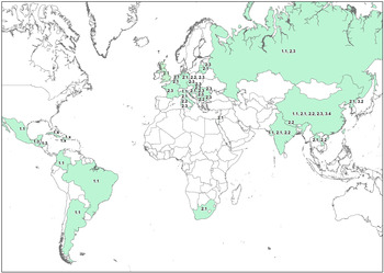
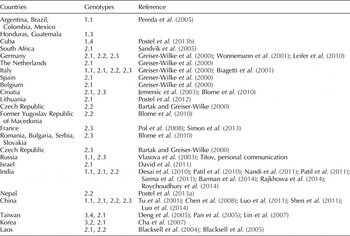
An overview of the above-mentioned genotype distribution is depicted in Fig. 1 and Table 1.
Global distribution of CSFV sub-genotypes. The figure covers reports that were published over the past two decades.

Global CSFV sub-genotype distribution

The table lists the countries from which published reports exist over the last two decades.
It has to be mentioned that despite the genetic diversity, no true serotypes exist and there are no indications for an impact on vaccine efficacy (Luo et al., Reference Luo, Li, Sun and Qiu2014). This assumption is supported by recent studies using challenge strains of different genotypes (Graham et al., Reference Graham, Everett, Haines, Johns, Sosan, Salguero, Clifford, Steinbach, Drew and Crooke2012; Blome et al., Reference Blome, Gabriel, Schmeiser, Meyer, Meindl-Bohmer, Koenen and Beer2014).
Virulence determinants
In general, there is no clear correlation of genotype and virulence. However, the most virulent strains such as ‘Shimen’ and ‘Koslov’ are found in genotype 1 (together with the vaccine strains), and most strains of genotype 2 are moderately virulent.
Although several authors sought to determine virulence factors (Van Gennip et al., Reference Van Gennip, Vlot, Hulst, De Smit and Moormann2004; Risatti et al., Reference Risatti, Borca, Kutish, Lu, Holinka, French, Tulman and Rock2005a, Reference Risatti, Holinka, Lu, Kutish, Tulman, French, Sur, Rock and Borcab, Reference Risatti, Holinka, Carrillo, Kutish, Lu, Tulman, Sainz and Borca2006, Reference Risatti, Holinka, Fernandez Sainz, Carrillo, Kutish, Lu, Zhu, Rock and Borca2007a, Reference Risatti, Holinka, Fernandez Sainz, Carrillo, Lu and Borcab; Sainz et al., Reference Sainz, Holinka, Lu, Risatti and Borca2008; Gladue et al., Reference Gladue, Holinka, Fernandez-Sainz, Prarat, O'Donell, Vepkhvadze, Lu, Rogers, Risatti and Borca2010, Reference Gladue, Holinka, Fernandez-Sainz, Prarat, O'Donnell, Vepkhvadze, Lu, Risatti and Borca2011; Tamura et al., Reference Tamura, Sakoda, Yoshino, Nomura, Yamamoto, Sato, Okamatsu, Ruggli and Kida2012, Reference Tamura, Nagashima, Ruggli, Summerfield, Kida and Sakoda2014), those determinants are still far from being understood.
Among the viral proteins that have been associated with virulence is, e.g. the autoprotease Npro. It was shown that virus mutants without Npro are avirulent (Mayer et al., Reference Mayer, Hofmann and Tratschin2004). Interestingly, mutations that abrogated the inhibitory effect on the interferon pathways did not result in full attenuation (Ruggli et al., Reference Ruggli, Summerfield, Fiebach, Guzylack-Piriou, Bauhofer, Lamm, Waltersperger, Matsuno, Liu, Gerber, Choi, Hofmann, Sakoda and Tratschin2009). The ribonuclease activity and dimerization of the Erns were also discussed as virulence factors (Meyers et al., Reference Meyers, Saalmuller and Buttner1999; Tews et al., Reference Tews, Schurmann and Meyers2009).
Furthermore, indications exist that the E2 plays a role in virulence (Risatti et al., Reference Risatti, Borca, Kutish, Lu, Holinka, French, Tulman and Rock2005a), probably in combination with the NS4B (Tamura et al., Reference Tamura, Sakoda, Yoshino, Nomura, Yamamoto, Sato, Okamatsu, Ruggli and Kida2012).
Direct comparison of the attenuated CSFV vaccine strain (GPE−) and its virulent ancestor (ALD) did not reveal a clear pattern (Ishikawa et al., Reference Ishikawa, Nagai, Katayama, Tsutsui, Tanabayashi, Takeuchi, Hishiyama, Saitoh, Takagi, Gotoh, Muramatsu and Yamada1995; Leifer et al., Reference Leifer, Ruggli and Blome2013). Attenuation through passaging had led to 225 nt exchanges in the genome of GPE−. However, forced reversion to virulence showed two amino acid changes in the NS4B region and one in the main immunogen E2 (Tamura et al., Reference Tamura, Sakoda, Yoshino, Nomura, Yamamoto, Sato, Okamatsu, Ruggli and Kida2012). These mutations could be confirmed as virulence determinants using reverse genetics. Interestingly, the underlying genetic profiles were already present in a minority of viruses from the viral quasispecies.
N-glycosylation sites of structural proteins were also shown to affect virulence as mutation of E2, Erns or E1 N-glycosylation sites resulted in attenuated viruses (Risatti et al., Reference Risatti, Holinka, Fernandez Sainz, Carrillo, Kutish, Lu, Zhu, Rock and Borca2007a, Reference Risatti, Holinka, Fernandez Sainz, Carrillo, Lu and Borcab; Sainz et al., Reference Sainz, Holinka, Lu, Risatti and Borca2008; Fernandez-Sainz et al., Reference Fernandez-Sainz, Holinka, Gavrilov, Prarat, Gladue, Lu, Jia, Risatti and Borca2009). In addition, the region around the O-glycosylation site in the E2-protein seems to play a role in CSFV virulence (Tang et al., Reference Tang, Pan and Zhang2008; Wu et al., Reference Wu, Wang, Feng, Liu, Teng, Yu and Chen2010; Leifer et al., Reference Leifer, Ruggli and Blome2013), although earlier studies with a mutated ‘Brescia’ strain did not result in an attenuated virus (Risatti et al., Reference Risatti, Holinka, Fernandez Sainz, Carrillo, Lu and Borca2007b).
Role of quasispecies
Quite recently, the quasispecies composition of CSFV has been discussed as a possible virulence factor (Töpfer et al., Reference Töpfer, Höper, Blome, Beer, Beerenwinkel, Ruggli and Leifer2013). As CSFV is an RNA virus, it harbors an RNA-dependent RNA polymerase. Due to the lack of proof-reading repair mechanisms of the viral RNA-dependent RNA polymerase, RNA viruses are in general considered to share high mutation rates during virus replication (Holland et al., Reference Holland, De La Torre and Steinhauer1992; Lauring and Andino, Reference Lauring and Andino2010). This error-prone replication often leads to a high genetic diversity of genomes within a virus population (‘quasispecies’; Eigen, Reference Eigen1971). Generally, a diverse virus could have a fitness advantage, e.g. through the presence of immune escape variants. In order to investigate this point, the next-generation sequencing data were analyzed. It was shown that a higher quasispecies diversity and nt variability was found in highly virulent strains such as ‘Koslov’ or ‘Brescia’ (Töpfer et al., Reference Töpfer, Höper, Blome, Beer, Beerenwinkel, Ruggli and Leifer2013). However, the higher replication rate of highly virulent strains could also lead to a higher diversity. Thus, source and consequence remain unclear. In addition, high virulence of cDNA-derived recombinant viruses with a homogenous genome composition shows that diversity and quasispecies is not a CSFV virulence factor per se.
Conclusions
CSFV-strains can be assigned to three genotypes and their sub-genotypes. Geographical pattern can be observed that are important for the understanding of disease dynamics and epidemiology. However, the genotypes do not result in serotypes and do not affect vaccine efficacy.
So far, universal virulence determinants have not been defined for CSFV. Indications exist that mutations in the E2 and the NS4B could play a role. However, most likely, virulence is a complex and multifactorial issue. Among the influencing factors could be the quasispecies composition. However, based on the current knowledge, the impact is probably limited.