




No CrossRef data available.
Article contents
An event-related potential study of the impact of institutional rearing on face recognition
Published online by Cambridge University Press: 01 November 2005
Abstract
Event-related potentials (ERPs) were recorded to brief images of caregivers' and strangers' faces for 72 institutionalized children (IG), ages 7–32 months, and compared with ERPs from 33 children, ages 8–32 months, who had never been institutionalized. All children resided in Bucharest, Romania. Prominent differences in four ERP components were observed: early negative (N170), early positive (P250), midlatency negative (Nc), and positive slow wave (PSW). For all but the P250, the amplitude of these components was larger in the never instituionalized group than the institutionalized group; this pattern was reversed for the P250. Typical effects of the Nc (amplitude greater to stranger vs. caregiver) were observed in both groups; in contrast, the IG group showed an atypical pattern in the PSW. These findings are discussed in the context of the role of experience in influencing the neural circuitry putatively involved in recognizing familiar and novel faces.
The work reported in this manuscript was supported by a research network (Early Experience and Brain Development) funded by the John D. and Catherine T. MacArthur Foundation (Charles A. Nelson, Network Chair). The Bucharest Early Intervention Core Group consists of Charles H. Zeanah, Anna T. Smyke, and Sebastian F. Koga (Tulane University); Charles A. Nelson (Harvard Medical School); Susan W. Parker (Randolph-Macon College); Nathan A. Fox (University of Maryland); Peter J. Marshall (Temple University); and Hermi R. Woodward (University of Pittsburgh/MacArthur Research Networks). The authors acknowledge the many invaluable contributions of their Romanian partner institutions, the SERA Romania Foundation, the Institute of Maternal and Child Health, and the Bucharest Departments of Child Protection. They are also deeply grateful to their Romanian team, whose hard work and dedication has made this study possible and to Dana Johnson and Mary Jo Spencer for conducting the pediatric screens.
Information
- Type
- Research Article
- Information
- Copyright
- © 2005 Cambridge University Press
The damaging effects of early institutional rearing on the development of young children have been well documented by both clinicians and researchers (for review, Gunnar, 2001; Johnson, 2000; Zeanah, Nelson, Fox, Smyke, Marshall, Parker, & Koga, 2003). To date, however, much of the literature on the effects of early institutional rearing has been concerned with global functions, such as IQ, attachment, peer relations, and the like. Although crucially important, it is difficult to relate such global functions to specific brain circuits. The focus of the current study, then, was to examine a fairly specific social–perceptual–cognitive function, the neural basis of which is reasonably well understood. Specifically, we sought to examine the neural correlates of recognizing familiar and novel faces. We targeted this ability for several reasons. First, responding to faces generally, and familiar faces specifically, may play a critically important function in social cognition; after all, prior to the onset of language most infant–caregiver communication is nonverbal in nature. Thus, understanding how infants respond to familiar faces, in this case, the face of the caregiver, may provide valuable insight into the development of higher level social cognitive functions. Second, one of us (Nelson, 2001) has proposed that many aspects of face processing represent an experience-expectant and activity-dependent function. As such, experience with faces drives the development of the circuitry that ultimately comes to be specialized for face processing. Third, it has now been reported in a number of studies that the development and/or function of this circuitry can be derailed by adverse early experiences. For example, event-related potentials (ERPs) in response to angry facial expressions differ in maltreated, compared with nonmaltreated, children (e.g., Pollak, Cicchetti, Klorman, & Brumaghim, 1997). Moreover, institutionalized and noninstitutionalized children's ERPs differ in response to a range of emotional expressions (e.g., Parker, Nelson, & The Bucharest Early Intervention Project Core Group, 2005). Based on these data, we expected to observe perturbations in the ERPs of infants reared in institutions, relative to children reared in their biological homes, when presented with pictures of their caregiver's face along with pictures of a stranger's face.
ERPs represent a measure of neural activity that is noninvasive and not dependent on language ability. By measuring the brain responses that occur during repeated presentation of a stimulus, reliable patterns of brain activity can be revealed. These patterns are believed to reflect the neural and cognitive processes involved in stimulus processing. Thus, ERPs can be used to compare the processing patterns of children affected by adverse situations with the patterns of typically developing children. Through this measure, the institutionalized children are expected to show electrophysiological evidence consistent with impairments in the circuitry involved in face recognition: specifically, the ERPs of the institutionalized group (IG) are expected to differ from the never institutionalized group (NIG).
Past work by de Hann and Nelson (1997, 1999) examined early face recognition in typically developing 6-month-olds using ERPs. Differences in the neural processing of the infant's mother's face and a dissimilar-looking female stranger's face were examined. This research revealed greater amplitudes for the middle-latency negative component (Nc) in response to the mother's face. De Hann and Nelson (1997) suggest that the larger Nc amplitude to the mother's face may reflect a greater allocation of attention (Courchesne, Ganz, & Norcia, 1981; Nelson & Collins, 1991; Nelson & Salapatek, 1986).
Conflicting findings were reported for the positive slow wave (PSW), however. One study revealed no differences in the PSW between the mother and stranger conditions (de Hann & Nelson, 1997). The second study, however, found the PSW to be larger in response to the stranger's face (de Hann & Nelson, 1999), a finding consistent with the idea that the PSW represents memory updating of a partially encoded stimulus.
In a design similar to that of de Hann and Nelson (1999), Dawson, Carver, Meltzoff, Panagiotides, McPartland, and Webb (2002) recorded ERPs from 3- and 4-year-old children in response to their mother's face and the face of a stranger. The ERP data revealed a trend toward a larger mean amplitude of the PSW in response to the stranger's face. Although this difference did not reach statistical significance, it is in line with the findings reported by de Hann and Nelson (1999) and supports the role of the PSW in memory updating and encoding of novel stimuli.
Dawson et al. (2002) also reported significantly larger amplitudes of the Nc in response to the stranger's face. This finding departs from those reported by de Hann and Nelson (1997, 1999). A possible explanation for this difference may be developmental changes, occurring between infancy and preschool age; specifically, significant developmental gains are made in cognitive and social functioning through the toddler and preschool years. Carver, Dawson, Panagiotides, Meltzoff, McPartland, Gray, and Munson (2003) suggest that the cognitive processing and recognition of faces may play an important role in the successful developmental of social relationships and peer interactions during this period in development. Specifically, Carver et al. (2003) maintains that the relative importance of the mother's and stranger's faces may evolve as children become more likely to seek out social exchanges with unfamiliar adults and peers.
This developmental shift was examined, using ERPs, in children ranging in age from 18 to 54 months (Carver et al., 2003). Analyses revealed a clear developmental trend in the peak amplitude of the Nc. Children aged 18–24 months showed a larger peak Nc amplitude in response to the mother's face than to a stranger's face; findings that are consistent with de Haan and Nelson's work (1997, 1999) with young infants. In contrast, data collected from children over the age of 45 months revealed a greater peak Nc amplitude in response to the stranger's face. For children between 24 and 45 months of age, however, there were no significant differences in the peak Nc amplitude found between the two conditions (Carver et al., 2003).
These developmental changes in neural processing may mirror changes in social development that are occurring simultaneously. Carver et al. (2003) suggests that the initial Nc amplitude difference likely reflects a greater allocation of attention to the mother's face, which may be especially important in the early development of the child–caregiver relationship. In contrast, by 4 years of age the child–caregiver relationship is generally firmly established and children must spend relatively more energy cultivating other social relationships. This shift may, in turn, be reflected in the greater Nc amplitude in response to a stranger's face. Moreover, typically developing children between 24 and 45 months may be in a transitional period where attention is equally allotted to the caregiver's face and faces of strangers. Therefore, there is no significant difference in the Nc amplitude observed in response to the two conditions (Carver et al., 2003).
As detailed, the existing body of literature utilizing ERP methodology in the study of face processing primarily emphasizes typical development. The study of normal development undoubtedly carries tremendous implications regarding the understanding and definition of psychopathology. The unification of typical and abnormal development within a single framework (Cicchetti & Cohen, 1995), however, would contribute significantly to our understanding of developmental psychopathology. Thus, the current study aims to examine current theories of normal development (Carver et al., 2003) within the context of an experiment of nature. Specifically, this study seeks to further elucidate the role of adverse early experience, specifically deprivation associated with early institutionalization, in the disruption of the normal development of face processing and the recognition of facial familiarity.
Based on previous research, children in the current study were expected to exhibit an early negative component (N1701
Although an N170 component is typically reported in the body of literature on face processing in adults, this component is typically prominent over occipital and lateral leads. Thus, based on its topographical distribution, the N170 reported in the current study is believed to represent a different component than the traditional N170.
Method
Sample
As we have reported in earlier work (Parker et al., 2005) participants in the study were 208 children aged between 5 and 31 months and their parents or institutional caregivers.
IG
The IG consisted of 136 children recruited from seven institutions in Bucharest, Romania, and who had spent at least half of their lives in institutional care. Screening for inclusion in the study consisted of a pediatric (including neurological) examination, growth measurements, and assessment of physical abnormalities. Exclusion criteria included genetic syndromes (e.g., Down syndrome), distinct signs of fetal alcohol syndrome, and microcephaly (more than 2.5 SD below the mean for occipitofrontal circumference), using standards from Tanner (1973). Subjects selected for inclusion fell within 2.5 SD from the mean for occipitofrontal circumference. Such screening made certain that the IG was comprised of children who were in fair health and suffered no obvious genetic abnormalities or anthropomorphic signs of fetal alcohol syndrome.
Of the 136 institutionalized children included in the study, 78 are of Romanian ethnicity (57.4% of sample), 36 are Rroma Gypsy (26.5%), 1 is Turkish (0.7%), 1 is of subcontinent Indian extraction (0.7%), and the remaining 20 (14.7%) could not be classified. At the time of baseline assessment, all subjects had spent between 51 and 100% of their lives in the institution (M = 89%). Of the entire group, 70 children (51.5% of sample) spent all of their lives in institutional care.
Gestational age data were available only for 112 subjects, and many of these estimates were of uncertain reliability (due to poor record keeping at maternity hospitals). There was a range of 30 to 42 weeks (M = 37.2 weeks, SD = 2.2 weeks). Birth weight (available for 117 cases) ranged from 900 to 4150 g (M = 2767 g, SD = 609 g) and was significantly different from the community control group (M = 3338 g, SD = 467 g).
In subsequent assessments related to the longitudinal nature of the overall study (Zeanah et al., 2003) it became evident that 11 children in the IG retrospectively failed to meet inclusion criteria due to surfacing medical disorders, the symptoms of which manifested following the initial screening. Data from these 11 children are excluded from further analysis in this paper.
NIG
Seventy-two children (41 males) made up the NIG and were recruited using birth records at the same maternity hospitals where the IG were born. The NIG were matched to the IG on age and gender. Screening included a pediatric examination, physical growth measurements, and a psychosocial interview with the family. All participants fell within 2 SD from the mean for physical growth (weight, length, occipitofrontal circumference). Of the 72 who consented to participate, 66 children (91.7%) were Romanian, 4 children (5.6%) were Rroma, 1 child was Spanish, and 1 child was Turkish. During baseline testing, families of seven children chose to exit the study, generally citing time pressures.
Additional participants
In the present study, eight of the children in the IG and two of the children in the NIG were not included in the longitudinal sequence of assessments outlined by Zeanah et al. (2003), but were tested in the laboratory for purposes of cross-sectional data collection. For details regarding the other domains of assessment, see Zeanah et al. (2003).
Participants for ERP analyses
Across the entire sample, ERP data were collected for 181 participants (125 IG, 56 NIG). ERPs were not collected from 8 institutionalized participants and 9 never institutionalized participants due to fussiness before or during placement of the EEG cap. In addition, the parents of 2 participants from the NIG refused to allow EEG data to be recorded from their children. The current analyses focus on the 177 participants who were between 7 and 32 months of age. Of these 177 participants, 46 (33 IG, 13 NIG) were excluded from further analyses because of problems with the ERP, including excessive noise in one or more channels, problems with the electrooculogram (EOG) signal, or other technical problems. Of the remaining 131 participants, 20 participants (13 IG, 7 NIG) had fewer than nine artifact-free trials and were excluded from further analyses.
Final analyses are reported for 75 (39 males) in the IG (55.1% of IG sample) between the ages of 7 and 32 months, and 35 (20 males) in the NIG (48.6% of NIG sample) between the ages of 8 and 32 months. The mean ages were was 22.3 months (range = 7.0–31.6 months, SD = 6.1 months) for the IG and 21.2 months (range = 8.0–31.7 months, SD = 6.7 months) for the NIG.
Division of participants into age groups
To examine the effect of age while maintaining acceptable group sizes, the IG and NIG were divided into age groups defined by splitting the groups based on their median ages, 23.5 and 22.5 months, respectively. These age groupings consisted of the following: 37 younger IG between the ages of 7 and 23.4 months (M = 17.5, SD = 4.9, 15 males), 38 older IG between the ages of 23.5 and 31.6 months (M = 26.9, SD = 2.5, 24 males), 18 younger NIG between the ages of 8 and 22.5 months (M = 15.7, SD = 4.5, 9 males), and 17 older NIG between the ages of 22.8 and 31.7 months (M = 26.9, SD = 2.3, 11 males). Neither the younger IG and younger NIG nor the older IG and older NIG differed significantly by age.
Electroencephalogram (EEG) recording
The EEG was recorded from midline (Pz, Fz) and lateral scalp (O1, O2, T7, T8, P3, P4, C3, C4, F3, F4) sites, as well as the left and right mastoids and the reference site at the vertex (Cz), using a Lycra Electro-Cap (Electro-Cap International Inc., Eaton, OH) with sewn-in tin electrodes. The ground electrode was represented by an anterior midline site (AFz).
Following cap placement, a small amount of abrasive gel was inserted into each of the electrode sites and the mastoid sites. Following gentle abrasion, a small amount of electrolytic conducting gel was added into each site. Impedances were measured at each electrode site and considered acceptable if they were at or below 10 kΩ. All channels were digitized at 512 Hz onto the hard drive of a PC using a 12-bit A/D converter (±2.5-V input range) and Snap-Master acquisition software (HEM Data Corporation, Southfield, MI). The vertical EOG was recorded from electrodes placed vertically above and below the left eye bisecting the midline to record blinks and other eye movement.
The EEG and EOG signals were amplified by factors of 5,000 and 2,500, respectively, using custom bioelectric amplifiers from SA Instrumentation Company (San Diego, CA). Amplifier filter settings for all channels were 0.1–100 Hz. Before the recording of EEG from each participant, a 50-μV 10-Hz signal was input into each of the channels and this amplified signal was recorded for calibration purposes.
ERP data reduction
All subsequent processing and analysis of the EEG signal was carried out using the ERP Analysis Systems from James Long Company (Caroga Lake, NY). The channels were rereferenced to an average mastoids configuration. Blinks in the EOG signal were regressed out of the EEG using a procedure based on methods described in the literature (e.g., Lins, Picton, Berg, & Scherg, 1993; Miller & Tomarken, 2001). Trials in which the EEG signal exceeded ±250 μV were eliminated from further analysis.
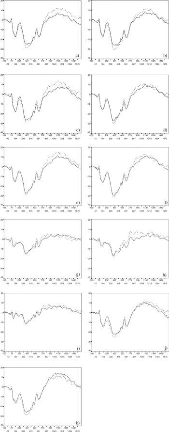
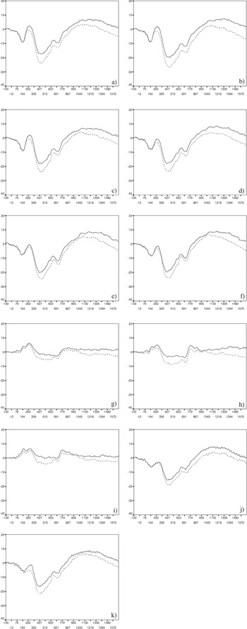
Individual subject averages were computed for each stimulus type (familiar, novel). Individuals who did not have a minimum of nine artifact-free trials for each electrode location for each average were excluded from further analysis. A grand average was then computed for each stimulus (see Figures 1, 2, 3, and 4).

Grand means of ERPs exhibited by the NIG. Amplitude (μV) is listed on the y axis and latency (ms) is listed on the x axis; (—) familiar face and (- - -) novel face. (a) Left frontal (F3) electrode site, (b) midline frontal (Fz) electrode site, (c) right frontal (F4) electrode site, (d) left central (C3) electrode site, (e) midline central (Cz) electrode site, (f) right central (C4) electrode site, (g) left parietal (P3) electrode site, (h) midline parietal (Pz) electrode site, (i) right parietal (P4) electrode site, (j) left temporal (T7) electrode site, and (k) right temporal (T8) electrode site.

Grand means of ERPs exhibited by the IG. Amplitude (μV) is listed on the y axis and latency (ms) is listed on the x axis; (—) familiar face and (- - -) novel face. (a) Left frontal (F3) electrode site, (b) midline frontal (Fz) electrode site, (c) right frontal (F4) electrode site, (d) left central (C3) electrode site, (e) midline central (Cz) electrode site, (f) right central (C4) electrode site, (g) left parietal (P3) electrode site, (h) midline parietal (Pz) electrode site, (i) right parietal (P4) electrode site, (j) left temporal (T7) electrode site, and (k) right temporal (T8) electrode site.

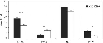
Amplitudes (μV) of the four ERP components at midline electrode sites. To illustrate the main effect of group, amplitudes are collapsed across condition and region and are reported as absolute values (***p = .001, **p < .01, *p < .05, +p < .1).

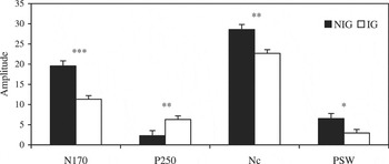
Amplitudes (μV) of the four ERP components at lateral electrode sites. To illustrate the main effect of group, amplitudes are collapsed across condition and region and are reported as absolute values (***p = .001, **p < .01, *p < .05).
On the basis of inspection of each of the individual subjects' averages and the grand averages, time windows containing ERP components of interest were identified. Four sets of windows were identified: 90–250 ms (N170), 150–400 ms (P250), 325–650 ms (Nc), and 900–1500 ms (PSW). Peak amplitudes relative to the 100-ms baseline and latency to peaks were determined for ERP activity within the first three windows and average amplitude was determined for the fourth window.
Procedure
Each infant was tested individually while sitting on the parent's or caregiver's lap and facing a computer monitor approximately 40 cm away. The monitor was surrounded by black panels that blocked the infant's view of the room behind the screen and to his/her sides to limit the possibility of distractions. A small hole in the screen was present directly above the computer monitor, allowing observers to monitor the infant's behavior.
Trials consisted of a 100-ms baseline, followed by a 500-ms presentation of the visual stimulus, followed by 1200 ms during which the screen was a blank blue background. The intertrial interval varied randomly between 500 to 1000 ms and during this time the screen displayed the blue background. The average duration of the task was 3 min.
An observer behind the screen watched the infant through the hole and if the infant looked away the observer signaled the computer, via push button, to delete the trial. Thus, brain activity from trials during which the infant was not looking at the stimuli was not used in further analyses. If needed, the observer tapped the screen or shook a toy rattle to attract the infant's attention to the screen. The session continued until the infant had seen the maximum number of trials (70) or became too fussy or bored to attend to the stimuli.
Stimuli
The stimuli were static, color images (8-bit color, 800 × 600 pixel resolution) of the participant's mother, in the case of the NIG, or preferred institutional caregiver, in the case of the IG, and a dissimilar looking woman who was a stranger to the participant (i.e., a caregiver from a different institution). Each image was presented randomly for 35 trials, for a maximum total of 70 trials. The pictures were taken against a gray background while the women wore a gray scarf around their necks to conceal any clothing.
Regarding the selection of stimuli, the familiar faces were represented by images of the mother of each NIG participant and the preferred institutional caregiver of each IG participant. The preferred caregiver of each child was determined based on surveys of the staff of caregivers working with the children. Novel faces were images of caregivers with whom the child would presumably have had little to no contact. For instance, in the case of the NIG, the novel faces depicted institutional caregivers and for the IG, images portraying caregivers from a different institutional from where the participant resided.
Results
For three of the ERP components (N170, P250, Nc) two repeated-measures analyses of variance (ANOVAs) were computed, one using peak amplitude as the dependent variable and the other using peak latency. The PSW was analyzed using a repeated-measures ANOVA, using average amplitude as the dependent variable. For each ANOVA, the within-subject factors were Region (frontal, central, parietal for the midline electrode analyses and frontal, central, parietal, temporal for the lateral electrode analyses) and Condition (familiar face, novel face). For the lateral electrode analyses, hemisphere (left, right) also served as a within-subject factor. The between-subject factors were Group (IG, NIG) and Age (younger, older; see Division of participants into age groups section). Because occipital leads typically do not exhibit evidence of face discrimination, these data will not be discussed further. Grand means in response to the familiar and novel faces are shown for the NIG and IG in Figures 1 and 2.
N170
Midline electrode sites.
Amplitude. There was a main effect of Group (see Figure 3), F (1, 106) = 18.62, p < .001, ηp2 = .149, indicating that, relative to the IG, the NIG exhibited larger amplitude N170 peaks.
There was also a main effect of Region, F (2, 212) = 61.508, p < .001, ηp2 = .367, suggesting that across the entire sample the N170 was maximal at frontal and central electrode sites.
Latency. There were no significant effects for latency to the N170 at midline electrode sites.
Lateral electrode sites.
Amplitude. There were main effects of Group (see Figure 4), F (1, 106) = 34.02, p < .001, ηp2 = .243, and Region, F (3, 318) = 96.85, p < .001, ηp2 = .477. These effects were qualified by an interaction of Region × Hemisphere × Group × Age, F (3, 318) = 4.57, p = .004, ηp2 = .041. Inspection of the means suggested hemispheric asymmetry in the parietal region for the older NIG, specifically, relative to the right, the left hemisphere exhibited a greater N170 amplitude. This pattern was not evident in the younger NIG or either age group of the IG.
The main effect of Region was further qualified by an interaction of Region × Hemisphere × Condition × Age, F (3, 318) = 3.43, p = .017, ηp2 = .031. Inspection of the means indicated that the N170 peaks exhibited in the temporal region are greater in amplitude in response to the novel face, in the right hemisphere of the younger group and in the left hemisphere of the older group.
Latency. There was a main effect of Hemisphere, F (1, 106) = 5.35, p = .023, ηp2 = .048, indicating that across the entire sample the latency to the N170 was shortest in the left hemisphere.
There was also a main effect of Region, F (3, 318) = 10.68, p < .001, ηp2 = .092, which was qualified by an interaction of Region × Group, F (3, 318) = 3.04, p = .052, ηp2 = .028. Follow-up repeated measures ANOVAs revealed a main effect of Region in the IG, F (3, 219) = 17.84, p < .001, ηp2 = .196, such that latency to the N170 is shortest at the parietal region and longest at central and temporal regions. No effects of Region were found in the NIG.
P250
Midline electrode sites.
Amplitude. There was a main effect of Group (see Figure 3), F (1, 106) = 7.13, p = .009, ηp2 = .063, suggesting that, relative to the NIG, the IG exhibited larger amplitude P250 peaks.
There was also a main effect of Region, F (2, 212) = 14.51, p < .001, ηp2 = .120, such that across the entire sample the P250 was maximal at parietal electrode sites.
Latency. There was a main effect of Region, F (2, 212) = 4.62, p = .011, ηp2 = .042, which was qualified by an interaction of Region × Age, F (2, 212) = 7.20, p = .001, ηp2 = .064. Follow-up repeated measures ANOVAs revealed a main effect of Region in the younger group, F (2, 106) = 11.71, p < .001, ηp2 = .181, such that latency to the P250 is shortest at the parietal and central regions and longest at the frontal region. No effects of Region were found in the older group.
Lateral electrode sites.
Amplitude. There were main effects of Group (see Figure 4), F (1, 106) = 8.56, p = .004, ηp2 = .075, and Region, F (3, 318) = 34.45, p < .001, ηp2 = .245. Both effects were qualified by an interaction of Region × Hemisphere × Group × Age, F (3, 318) = 2.92, p = .034, ηp2 = .027. Inspection of the means indicated that hemispheric asymmetry was exhibited by the older NIG, specifically greater P250 amplitudes were found in the left hemisphere of the central region and greater P250 amplitudes were found in the right hemisphere of the parietal region. Similar patterns were absent in the other groups.
Moreover, the effect of Region was further qualified by an interaction of Region × Condition, F (3, 318) = 2.78, p = .041, ηp2 = .026. Follow-up analyses of the interaction were inconclusive.
Latency. There was a main effect of Hemisphere, F (1, 106) = 7.57, p = .007, ηp2 = .067, indicating that across the entire sample the latency to the P250 was shortest in the right hemisphere.
There was also a main effect of Region, F (3, 318) = 2.84, p = .038, ηp2 = .026, which was qualified by an interaction of Region × Age, F (3, 318) = 3.42, p = .018, ηp2 = .031. Follow-up repeated measures ANOVAs revealed a main effect of Region in the younger group, F (3, 159) = 7.26, p < .001, ηp2 = .120, such that latency to the P250 is longest at the frontal region. No effects of Region on latency to the P250 were found in the older group.
Nc
Midline electrode sites.
Amplitude. There was a main effect of Condition (see Figure 5), F (1, 106) = 7.30, p = .008, ηp2 = .064, indicating that across the entire sample the Nc amplitude in response to the novel face was greater than the Nc in response to the familiar face.

Amplitudes (μV) of the Nc amplitude at midline and lateral electrode sites. To illustrate the main effect of condition, amplitudes are collapsed across group and region and are reported as absolute values (**p < .01).
There was also a main effect of Region, F (2, 212) = 95.62, p < .001, ηp2 = .474, which was qualified by an interaction of Region × Group × Age, F (2, 212) = 3.80, p = .024, ηp2 = .035. Inspection of the means indicate an age-related decrease in the amplitude of the Nc for the NIG, specifically at the central region. A similar age-related decrease in amplitude was not noted for the IG.
Latency. There was a main effect of Group, F (1, 106) = 10.06, p = .002, ηp2 = .087, which was qualified by an interaction of Region × Group, F (2, 212) = 3.09, p = .048, ηp2 = .028. Follow-up repeated measures ANOVAs revealed a main effect of Region in the IG, F (2, 146) = 6.14, p = .003, ηp2 = .078, such that latency to the Nc was longest at the parietal region. No effects of Region on latency to the Nc were found in the NIG.
Lateral electrode sites.
Amplitude. There was a main effect of Group (see Figure 4), F (1, 106) = 7.87, p = .006, ηp2 = .069, indicating that, relative to the IG, the NIG exhibited larger amplitude Nc peaks.
There was also a main effect of Condition (see Figure 5), F (1, 106) = 8.69, p = .004, ηp2 = .076, suggesting that across the entire sample, a greater Nc amplitude was displayed in response to the novel face, relative to the familiar face.
Finally, there was a main effect of Region, F (3, 318) = 104.09, p < .001, ηp2 = .495, which was qualified by a Region × Hemisphere interaction, F (3, 318) = 9.09, p < .001, ηp2 = .079. Follow-up repeated measures ANOVAs revealed a main effect of Hemisphere at the central, F (1, 106) = 6.52, p = .012, ηp2 = .058, and parietal regions, F (1, 106) = 13.66, p < .001, ηp2 = .114, such that greater Nc amplitudes were exhibited in the right hemisphere of the central region and in the left hemisphere of the parietal region. No effects of Hemisphere were identified in the frontal or temporal regions.
Latency. There were main effect of Group, F (1, 106) = 14.41, p < .001, ηp2 = .120, and Region, F (3, 318) = 14.19, p < .001, ηp2 = .118. Both effects were qualified by an interaction of Region × Group × Age, F (3, 318) = 3.29, p = .021, ηp2 = .030. Inspection of the means indicate that whereas the IG exhibited a decrease in latency with age in all except the parietal regions, the NIG appeared to display an age-related change in the latency to the Nc only in the parietal region.
An interaction of Condition × Age, F (1, 106) = 6.97, p = .010, ηp2 = .062, was also noted. Follow-up repeated measures ANOVAs revealed a main effect of Condition in the older group, F (1, 53) = 8.58, p = .005, ηp2 = .139, such that a shorter latency to the Nc was displayed in response to the novel, as compared to the familiar, face. No effects of Condition were identified in the younger group.
PSW
Midline electrode sites
There was a main effect of Region, F (2, 212) = 18.52, p < .001, ηp2 = .149, indicating that across the entire sample the PSW was maximal at frontal and central electrode sites.
There was also a main effect of Group (see Figure 3), F (1, 106) = 5.72, p = .019, ηp2 = .051, which was qualified by an interaction of Group × Condition (see Figure 6), F (1, 106) = 4.27, p = .041, ηp2 = .039. Follow-up repeated measures ANOVAs revealed a main effect of Condition in the IG, F (1, 73) = 5.61, p = .020, ηp2 = .71, such that a greater PSW was displayed in response to the familiar, compared to the novel, face. No effects of Condition on the PSW were identified in the NIG.

Amplitudes (μV) of the PSW average amplitude at midline electrode sites. To illustrate the interaction of Group × Condition, amplitudes are collapsed across region (**p < .01).
Lateral electrode sites
There was a main effects of Group (see Figure 4), F (1, 106) = 6.21 p = .014, ηp2 = .055, suggesting that, relative to the IG, the NIG exhibited larger PSW amplitudes.
There was also a main effect of Region, F (3, 318) = 18.55, p < .001, ηp2 = .149, indicating that across the entire sample the PSW was maximal at temporal and central electrode sites.
In addition, there was a main effect of Hemisphere, F (1, 106) = 5.41, p = .022, ηp2 = .049, such that across the entire sample the PSW was maximal in the right hemisphere.
Summary of Results
A dramatic group difference in amplitude was evident across all four components. Relative to the IG, the NIG exhibited greater amplitudes of the N170, Nc, and PSW components. In contrast, relative to the NIG, the IG exhibited a greater amplitude P250. Age was also found to play a principal role in the findings. Specifically, the NIG and IG appear to show different developmental trajectories in a number of measures.
Both groups exhibited evidence of discrimination between the familiar and novel faces as revealed in the amplitudes of the Nc at midline and lateral electrode sites. Specifically, both groups displayed a greater amplitude Nc in response to the novel face. In contrast, group differences in face discrimination were evident for the PSW from midline electrode sites: the IG displayed a greater PSW in response to the familiar face, whereas the NIG showed no evidence of discrimination at the PSW.
Regarding the topographical distribution of the components, regional effects indicated that the N170, Nc, and PSW were all maximal at anterior regions. The P250, however, appeared to be maximal over more posterior regions.
Discussion
The current study sought to examine the impact of early institutionalization on the neural correlates of face processing. As expected, participants exhibited N170, P250, Nc, and PSW in response to the images of the caregiver's and stranger's faces. Regarding face differentiation, both the NIG and IG displayed evidence of discrimination between the caregiver and stranger faces. Specifically, both groups exhibited greater Nc amplitudes in response to the novel face. A group difference in face discrimination, however, was noted for the PSW activity. Moreover, a dramatic group difference in amplitude was evident across all four ERP components.
Previous ERP research regarding young children's processing of familiar and unfamiliar faces (Carver et al., 2003; Dawson et al., 2002; de Hann & Nelson, 1997, 1999) suggests developmental changes in the neural activity involved in face processing. Specifically, Carver et al. (2003) suggest that the amplitude of the Nc, relative to familiar and novel faces, shifts between infancy and preschool. During infancy, the Nc is maximal to the mother's face (de Hann & Nelson, 1997, 1999); whereas, by preschool, the Nc is maximal to the face of a stranger (Carver et al., 2003; Dawson et al., 2002). This shift is believed to reflect changes in the salience of children's different social relationships. In particular, during formation of the attachment relationship, the face of the primary caregiver is likely the most significant social stimulus for the child. By preschool age, however, children's interest in and approach of peers and other adults become more prominent (Carver et al., 2003).
Reflecting a similar pattern, the current study reports a greater Nc amplitude in response to the stranger's face, relative to the caregiver's face. These findings, however, are evident across both age groups (age range = 7–32 months) and within both the NIG and IG; thus, no developmental change, as noted in the Carver and Dawson work (Carver et al., 2003; Dawson et al., 2002), was evident. Although the lack of group difference is significant, the absence of evidence for developmental shift in responding is equally noteworthy. It is doubtful that the development of early social relationships is markedly different between Romanian and American infants and toddlers. More likely, the wide age range of the current sample, though considered in separate age groups, may have concealed any age-related variations in the pattern of responding.
In contrast to the findings for the Nc amplitude, the pattern of responding observed at the PSW differed strikingly between the IG and NIG. Specifically, the IG exhibited a greater PSW in response to the caregiver's face, relative to the stranger's face. The NIG, however, did not exhibit a difference in PSW activity based on facial familiarity. Notably, visual depictions of the ERPs (see Figure 1) suggest a pattern of greater PSW activity in response to the stranger's face; however, the difference is not significant. Based on past research indicating that the PSW represents memory updating of stimuli that are not fully encoded (de Hann & Nelson, 1997; Nelson & Monk, 2001), the current findings indicate that the faces of primary caregivers in institutional settings may not be fully encoded by the children in their care. Due to the limited amount of individual contact between the caregiver and child in such settings, and a corresponding lack of close caregiver–child relationships (Zeanah et al., 2003), a fragile representation of the caregiver's face may be expected. Thus, the current findings may provide a subtle indication of the dramatic effects of early institutional rearing on brain development.
As expected, another primary finding from the current study is a marked group difference in amplitude at all four ERP components. Specifically, the NIG exhibited larger amplitudes for the N170, Nc, and PSW, whereas the IG exhibited larger amplitudes for the P250. As discussed previously, the Nc is believed to represent some aspect of obligatory or automatic attention allocation (Nelson & Monk, 2001; Courchesne, 1978). Consequently, amplitude differences in the Nc in response to specific stimuli are generally interpreted as differences in attention allotted to each of the stimuli. Group differences in amplitude of ERP components, as noted in the current study, however, are more difficult to interpret.
Similarities can be drawn, however, between the current findings and those reported in other tasks involving the same sample of children. In an ERP study of emotion discrimination (Parker et al., 2005), the IG and NIG exhibited similar patterns of ERP amplitudes. Likewise, electroencelphalogram (EEG) measures report that, relative to the NIG, the IG displayed a pattern of increased low-frequency (theta) power in posterior scalp regions and decreased high-frequency (alpha and beta) power, particularly at frontal and temporal electrode regions (Marshall, Fox, & The Bucharest Early Intervention Project Core Group, 2004).
When viewed in concert, such findings provide support for a general cortical hypoactivation resulting from early institutional rearing (Marshall et al., 2004; Parker et al., 2005). Research involving a sample of children adopted from Romanian institutions provides further support for the possibility that the brains of children who have experienced early institutionalization are, in a general sense, underpowered (Chugani, Behen, Muzik, Juhász, Nagy, & Chugani, 2001). Positron emission tomography scans of this sample of children revealed significantly decreased metabolic activity in multiple brain regions. Remarkably, these results were evident even following an average of 5.5 years in adoptive homes. Although the current work focused on infants and toddlers, Chugani's research involved children between 7 and 11 years old. Thus, these findings may suggest an effect of early institutional care that persists through early and late childhood. Although the findings from these studies provide tentative support for the possibility of a type of hypoactivation resulting from early institutional rearing, none speak to the potential mechanism underlying this effect on neural activity. Likewise, the implications of such deficiencies on function are unclear.
Speculation by Critchley et al. (2000) suggests that if social stimuli do not acquire special meaning or importance, as a result of atypical experience or brain injury, circuitry in the medial temporal lobe may be marked by abnormal connectivity (Bauman & Kemper, 1985). When applied to the current work, the attenuated ERP amplitudes may reflect insults resulting from the absence of experience with a primary attachment figure early in life. Similarly, such effects may manifest in social and behavioural abnormalities later in life.
The possible role of early institutional rearing in the disruption of the neural circuitry involved in the processing of social information is further supported by the current study. The group differences in overall ERP amplitudes and PSW response to facial familiarity suggest dysfunction pertaining to brain function regulating face processing in the IG. Specifically, early deprivation may negatively impact medial temporal lobe circuitry, resulting in deficits in processing social information, namely faces. When viewed in conjunction with the developmental patterns displayed by typically developing populations (Carver et al., 2003), these data suggest a disruption in the normal development of social relationships. This potential dysfunction is further supported by past research documenting the social and behavioral abnormalities typical of children reared in institutional settings (Fisher, Ames, Chisholm, & Savoie, 1997; O'Connor, Bredenkamp, & Rutter, 1999). Indeed, the social deficits exhibited by institutionalized children include the inappropriate recognition and/or use of social cues. Thus, the current findings indicate the need for future work directed at clarifying the association between early rearing experience, processing of socioemotional information, and developmental psychopathology.
Because the data presented in this manuscript represent a larger longitudinal intervention study of foster care, the current work will be invaluable in exploring the long-term effects of deprivation on neural function, face recognition, and social behavior. In addition, because the present data represent the participants' baseline data function, they are an important benchmark of behavior and neural processing during institutionalization. Evaluation following foster care intervention will allow for a rare view regarding the potential for plasticity.
References
REFERENCES
Bauman, M., &
Kemper, T. L.
(1985).
Histoanatomic observations of the brain in early infantile
autism.
Neurology
35,
866–874.Google Scholar
Carver, L. J.,
Dawson, G.,
Panagiotides, H.,
Meltzoff, A. N.,
McPartland, J.,
Gray, J., &
Munson, J.
(2003).
Age-related differences in neural correlates of face recognition
during the toddler and preschool years.
Developmental Psychobiology
42,
148–159.Google Scholar
Cicchetti, D., &
Cohen, D. J.
(1995).
Perspectives in developmental psychopathology. In
D. Cicchetti &
D. J. Cohen (Eds.),
Developmental psychopathology.
New York:
Wiley.
Chugani, H. T.,
Behen, M. E.,
Muzik, O.,
Juhász, C.,
Nagy, F., &
Chugani, D.
(2001).
Local brain functional activity following early deprivation: A study
of postinstitutionalized Romanian orphans.
NeuroImage
14,
1290–1301.Google Scholar
Courchesne, E.
(1978).
Neurophysiological correlates of cognitive development: Changes in
long-latency event-related potentials from childhood to adulthood.
Electroencephalography Clinical Neurophysiology
45,
468–482.Google Scholar
Courchesne, E.,
Ganz, L., &
Norcia, A. M.
(1981).
Event-related brain potentials to human faces in infants.
Child Development
52,
804–811.Google Scholar
Critchley, H. D.,
Daly, E. M.,
Bullmore, E. T.,
Williams, S. C. R.,
Van Amelsvoort, T.,
Robertson, D. M.,
Rowe, A.,
Phillips, M.,
McAlonan, G.,
Howlin, P., &
Murphy, D. G. M.
(2000).
The functional neuroanatomy of social behaviour: Changes in cerebral
blood flow when people with autistic disorder process facial
expressions.
Brain
123,
2203–2212.Google Scholar
Dawson, G.,
Carver, L.,
Meltzoff, A. N.,
Panagiotides, H.,
McPartland, J., &
Webb, S.
(2002).
Neural correlates of face and object recognition in young children
with autism spectrum disorder, developmental delay, and typical
development.
Child Development
73,
700–717.Google Scholar
de Hann, M., &
Nelson, C. A.
(1997).
Recognition of the mother's face by six-month-old infants: A
neurobehavioral study.
Child Development
68,
187–210.Google Scholar
de Hann, M., &
Nelson, C. A.
(1999).
Brain activity differentiates face and object processing in
6-month-old infants.
Developmental Psychology
35,
1113–1121.Google Scholar
Fisher, L.,
Ames, E. W.,
Chisholm, K., &
Savoie, L.
(1997).
Problems reported by parents of Romanian orphans adopted to British
Columbia.
International Journal of Behavioral Development
20,
6–82.Google Scholar
Gunnar, M. R.
(2001).
Effects of early deprivation: Findings from orphanage-reared
infants and children. In
C. A. Nelson &
M. Luciana (Eds.),
Handbook of developmental cognitive neuroscience.
Cambridge, MA:
MIT Press.
Johnson, D. E.
(2000).
Medical and developmental sequelae of early childhood
institutionalization in Romania. In
C. A. Nelson (Vol. Ed.),
The Minnesota symposia on child psychology: Vol. 31. Effects of
early adversity on neurobehavioral development (pp.
113–162).
Mahwah, NJ:
Erlbaum.
Lins, O. G.,
Picton, T. W.,
Berg, P., &
Scherg, M.
(1993).
Ocular artifacts in EEG and event-related potentials. I: Scalp
topography.
Brain Topography
6,
51–63.Google Scholar
Marshall, P. J.,
Fox, N. A., &
The Bucharest Early Intervention Project Core Group.
(2004).
A comparison of the electroencephalogram between institutionalized
and community children in Romania.
Journal of Cognitive Neuroscience
16,
1327–1338.Google Scholar
Miller, A., &
Tomarken, A. J.
(2001).
Task-dependent changes in frontal brain asymmetry: Effects of
incentive cues, outcome expectancies, and motor responses.
Psychophysiology
38,
500–511.Google Scholar
Nelson, C. A.
(2001).
The development and neural bases of face recognition.
Infant and Child Development
10,
3–18.Google Scholar
Nelson, C. A., &
Collins, P. F.
(1991).
Event-related potential and looking-time analysis of infants'
responses to familiar and novel event: Implications for visual recognition
memory.
Developmental Psychology
27,
50–58.Google Scholar
Nelson, C. A., &
Monk, C. S.
(2001).
The use of event related potentials in the study of cognitive
development. In
C. A. Nelson &
M. Luciana (Eds.),
Handbook of developmental cognitive neuroscience (pp.
125–136).
Cambridge, MA:
MIT Press.
Nelson, C. A., &
Salapatek, P.
(1986).
Electrophysiological correlates of infant recognition memory.
Child Development
57,
1483–1497.Google Scholar
O'Connor, T. G.,
Bredenkamp, D., &
Rutter, M.
(1999).
Attachment disturbances and disorders in children exposed to early
severe deprivation.
Infant Mental Health Journal
20,
10–29.Google Scholar
Parker, S. W.,
Nelson, C. A., &
The Bucharest Early Intervention Project Core Group.
(2005).
The impact of early institutional rearing on the ability to
discriminate facial expressions of emotion: An event-related potential
study.
Child Development
76,
54–72.Google Scholar
Pollak, S. D.,
Cicchetti, D.,
Klorman, R., &
Brumaghim, J. T.
(1997).
Cognitive brain event-related potentials and emotion processing in
maltreated children.
Child Development
68,
773–787.Google Scholar
Tanner, J. M.
(1973).
Physical growth and development. In
J. O. Forfar &
G. C. Arneil (Eds.),
Textbook of pediatrics.
London:
Churchill Livingstone.
Zeanah, C. H.,
Nelson, C. A.,
Fox, N. A.,
Smyke, A. T.,
Marshall, P.,
Parker, S. W., &
Koga, S.
(2003).
Designing research to study the effects of institutionalization on
brain and behavioral development: The Bucharest Early Intervention
Project.
Development and Psychopathology
15,
885–907.Google Scholar
Grand means of ERPs exhibited by the NIG. Amplitude (μV) is listed on the y axis and latency (ms) is listed on the x axis; (—) familiar face and (- - -) novel face. (a) Left frontal (F3) electrode site, (b) midline frontal (Fz) electrode site, (c) right frontal (F4) electrode site, (d) left central (C3) electrode site, (e) midline central (Cz) electrode site, (f) right central (C4) electrode site, (g) left parietal (P3) electrode site, (h) midline parietal (Pz) electrode site, (i) right parietal (P4) electrode site, (j) left temporal (T7) electrode site, and (k) right temporal (T8) electrode site.
Grand means of ERPs exhibited by the IG. Amplitude (μV) is listed on the y axis and latency (ms) is listed on the x axis; (—) familiar face and (- - -) novel face. (a) Left frontal (F3) electrode site, (b) midline frontal (Fz) electrode site, (c) right frontal (F4) electrode site, (d) left central (C3) electrode site, (e) midline central (Cz) electrode site, (f) right central (C4) electrode site, (g) left parietal (P3) electrode site, (h) midline parietal (Pz) electrode site, (i) right parietal (P4) electrode site, (j) left temporal (T7) electrode site, and (k) right temporal (T8) electrode site.
Amplitudes (μV) of the four ERP components at midline electrode sites. To illustrate the main effect of group, amplitudes are collapsed across condition and region and are reported as absolute values (***p = .001, **p < .01, *p < .05, +p < .1).
Amplitudes (μV) of the four ERP components at lateral electrode sites. To illustrate the main effect of group, amplitudes are collapsed across condition and region and are reported as absolute values (***p = .001, **p < .01, *p < .05).
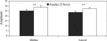
Amplitudes (μV) of the Nc amplitude at midline and lateral electrode sites. To illustrate the main effect of condition, amplitudes are collapsed across group and region and are reported as absolute values (**p < .01).
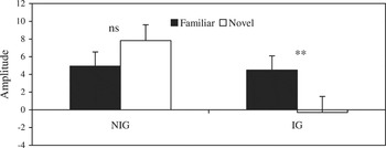
Amplitudes (μV) of the PSW average amplitude at midline electrode sites. To illustrate the interaction of Group × Condition, amplitudes are collapsed across region (**p < .01).
 You have
Access
- 62
- Cited by