

The representation of multiple languages in the brain of a bilingual has been a subject of research for more than a century. Many theories exist about how the brain processes first language (L1) and second language (L2). Two general approaches were adopted in the study of the bilingual brain. The first approach has examined the effect of brain damage on language processing in individuals with bilingual aphasia (Fabbro, Reference Fabbro1999). Clinical studies have enhanced our knowledge about the patterns of language recovery after stroke in bilinguals. Paradis (Reference Paradis, Whitaker and Whitaker1977) proposed five patterns of cross-linguistic recovery in bilingual individuals with aphasia. The five types of classic recovery patterns are (a) parallel recovery, (b) differential recovery, (c) selective recovery, (d) successive recovery, and (e) antagonistic recovery. The recovery patterns provide evidence for distinct and overlapping neuroanatomical representation of the languages in the brain. For example, research on parallel recovery after stroke in bilinguals suggests that for many bilinguals and polyglots, the areas involved in processing language may be the same (Albert & Obler, Reference Albert and Obler1978; Fabbro, Reference Fabbro1999). However, many researchers interpret selective or differential recovery of one or more language in bilingual or polyglots after stroke as suggesting nonoverlapping cortical representation (Gomez-Tortosa, Martin, Gaviria, Charbel, & Ausman, Reference Gomez-Tortosa, Martin, Gaviria, Charbel and Ausman1995; Junque, Vendrell, & Vendrell, Reference Junque, Vendrell, Vendrell and Paradis1995, pp. 139–176; Nilipourn & Ashayeri, Reference Nilipour and Ashayeri1989). These studies, however, do not provide any conclusions about specific regions in the brain that are responsible for processing one language versus the other.
Another approach to the study of the bilingual brain is of the experimental type, such as electrophysiological investigations (electrocortico stimulation during brain surgery and event-related potentials) and functional neuroimaging studies. Neuroimaging techniques such as position emission tomography (PET) and functional magnetic resonance imaging (fMRI) allow a more direct and noninvasive study of the cortical representation of languages in the brain and can provide a clear indication of the neural regions that may be involved in processing of two languages. A large body of functional neuroimaging studies has been devoted to the investigation of language organization in the intact human brain. The application of these techniques to the study of bilingualism may enable us to investigate the role of several factors that have been considered to influence the neural organization of languages, such as age of L2 acquisition and proficiency in L2.
A few reviews have examined the bilingual neuroimaging literature (Abutalebi, Reference Abutalebi2008; Abutalebi, Cappa, & Perani, Reference Abutalebi, Cappa and Perani2001, Reference Abutalebi, Cappa, Perani, Kroll and De Groot2005; Indefrey, Reference Indefrey2006; Stowe & Sabourin, Reference Stowe and Sabourin2005). Abutalebi et al. (Reference Abutalebi, Cappa, Perani, Kroll and De Groot2005, pp. 497–515) reviewed several bilingual neuroimaging studies focusing on the role of age of L2 acquisition and degree of proficiency attained in each of the languages. Using a traditional tabulation of label-reported regions, the authors found that when the degree of proficiency in bilinguals was high, a common language system appeared to be responsible for the processing of both languages. This included the left inferior frontal gyrus, superior and middle temporal gyri, the angular gyrus, and the temporal pole. Lower proficiency in L2 was associated with a more extended network of activation, including foci in the right hemisphere. The authors concluded that, when language proficiency is kept constant, age of acquisition does not seem to have a major impact on neural representation of L2.
In another study, Stowe and Sabourin (Reference Stowe and Sabourin2005) reviewed eight bilingual neuroimaging studies to explore whether L1 and L2 activated similar neural regions and the extent to which it depended on the age of acquisition. They examined three different language tasks: lexical semantics, syntactic comprehension, and phonological processing. The results indicated that L1 and L2 activated the same typical language areas in the left frontal and temporoparietal regions. In addition, late language learners used the same neural substrates for L2 processing as used for L1 processing. However, the neurological system that underlies language appeared to be used less efficiently for L2 as demonstrated by more extensive activations for L2 compared to L1. They concluded that the subprocesses involved in normal language processing are not utilized optimally in some aspects of L2 processing, at least for the less proficient speakers.
The conclusions draw by Stowe and Sabourin (Reference Stowe and Sabourin2005) were similar to that drawn by Indefrey (Reference Indefrey2006). Indefrey (Reference Indefrey2006) analyzed the results of 30 hemodynamic experiments comparing L1 processing and L2 processing in a range of tasks. The results of the review indicated differences between L1 and L2, but only for subgroups of bilingual speakers. During word production, L1 and L2 engaged the same cortical areas, whereas L2 speakers with late L2 onset or lower proficiency recruited the left inferior frontal cortex more strongly than L1. For word-level semantic processing in comprehension, L2 onset and exposure did not play a major role. By contrast, L2 onset played an important factor for activation differences related to syntactic processing in sentence comprehension.
In a recent review, Abutalebi (Reference Abutalebi2008) examined the existing bilingual literature with a narrower focus on the neural correlates of grammatical and lexicosemantic processing in bilinguals. According to the author, neural differences between L1 and L2 existed for grammatical processing and lexicosemantic processing. These differences are particularly prominent in the initial stages of L2 acquisition and/or when L2 is processed with a “nonnativelike” proficiency. The differences between L1 and L2 disappear once a more “nativelike” proficiency is established in L2, reflecting a change in language processing mechanism: from controlled processing for a less proficient L2 to more automatic processing.
The above-mentioned reviews suggest that the neural representation of languages in bilinguals is modulated by a number of factors, of which proficiency appears to be the most important factor. However, all these reviews have attempted to synthesize the literature using a descriptive/qualitative approach that inherently relies on author-supplied anatomical labels that may be unduly broad (e.g., left prefrontal cortex). Comparison of reported coordinates across studies can also prove challenging in that localization of a given set of coordinates to a particular neuroanatomical location is dependent on the target brain atlas and corresponding stereotaxic space in which the data set was registered.
In the present study, we build upon these prior efforts by using the recently developed activation likelihood estimation (ALE; Eickhoff et al., Reference Eickhoff, Laird, Grefkes, Wang, Zilles and Fox2009; Laird, Fox, et al., Reference Laird, Fox, Price, Glahn, Uecker and Lancaster2005; Turkeltaub, Eden, Jones, & Zeffiro, Reference Turkeltaub, Eden, Jones and Zeffiro2002) technique to carry out a quantitative, voxelwise meta-analysis of published functional neuroimaging studies on bilingualism. A quantitative meta-analysis can provide a useful method to assess the state of the field and to provide a plan for future research. This technique provides an unbiased, statistically based approach to examine findings across studies, as opposed to the traditional “box score” or label-based qualitative methods (Laird, McMillan, et al., Reference Laird, McMillan, Lancaster, Kochunov, Turkeltaub and Pardo2005). Activation likelihood estimation was originally developed to identify the brain regions that were consistently activated during speech production (Turkeltaub et al., Reference Turkeltaub, Eden, Jones and Zeffiro2002) using coordinates reported by different functional imaging studies. It assumes that each study reports specific coordinates of activations, which vary due to differences associated with scanners, analyses, paradigms, and intersubject variability. The spatial distribution of these foci is analyzed to determine the regions in which activations are consistently observed, regardless of variability in study design. In activation likelihood estimation, the foci reported by each study are modeled as a probability distribution. Then a map of the whole brain is constructed, assigning to each voxel a value equal to the probability that an activation lies within the voxel. Significance of these values are determined by computing a null distribution obtained by permutation testing, correcting for multiple comparisons by controlling the false discovery rate (FDR). For example, an FDR correction guarantees that in a set of voxels deemed significant for a test of p = .05, the expected proportion of false positives is controlled (Laird, Fox, et al., Reference Laird, Fox, Price, Glahn, Uecker and Lancaster2005).
The ALE technique has been applied in the study of a variety of imaging areas such as of specific cognitive functions (Derrfuss, Brass, Neumann, & Yves von Cramon, Reference Derrfuss, Brass, Neumann and Yves von Cramon2005; Krain, Wilson, Arbuckle, Castellanos, & Milham, Reference Krain, Wilson, Arbuckle, Castellanos and Milham2006; Laird, McMillan, et al., Reference Laird, McMillan, Lancaster, Kochunov, Turkeltaub and Pardo2005; McMillan, Laird, Witt, & Meyerand, Reference McMillan, Laird, Witt and Meyerand2007; Owen, McMillan, Laird, & Bullmore, Reference Owen, McMillan, Laird and Bullmore2005; Price, Devlin, Moore, Morton, & Laird, Reference Price, Devlin, Moore, Morton and Laird2005; Tan, Laird, Li, & Fox, Reference Tan, Laird, Li and Fox2005), major depressive disorder (Fitzgerald et al., Reference Fitzgerald, Oxley, Laird, Kulkarni, Egan and Daskalakis2006), attention-deficit/hyperactivity disorder (Ellison-Wright, Glahn, Laird, Thelen, & Bullmore, Reference Ellison-Wright, Glahn, Laird, Thelen and Bullmore2008), stuttering (Brown, Lasird, Ingham, Ingham, & Fox, Reference Brown, Laird, Ingham, Ingham and Fox2005), schizophrenia (Glahn et al., Reference Glahn, Ragland, Abramoff, Barrett, Laird and Bearden2005), and obsessive–compulsive disorder (Menzies et al., Reference Menzies, Chamberlain, Laird, Thelen, Sahakian and Bullmore2008).
In this study we quantitatively analyze the role of L2 proficiency in bilingual neural representation using the ALE technique. The studies are divided into two groups based on the L2 proficiency of the subjects: high proficiency group and low/moderate proficiency group. Our hypothesis is that greater proficiency in a language will be associated with the recruitment of a common overlapping network and focal activation in core regions in the language network, whereas lesser proficiency in a language will be associated with a more distributed network of regions. This same observation, that proficiency induces efficiency (i.e., a more circumscribed network of activation), has been made in numerous neural systems, perhaps most convincingly in studies of motor learning (Ma et al., Reference Ma, Wang, Narayana, Hazeltine, Chen and Robin2010). The present experiment aimed to examine this issue in the context of functional neuroimaging studies of bilingualism.
METHODS
Literature search
Multiple PubMed literature searches (www.pubmed.com) were conducted to find all fMRI and PET studies on bilingualism. The search included keywords such as “bilingualism,” “first language,” “second language,” “neuroimaging,” “fMRI,” and “PET.” In addition, the reference lists of these articles were reviewed for relevant studies not identified by the initial database search. This approach identified 33 potential articles. We individually screened all the articles for the presence of Talairach or MNI coordinates and tabulated the studies into two groups based on the subjects’ proficiency in the L2: (a) high proficiency group, and (b) low/moderate proficiency group. The subjects’ proficiency in the L2 was determined based on the information provided in the methods section in each of the screened articles. Some studies used language screening questionnaire/proficiency rating scales to determine the degree of L2 proficiency (e.g., Chee, Tan, & Thiel, Reference Chee, Tan and Thiel1999; Luke, Liu, Wai, Wan, & Tan, Reference Luke, Liu, Wai, Wan and Tan2002; Meschyan & Hernandez, Reference Meschyan and Hernandez2006), whereas other studies used standardized national examination scores (e.g., Ding et al., Reference Ding, Perry, Peng, Ma, Li and Xu2003; Golestani et al., Reference Golestani, Alario, Meriaux, Le Bihan, Dehaene and Pallier2006). In order to ensure comparability, all of the studies that classified the bilingual subjects as “fluent,” “proficient,” were grouped in the high proficiency group, and all the studies that classified the bilingual subjects as “less proficient than L1,” “moderately proficient,” “low proficient” were grouped in the low/moderate proficiency group.
Of the 33 neuroimaging studies, 19 neuroimaging studies were not included in the meta-analysis study for the following reasons: (a) studies that did not report activation foci as three-dimensional (3-D) coordinates (x, y, z) in stereotactic space (Dehaene et al., Reference Dehaene, Dupoux, Mehler, Cohen, Paulesu and Perani1997; Hasegawa, Carpenter, & Just, Reference Hasegawa, Carpenter and Just2002; Illes et al., Reference Illes, Francis, Desmond, Gabrieli, Glover and Poldrack1999; Kim, Relkin, Lee, & Hirsch, Reference Kim, Relkin, Lee and Hirsch1997; Klein, Zatorre, Milner, Meyer, & Evans, Reference Klein, Zatorre, Milner, Meyer and Evans1994; Mahendra, Plante, Magloire, Milman, & Trouard, Reference Mahendra, Plante, Magloire, Milman and Trouard2003; Marian, Spivery, & Hirsch, Reference Marian, Spivery and Hirsch2003; Pillai et al., Reference Pillai, Araque, Allison, Sethuraman, Loring and Thiruvaiyaru2003), (b) studies that reported results that were based on regions of interest analysis (Chee et al., Reference Chee, Weekes, Lee, Soon, Schreiber and Hoon2000; Pillai et al., Reference Pillai, Allison, Sethuraman, Araque, Thiruvaiyaru and Ison2004), (c) studies that used bilingual switching and translation tasks (Hernandez, Dapretto, Mazziotta, & Bookheimer, Reference Hernandez, Dapretto, Mazziotta and Bookheimer2001; Hernandez, Martinez, & Kohnert, Reference Hernandez, Martinez and Kohnert2000; Price, Green, & von Studnitz, Reference Price, Green and von Studnitz1999; Wang, Xue, Chen, Xue, & Dong, Reference Wang, Xue, Chen, Xue and Dong2007), (d) studies that did not compare the activation patterns of L1 and L2 processing on the same subjects (Rüschemeyer, Fiebach, Kempe, & Friederici, Reference Rüschemeyer, Fiebach, Kempe and Friederici2005; Ruschemeyer, Zysset, & Friderici, Reference Ruschemeyer, Zysset and Friederici2006), (e) studies that did not present data for contrasts examining L1 versus baseline and L2 versus baseline (Wartenburger et al., Reference Wartenburger, Heekeren, Abutalebi, Cappa, Villringer and Perani2003), and (f) studies that focused on suprasegmental processing rather than linguistic processing (Callan, Jones, Callan, & Akalhane-Yamada, Reference Callan, Jones, Callan and Akahane-Yamada2004; Gandour et al., Reference Gandour, Tong, Talavage, Wong, Dzemidzic and Xu2007). This filtering of publications resulted in a relatively homogenous set of studies with similar fMRI methodology.
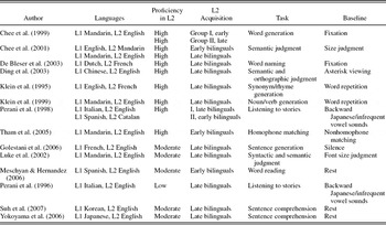
Fourteen neuroimaging studies of bilingual language processing are summarized in Table 1. Among these studies, 8 studies investigated language processing in bilinguals with high L2 proficiency (Chee et al., Reference Chee, Tan and Thiel1999; Chee, Hon, Lee, & Soon, Reference Chee, Hon, Lee and Soon2001; De Bleser et al., Reference De Bleser, Dupont, Postler, Bormans, Speelman and Mortelmans2003; Ding et al., Reference Ding, Perry, Peng, Ma, Li and Xu2003; Klein et al., Reference Klein, Milner, Zatorre, Zhao and Nikelski1999; Klein, Milner, Zatorre, Meyer, & Evans, Reference Klein, Milner, Zatorre, Meyer and Evans1995; Perani et al., Reference Perani, Paulesu, Galles, Dupoux, Dehaene and Bettinardi1998; Tham et al., Reference Tham, Liow, Rajapakse, Leong, Ng and Lim2005), and other 6 studies investigated language processing in bilinguals with moderate/low L2 proficiency (Golestani et al., Reference Golestani, Alario, Meriaux, Le Bihan, Dehaene and Pallier2006; Luke et al., Reference Luke, Liu, Wai, Wan and Tan2002; Meschyan & Hernandez, Reference Meschyan and Hernandez2006; Perani et al., Reference Perani, Dehaene, Grassi, Cohen, Cappa and Dupoux1996; Suh et al., Reference Suh, Yoon, Lee, Chung, Cho and Park2007; Yokoyama et al., Reference Yokoyama, Okamoto, Miyamoto, Yoshimoto, Kim and Iwata2006). The tasks included word or sentence generation (Chee et al., Reference Chee, Tan and Thiel1999; Golestani et al., Reference Golestani, Alario, Meriaux, Le Bihan, Dehaene and Pallier2006; Klein et al., Reference Klein, Milner, Zatorre, Meyer and Evans1995, Reference Klein, Milner, Zatorre, Zhao and Nikelski1999), picture naming (De Bleser et al., Reference De Bleser, Dupont, Postler, Bormans, Speelman and Mortelmans2003), word reading (Meschyan & Hernandez, Reference Meschyan and Hernandez2006), semantic judgment (Chee et al., Reference Chee, Hon, Lee and Soon2001; Ding et al., Reference Ding, Perry, Peng, Ma, Li and Xu2003; Luke et al., Reference Luke, Liu, Wai, Wan and Tan2002), listening to stories (Perani et al., Reference Perani, Dehaene, Grassi, Cohen, Cappa and Dupoux1996, Reference Perani, Paulesu, Galles, Dupoux, Dehaene and Bettinardi1998), sentence comprehension (Suh et al., Reference Suh, Yoon, Lee, Chung, Cho and Park2007; Yokoyama et al., Reference Yokoyama, Okamoto, Miyamoto, Yoshimoto, Kim and Iwata2006), and homophone matching (Tham et al., Reference Tham, Liow, Rajapakse, Leong, Ng and Lim2005). All studies reported data for several contrasts. Of these, only contrasts examining L1 versus baseline and L2 versus baseline were included in the present study.
Fourteen neuroimaging studies included in the meta-analyses of L1 and L2 processing in high proficient bilinguals and low/moderate proficient bilinguals

Note: L1, First language; L2, second language.
Language pairs included orthographically similar languages like Spanish–English (Meschyan & Hernandez, Reference Meschyan and Hernandez2006), Italian–English (Perani et al., Reference Perani, Dehaene, Grassi, Cohen, Cappa and Dupoux1996, Reference Perani, Paulesu, Galles, Dupoux, Dehaene and Bettinardi1998), and Catalan–English (Perani et al., Reference Perani, Paulesu, Galles, Dupoux, Dehaene and Bettinardi1998) and orthographically distant languages like Mandarin–English (Chee et al., Reference Chee, Tan and Thiel1999, Reference Chee, Weekes, Lee, Soon, Schreiber and Hoon2000; Ding et al., Reference Ding, Perry, Peng, Ma, Li and Xu2003; Tham et al., Reference Tham, Liow, Rajapakse, Leong, Ng and Lim2005), Korean–English (Suh et al., Reference Suh, Yoon, Lee, Chung, Cho and Park2007), and Japanese–English (Yokoyama et al., Reference Yokoyama, Okamoto, Miyamoto, Yoshimoto, Kim and Iwata2006).
Meta-analysis procedures
All meta-analyses were carried out using the ALE technique (Turkeltaub et al., Reference Turkeltaub, Eden, Jones and Zeffiro2002) implemented in Brainmap (Eickhoff et al., Reference Eickhoff, Laird, Grefkes, Wang, Zilles and Fox2009; Laird, Fox, et al., Reference Laird, Fox, Price, Glahn, Uecker and Lancaster2005), which was developed at the Research Imaging Center of the University of Texas Health Science Center San Antonio (http://www.brainmap.org/ale). The results reported in MNI coordinates (De Bleser et al., Reference De Bleser, Dupont, Postler, Bormans, Speelman and Mortelmans2003; Tham et al., Reference Tham, Liow, Rajapakse, Leong, Ng and Lim2005) were converted to Talairach space using the icbm2tal transform (Lancaster et al., Reference Lancaster, Tordesillas-Gutierrez, Martinez, Salinas, Evans and Zilles2007). Four separate meta-analyses were conducted: (a) L1 processing in the high proficiency group, (b) L2 processing in the high proficiency group, (c) L1 processing in the low/moderate proficiency group, and (d) L2 processing in the low/moderate proficiency group. In the present study, activation foci for contrasts examining L1 versus baseline and L2 versus baseline were included.
Each of the meta-analysis was conducted using the activation foci defined as the x, y, and z coordinate in a standard stereotactic space. Using ALE, the coordinates identified in the literature search were modeled with a 3-D Gaussian distribution, and their convergence across experiments was quantitatively assessed. A prespecified full width at half maximum was not used in these analyses as in the original ALE approach (Laird, Fox, et al., Reference Laird, Fox, Price, Glahn, Uecker and Lancaster2005). Instead, a new algorithm (Eickhoff et al., Reference Eickhoff, Laird, Grefkes, Wang, Zilles and Fox2009) was used to model the spatial uncertainty of each focus using an estimation of the intersubject and interlaboratory variability typically observed in neuroimaging experiments. This algorithm limits the meta-analysis to an anatomically constrained space specified by a gray matter mask, and includes a method that calculates the above-chance clustering between experiments (i.e., random-effects analysis), rather than between foci (i.e., fixed-effects analysis) The test was corrected for multiple comparisons using the FDR algorithm (Genovese, Laxar, & Nochols, Reference Genovese, Laxar and Nochols2002). Finally, the thresholded ALE map was created (p < .05), and overlaid onto the “colinbrain” anatomical template normalized to Talairach space (Kuchonov et al., Reference Kuchonov, Lancaster, Thompson, Toga, Brewer and Hardies2002) using the MRIcron software (http://www.sph.sc.edu/comd/rorden/mricron). A minimum cluster size of 150 mm3 was applied. Identification of the anatomical location of the voxels with peak probabilities was manually determined by reference to the Talairach and Tournoux atlas (Talairach & Tournoux, Reference Talairach and Tournoux1988).
RESULTS
High proficiency group
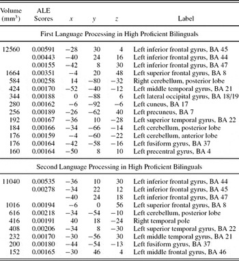
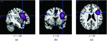
The results from the two ALE analyses of L1 and L2 processing in the high proficiency group are shown in Table 2 and Figure 1. Eight studies (Chee et al., Reference Chee, Tan and Thiel1999, Reference Chee, Hon, Lee and Soon2001; De Bleser et al., Reference De Bleser, Dupont, Postler, Bormans, Speelman and Mortelmans2003; Ding et al., Reference Ding, Perry, Peng, Ma, Li and Xu2003; Klein et al., Reference Klein, Milner, Zatorre, Meyer and Evans1995, Reference Klein, Milner, Zatorre, Zhao and Nikelski1999; Perani et al., Reference Perani, Paulesu, Galles, Dupoux, Dehaene and Bettinardi1998; Tham et al., Reference Tham, Liow, Rajapakse, Leong, Ng and Lim2005) were included in the high proficiency bilingual group. The language tasks included noun or verb generation (Chee et al., Reference Chee, Tan and Thiel1999; Klein et al., Reference Klein, Milner, Zatorre, Meyer and Evans1995, Reference Klein, Milner, Zatorre, Zhao and Nikelski1999), picture naming (De Bleser et al., Reference De Bleser, Dupont, Postler, Bormans, Speelman and Mortelmans2003), semantic judgment (Chee et al., Reference Chee, Hon, Lee and Soon2001; Ding et al., Reference Ding, Perry, Peng, Ma, Li and Xu2003), listening to stories (Perani et al., Reference Perani, Paulesu, Galles, Dupoux, Dehaene and Bettinardi1998), and homophone matching (Tham et al., Reference Tham, Liow, Rajapakse, Leong, Ng and Lim2005). In total, 141 activation foci were analyzed for L1 processing and 156 activation foci were analyzed for L2 processing. Our meta-analyses detected significantly elevated probabilities of activation in a distributed network of brain regions primarily in the left hemisphere for both L1 and L2 processing.
Major cluster volumes and activation likelihood estimation (ALE) scores for first and second language processing in high proficiency second language group

Note: BA, Brodmann area.
Activation likelihood estimation results for eight studies in the high proficiency second language (L2) group. Representative slices in (a) sagittal, (b) coronal, and (c) axial views (p < .05; false discovery rate corrected). In the online color version, red represents first language (L1), blue represents L2, and purple represents the overlap in activation. The analysis demonstrated a primarily left-lateralized network, with an overlap in the frontal region and temporal region for L1 and L2. [A color version of this figure can be viewed online at journal.cambridge.org/aps]

The areas of activation included the left inferior frontal gyri (Brodmann area [BA] 44, 45), left superior frontal gyrus (BA 8), left precentral gyrus (BA 4), left middle and superior temporal gyri (BA 21, 22), left fusiform gyrus, (BA 37), left precuneus gyrus (BA 7), and left cerebellum. Additional activation was present in the right cerebellum. For L1 and L2 processing in the high proficient group, the clusters identified in the left frontal regions were larger than the clusters identified in the ALE maps from the left temporal and parietal regions.
Low/moderate proficiency group
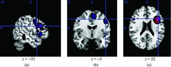
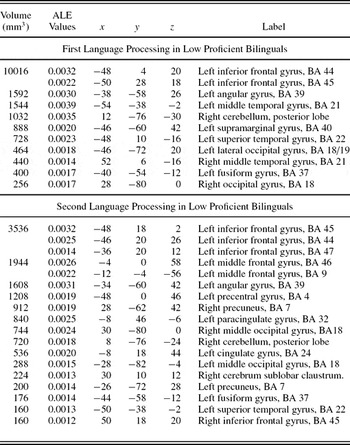
The results from the two ALE analyses of L1 and L2 processing in the low proficiency group are shown in Table 3 and Figure 2. Six studies (Golestani et al., Reference Golestani, Alario, Meriaux, Le Bihan, Dehaene and Pallier2006; Luke et al., Reference Luke, Liu, Wai, Wan and Tan2002; Meschyan & Hernandez, Reference Meschyan and Hernandez2006; Perani et al., Reference Perani, Dehaene, Grassi, Cohen, Cappa and Dupoux1996; Suh et al., Reference Suh, Yoon, Lee, Chung, Cho and Park2007; Yokoyama et al., Reference Yokoyama, Okamoto, Miyamoto, Yoshimoto, Kim and Iwata2006) were included in the low/moderate proficiency bilingual group. The language tasks included word reading (Meschyan & Hernandez, Reference Meschyan and Hernandez2006), sentence generation (Golestani et al., Reference Golestani, Alario, Meriaux, Le Bihan, Dehaene and Pallier2006), sentence comprehension (Suh et al., Reference Suh, Yoon, Lee, Chung, Cho and Park2007; Yokoyama et al., Reference Yokoyama, Okamoto, Miyamoto, Yoshimoto, Kim and Iwata2006), listening to stories (Perani et al., Reference Perani, Dehaene, Grassi, Cohen, Cappa and Dupoux1996), and semantic and syntactic judgment (Luke et al., Reference Luke, Liu, Wai, Wan and Tan2002). In total, 147 activation foci were analyzed for L1 processing and 122 activation foci were analyzed for L2 processing.
Activation likelihood estimation results for six studies in the low/moderate proficiency second language group. Representative slices in (a) sagittal, (b) coronal, and (c) axial views (p < .05; false discovery rate corrected). In the online color version, red represents first language, blue represents second language, and purple represents the overlap in activation. [A color version of this figure can be viewed online at journal.cambridge.org/aps]

Major cluster volumes and activation likehood estimation (ALE) scores for first and second language processing in low/moderate proficiency second language group

Note: BA, Brodmann area.
The activation for L1 processing was observed in a predominantly left lateralized network. For L1 processing, frontal regions showed significantly elevated probabilities of activation in areas of left inferior frontal gyrus and premotor area (BA 4, 6, 44, 45, 47). In addition, significant elevated probabilities of activation were also identified bilaterally in the middle and superior temporal gyri (BA 21, 22), left fusiform gyrus (BA 37), left angular gyrus, (BA 39), left supramarginal gyrus (BA 40), left precuneus (BA 7), bilateral occipital lobe (BA 17), and left and right cerebellum.
Activation for L2 processing was observed bilaterally. Significant elevated probabilities of activation were observed bilaterally in the left and right inferior frontal gyrus (BA 44, 45), left middle frontal gyrus, dorsolateral prefrontal cortex (46/9), left parietal (BA 7), bilateral occipital regions (BA 17, 18), bilateral cerebellum, left cingulate gyrus (BA 24), left paracingulate gyrus (BA 32), and left superior temporal gyrus (BA 22). Activation was observed in the cingulate and paracingulate gyrus for L2 processing only in the low proficiency group. In addition, activation in the superior temporal gyrus (BA 22) barely reached significance for L2 processing in the low proficiency group. Further, the clusters were generally smaller than the clusters identified in the other three meta-analyses.
Overlap in activation
An overlap in activation was observed for L1 and L2 in the high proficiency group and L1 in the low/moderate proficiency group. It should be noted that (see Tables 2 and 3) the Talairach coordinates of activation in the frontotemporal, temporoparietal, and occipital regions for L1 processing in the moderate/low proficient group were very similar to that of the high proficient group.
DISCUSSION
Given the importance of language proficiency in bilingual neural representation, the present study conducted a meta-analysis focusing on low/moderate and high proficient bilinguals. The aim of this study was to quantitatively analyze the results of a number of neuroimaging studies done on bilingualism. We were particularly interested in the role of language proficiency on the neural representation of L1 and L2. Our findings suggest that despite varying experimental tasks, there appears to be a similar pattern of regions involved in bilingual neural representation in high proficient bilinguals. In low proficient bilinguals, however, there are several differences between L1 and L2 processing.
Language processing in high proficiency group
Eight studies were included in our meta-analyses study of language processing in the high proficiency group. The Talairach coordinates of activation in the frontotemporal, temporoparietal, and occipital regions in L1 processing were very similar to that of L2 processing. An interesting finding of these meta-analyses was the presence of large cluster volumes on the ALE maps in the left frontal region for L1 and L2 processing. The frontal region appears to be one of those regions where different aspects of language processing interact with each other to deliver highly complex and interactive human language processing. Specifically, the frontal lobe is assumed to be related to semantic, syntactic, and phonological processing (Bookheimer, Reference Bookheimer2002; Hagoort, Reference Hagoort2005; Nakai et al., Reference Nakai, Matsuo, Kato, Matsuzawa, Okada and Glover1999; Siok, Perfetti, Jin, & Tan, Reference Siok, Perfetti, Jin and Tan2004; Tan et al., Reference Tan, Liu, Perfetti, Spinks, Fox and Gao2001, Reference Tan, Spinks, Feng, Siok, Perfetti and Xiong2003).
Increased probabilities of activation in the frontal region might be related to the tasks used in the studies included in the meta-analyses. Four out of the eight studies in the high proficient group used production-based tasks that included word/verb generation (Chee et al., Reference Chee, Tan and Thiel1999; Klein et al., Reference Klein, Milner, Zatorre, Meyer and Evans1995, Reference Klein, Milner, Zatorre, Zhao and Nikelski1999) and word naming (De Bleser et al., Reference De Bleser, Dupont, Postler, Bormans, Speelman and Mortelmans2003). This argument is consistent with findings of a meta-analysis study on word production. Indefrey and Levelt (Reference Indefrey and Levelt2004) identified the inferior frontal gyrus and the precentral cortex as the main regions that were activated during a variety of naming and word generation tasks, suggesting that the left frontal regions contribute to the core processes of language production. Alternatively, increased activation in this region might reflect comprehension processes as the other four studies used comprehension-based tasks such as semantic judgment (Chee et al., Reference Chee, Hon, Lee and Soon2001; Ding et al., Reference Ding, Perry, Peng, Ma, Li and Xu2003), listening to stories (Perani et al., Reference Perani, Paulesu, Galles, Dupoux, Dehaene and Bettinardi1998), and homophone matching (Tham et al., Reference Tham, Liow, Rajapakse, Leong, Ng and Lim2005). Several studies support the notion that the left frontal lobe, specifically the left inferior frontal gyrus, supports language comprehension in addition to language production (Bookheimer, Reference Bookheimer2002; Moro et al., Reference Moro, Tettamanti, Perani, Donati, Cappa and Fazio2001; Zatorre, Meyer, Gjedde, & Evans, Reference Zatorre, Meyer, Gjedde and Evans1996).
Another interesting finding was the increased probabilities of activation in the cerebellum. Although most studies in the fMRI evaluation of language processing (semantic, phonological, and other) have demonstrated predominantly left hemispheric areas of supratentorial activation (inferior frontal gyrus, dorsolateral prefrontal cortex, superior temporal gyrus, supramarginal gyrus, etc.), several studies show cerebellum is also involved in many cognitive processes including language processing (Booth et al., Reference Booth, Wood, Lu, Houk and Bitan2007; Desmond & Fiez, Reference Desmond and Fiez1998; Fulbright et al., Reference Fulbright, Jenner, Mencl, Pugh, Shaywitz and Shaywitz1999). The finding of significant activation in cerebellum is consistent with other bilingual studies that have found activation in this region during language tasks (Chee et al., Reference Chee, Hon, Lee and Soon2001; De Bleser et al., Reference De Bleser, Dupont, Postler, Bormans, Speelman and Mortelmans2003; Marian et al., Reference Marian, Spivery and Hirsch2003; Pillai et al., Reference Pillai, Araque, Allison, Sethuraman, Loring and Thiruvaiyaru2003, Reference Pillai, Allison, Sethuraman, Araque, Thiruvaiyaru and Ison2004).
The other regions that were activated included the fusiform gyrus (BA 37), precuneus (BA 7), and the occipital cortex (BA 17/18). Previous bilingual neuroimaging studies have shown strong activation in the left fusiform gyrus when viewing visually presented words (Chee et al., Reference Chee, Tan and Thiel1999, Reference Chee, Hon, Lee and Soon2001; DeBleser et al., Reference De Bleser, Dupont, Postler, Bormans, Speelman and Mortelmans2003; Ding et al., Reference Ding, Perry, Peng, Ma, Li and Xu2003; Tham et al., Reference Tham, Liow, Rajapakse, Leong, Ng and Lim2005). These studies suggest that the fusiform region may participate in phonological decoding and semantic processing for visually presented stimuli.
In general, although there were differences in the probabilities of activation in terms of the cluster volumes, the results of our meta-analyses confirmed our prediction that similar areas will be recruited during L1 and L2 processing in high proficient bilinguals. This corroborates the findings of previous bilingual neuroimaging studies that suggest both languages of a proficient bilingual recruit a common neural network (Chee et al., Reference Chee, Hon, Lee and Soon2001; Illes et al., Reference Illes, Francis, Desmond, Gabrieli, Glover and Poldrack1999, Perani et al., Reference Perani, Paulesu, Galles, Dupoux, Dehaene and Bettinardi1998).
Language processing in low/moderate proficiency group
Six studies were included in our meta-analyses of language processing in the low/moderate proficiency group. The results of the meta-analyses suggest differences in probabilities of activation observed between L1 and L2 processing in the low proficient bilinguals. For L2 processing, the clusters were generally smaller and distributed over wider areas than the clusters identified in the ALE maps from L1 processing (see Figure 2). In addition, more regions were activated in the right hemisphere and in the left hemisphere for the low proficient group compared to the high proficient group. This finding is consistent with the view that low proficient bilinguals recruit additional areas to compensate for reduced language proficiency (Dehaene et al., Reference Dehaene, Dupoux, Mehler, Cohen, Paulesu and Perani1997; Perani et al., Reference Perani, Dehaene, Grassi, Cohen, Cappa and Dupoux1996).
An interesting finding observed in this study was the presence of increased activation in the right inferior frontal gyrus for L2 processing. Clinical evidence has suggested that impairment of the left frontal regions might induce contralesional areas to take over functions previously carried out by the homologous left-sided brain structures, suggesting a compensatory role for right frontal regions (Fernandez et al., Reference Fernandez, Cardebat, Demonet, Joseph, Mazaux and Barat2004; Thulborn, Carpenter, & Just, Reference Thulborn, Carpenter and Just1999; Weiller et al., Reference Weiller, Isensee, Rijntjes, Huber, Muller and Bier1995; Winhuisen et al., Reference Winhuisen, Thiel, Schumacher, Kessler, Rudolf and Haupt2007). Bush et al. (Reference Bush, Frazier, Rauch, Seidman, Whalen and Jenike1999) also observed bilateral inferior frontal activation during the counting Stroop test in adults with attention-deficit/hyperactivity disorder. This increased activation was attributed to enhanced cognitive effort required for subjects with attention-deficit/hyperactivity disorder. Similarly, low proficient bilinguals might recruit the right frontal regions to compensate for reduced language proficiency in L2. This argument is in line with previous findings, which have indicated the recruitment of the right hemisphere during processing of weaker L2 (Luke et al., Reference Luke, Liu, Wai, Wan and Tan2002; Meschyan & Hernandez, Reference Meschyan and Hernandez2006; Yokohama et al., Reference Yokoyama, Okamoto, Miyamoto, Yoshimoto, Kim and Iwata2006).
Another interesting finding was that the activation in the left superior temporal gyrus barely reached significance in the low proficient group. The size of the cluster was 160 for L2 processing compared to 728 for L1 processing. Stronger activation of this area might be related to a more extensive conceptual processing associated with the larger amount of retrieved semantic information (Vandenberghe, Price, Wise, Josephs, & Frackowiak, Reference Vandenberghe, Price, Wise, Josephs and Frackowiak1996). It is possible that the reduced activation in the temporal region for L2 processing in the low proficient group may reflect poor semantic representation in the less proficient L2. This meta-analysis reflects the lack of temporal activation in many of the studies in the low proficient group. For example, Yokohama et al. (Reference Yokoyama, Okamoto, Miyamoto, Yoshimoto, Kim and Iwata2006) did not find activation in the temporal region during L2 processing in their low proficient L2 subjects during sentence comprehension. Similarly, Suh et al. (Reference Suh, Yoon, Lee, Chung, Cho and Park2007) did not find activation in their subjects who were moderately proficient in L2 during sentence comprehension.
Increased probabilities of activation were also observed in the dorsolateral prefrontal cortex (BA 46, 9) and the anterior cingulate cortex (BA 24, 32) during the less proficient L2 processing. According to Abutalebi and Green (Reference Abutalebi and Green2007), functional neuroimaging studies investigating the aspects of cognitive control confirm that cognitive control emerges from the integration of separable neural systems (Botvinick, Nystrom, Fissell, Carter, & Cohen, Reference Botvinick, Nystrom, Fissell, Carter and Cohen1999; MacDonald, Cohen, Stenger, & Carter, Reference MacDonald, Cohen, Stenger and Carter2000; Petrides, Alivasatos, Meyer, & Evans, Reference Petrides, Alivasatos, Meyer and Evans1993). According to Braver, Barch, Gray, Molfesek, and Avraham (Reference Braver, Barch, Gray, Molfese and Avraham2001), these systems include prefrontal, inferior parietal cortex, and anterior cingulate cortex. The prefrontal cortex is a collection of interconnected neocortical areas that sends and receives projections from virtually all cortical sensory systems, motor systems, and many subcortical structures. Models of cognitive control conceptualize the prefrontal cortex functions as top-down bias mechanisms that facilitate the processing of task-relevant representations even in the presence of prepotent, irrelevant ones (Dehaene & Changeux, Reference Dehaene and Changeux1991; Desimone & Duncan, Reference Desimone and Duncan1995; Miller & Cohen, Reference Miller and Cohen2001). In the bilingual case, such a situation could ideally be when a bilingual has to produce words in a weaker L2, and hence, to block potential prepotent responses from the stronger L1. Thus, increased probabilities of activation observed during L2 processing in the prefrontal cortex might be interpreted as blocking potential prepotent responses from the stronger language (L1) while processing the weaker language (L2).
Another region that is involved in cognitive control is the anterior cingulate cortex (ACC). In contrast to the prefrontal cortex that guides response selection under conditions of response, the ACC modulates cognitive control (Bush, Luu, & Posner, Reference Bush, Luu and Posner2000; Cabeza & Nyberg, Reference Cabeza and Nyberg1997). Functional neuroimaging studies have shown ACC activity during tasks that engage working memory, language generation, and controlled information processing (Cabeza & Nyberg, Reference Cabeza and Nyberg1997). One of its most important roles of the ACC is detecting and signaling the occurrence of conflicts in information processing (Botvinick et al., Reference Botvinick, Nystrom, Fissell, Carter and Cohen1999; Carter et al., Reference Carter, Braver, Barch, Botvinick, Noll and Cohen1998). Researchers indicate that the ACC activation is related to the degree of response conflict present in a given cognitive task (Botvinick et al., Reference Botvinick, Nystrom, Fissell, Carter and Cohen1999; Carter et al., Reference Carter, Braver, Barch, Botvinick, Noll and Cohen1998). Further, activation in the anterior cingulate cortex is also linked to attention. Kerns et al. (Reference Kerns, Cohen, MacDonald, Cho, Stenger and Carter2004) suggests that there is a link between ACC activation and subsequent performance. Specifically, strong ACC engagement should be followed by behavior reflecting relatively focused attention and weak ACC engagement by less focused attention. Kerns et al. (Reference Kerns, Cohen, MacDonald, Cho, Stenger and Carter2004) also observed ACC engagement in association with errors. The magnitude of error responses was related to posterror performance, with higher activation associated with a relatively large slow-down in reaction times. This finding is consistent with the proposal put forth by Botvinick et al. (Reference Botvinick, Nystrom, Fissell, Carter and Cohen1999) that posterror slowing might be a consequence of conflict monitoring.
Increased probabilities of activation observed during L2 processing in the ACC might be interpreted not only due to an intrusion from the proficient L1 during language tasks in the less proficient L2 but also due to increased attentional demand in L2 processing. This premise is supported by several behavioral studies that show the nontarget language is activated and cross-language effects appear even in situations and tasks that are purely monolingual. For example, in unbalanced bilinguals, cross-language effects of L2 on L1 were found in a purely L1 context (van Hell & Dijkstra, Reference van Hell and Dijkstra2002; van Wijnendaele & Brysbaert, Reference van Wijnendaele and Brysbaert2002).
Taken together, these findings suggest that processing the less proficient language is demanding and is associated with a more extended network of activation, including foci in the right hemisphere. More specifically, the results of these meta-analyses suggest that lower L2 proficiency is associated with increased probabilities of activation in the dorsolateral prefrontal cortex, anterior cingulate cortex, and right inferior frontal gyrus. Notably, the results of this study are consistent even with the bilingual neuroimaging studies that were not included in this analysis. Several of these studies found that bilinguals with less proficient L2 showed more extensive activation in L2 than in L1 (Dehaene et al., Reference Dehaene, Dupoux, Mehler, Cohen, Paulesu and Perani1997; Hasegawa et al., Reference Hasegawa, Carpenter and Just2002; Kim et al., Reference Kim, Relkin, Lee and Hirsch1997; Pillai et al., Reference Pillai, Araque, Allison, Sethuraman, Loring and Thiruvaiyaru2003, Reference Pillai, Allison, Sethuraman, Araque, Thiruvaiyaru and Ison2004) and bilinguals who were equally proficient in both languages consistently showed similar patterns of activation for both languages (Hernandez et al., Reference Hernandez, Martinez and Kohnert2000, Reference Hernandez, Dapretto, Mazziotta and Bookheimer2001; Illes et al., Reference Illes, Francis, Desmond, Gabrieli, Glover and Poldrack1999; Klein et al., Reference Klein, Zatorre, Milner, Meyer and Evans1994; Marian et al., Reference Marian, Spivery and Hirsch2003, Price et al., Reference Price, Green and von Studnitz1999).
Our ALE meta-analysis is also in agreement with previous reviews of bilingual brain activation studies (Abutalebi, Reference Abutalebi2008; Abutalebi et al., Reference Abutalebi, Cappa and Perani2001, Reference Abutalebi, Cappa, Perani, Kroll and De Groot2005; Indefrey, Reference Indefrey2006; Stowe & Sabourin, Reference Stowe and Sabourin2005). However, there are a number of limitations to be considered in the interpretation of these results. First, the present study reflects analysis of heterogeneous tasks. The eight studies in the “high proficient group” included tasks like noun or verb generation, picture naming, semantic judgment, listening to stories, and homophone matching, whereas the six studies in the “low/moderate proficient group” included tasks like sentence generation, sentence comprehension, listening to stories, and semantic and syntactic judgment. The heterogeneity of the tasks could have resulted in the differences in activation patterns between the “low/moderate” and “high” proficient group. Nonetheless, the inclusion of heterogeneous tasks in this meta-analysis in order to identify brain regions recruited during processing of less and more proficient language is consistent with the viewpoint that irrespective of task, the brain regions involved in processing linguistic elements during semantic, syntactic, and phonological aspects are essentially the same. For instance, Hagoort (Reference Hagoort2005) and Bookheimer (Reference Bookheimer2002) have convincingly argued that the regions within left inferior frontal gyrus are involved in processing phonological, semantic, and syntactic information and are not modality or content specific. Our view is consistent with this premise that a network of highly interactive nodes (inferior frontal gyrus, dorsolateral prefrontal cortex, ACC, superior temporal gyrus) are routinely involved in selecting, analyzing, retrieving, comparing and modulating linguistic information that is independent of task, modality and perhaps even the language structure.
Second, the present study only focused on the role of language proficiency in bilingual neural representation. It should be noted that other factors such as L2 onset, exposure, and task differences might also account for differences in bilingual neural representation. For example, Indefrey (Reference Indefrey2006) carried out a systematic review of 30 neuroimaging experiments to examine the relative influence of the three factors (onset, proficiency, and exposure) on bilingual neural representation. The author also analyzed the variations in subject groups and tasks that might explain why some studies found differences and others did not. The results indicated that for word-level production all the three factors played a role, whereas for word-level semantic processing in comprehension only proficiency played a role. By contrast, L2 onset seemed to be the most important factor for activation differences related to syntactic processing in sentence comprehension.
Third, only a limited number of studies were included in the present study. Several studies in the literature could not be included in this analysis as they did not report their results in standard 3-D stereotactic coordinates, and only reported their findings in terms of anatomically determined regions of interest. Without the use of standardized 3-D coordinates, comparisons between the studies must rely on more subjective approaches and cannot be included in voxel-based, statistical meta-analyses such as the present study. Despite exclusion of these studies, our results are broadly consistent with those of qualitative reviews that included both studies with and without stereotactic coordinates.
In summary, functional neuroimaging techniques, such as fMRI and PET, provide a unique window into the organization of language in neurologically intact bilinguals. How a later learned L2 becomes organized in the brain, relative to the first, is an intriguing question for theoretical and practical reasons. These results further confirm the premise that greater proficiency in language recruits a more focal core language network, whereas lower proficiency in language recruits a more distributed network of regions. This observation of an inverse relationship between proficiency and activation has been made in numerous neural systems, perhaps most convincingly in studies of motor learning (Ma et al., Reference Ma, Wang, Narayana, Hazeltine, Chen and Robin2010; Xiong et al., Reference Xiong, Ma, Wang, Narayana, Duff and Egan2009) indicating that proficiency induces efficiency. This premise is consistent with our previous work in bilingual aphasia rehabilitation (Edmonds & Kiran, Reference Edmonds and Kiran2006) that has shown that training the less proficient language (i.e., a more diffuse network) in stroke patients results in cross language transfer to the more proficient language, whereas training the proficient language (i.e., a highly specialized network) does not result in cross-language transfer to the weaker language. Therefore, it could be argued that training the less proficient and distributed L2 language network may intrinsically strengthen the dense L1 language network because of the overlapping nodes in the two networks. This conjecture is currently being examined in ongoing work in our laboratory. In addition, these results have clinical implications in presurgical planning affecting the frontal and temporal lobe in high and low proficient bilinguals. It is critical to assess language proficiency in L1 and L2 prior to surgical planning in bilingual patients with lesions in close spatial proximity to essential language cortex. For example, surgical resection of the right frontal lobe could have a detrimental effect on language processing in low proficient bilinguals and not high proficient bilinguals. Further studies should also address the role of practice and the frequency of usage of the L2 in bilingual neural representation.
CONCLUSION
A number of functional imaging studies have addressed the issue of cerebral representation of the two languages. The present study is the first ALE meta-analysis study on bilingual neuroimaging. Our findings draw attention to the presence of consistent differences in the bilingual neural representation between high proficient bilinguals and low/moderate proficient bilinguals. Specifically, in the low proficient group, clusters were generally smaller and distributed over wider areas in both the hemispheres than the clusters identified in the ALE maps from the high proficient group. Another important finding of the present study was reduced activation in the temporal region during L2 processing in low proficient bilinguals. This may reflect poor semantic representation in the less proficient L2. In addition, greater probabilities of activation were found not only in regions traditionally involved in language processing, but also in regions known to sustain the cognitive control, such as the prefrontal cortex and the anterior cingulate cortex.
ACKNOWLEDGMENTS
This work was supported in part by NIMH Grant R01-MH074457. The authors thank Allison Darling and Sarah Thelen for help during data entry and analysis.