




No CrossRef data available.
Structure and synthesis of nematode phosphorylcholine-containing glycoconjugates
Published online by Cambridge University Press: 18 November 2004
Abstract
Infection with filarial nematodes produces a chronic, long-lasting illness with adult worms able to survive within human hosts for up to 15 years. A contributor to the longevity of these parasites is the presence of phosphorylcholine (PC) on components of the worms' molecular secretions (ES). PC on ES modulates host immune responses towards an anti-inflammatory phenotype thereby generating an environment favourable for parasite survival. PC is attached to nematode ES via a covalent association with carbohydrate, which, although well-documented in bacteria and fungi, is absent from humans, making it an ideal target for the development of novel drugs. In order to produce such drugs it is first necessary to understand the structure and synthesis of nematode PC-glycans. ES-62 is the major PC-ES-product of Acanthocheilonema viteae and is a homologue of PC-ES found in human filarial nematodes. We have studied the structure and biosynthesis of PC-glycans of ES-62 by a combination of pulse-chase experiments, experiments involving the use of inhibitors of each of intracellular trafficking, oligosaccharide processing and phospholipid biosynthesis and various forms of mass spectrometry. Our indications indicate that PC is transferred in the lumen of the medial Golgi to an N-type glycan consisting of a trimannosyl core with or without core fucosylation bearing between 1 and 4 N-acetyl glucosamine residues. The structure of the PC-N-glycans found in ES-62 appears to be conserved amongst filarial nematodes in that it has additionally been identified in Onchocerca volvulus and O. gibsoni. Also, similar structures have been found in non-filarial parasitic nematodes and in the free-living nematode Caenorhabditis elegans. Finally, PC has also been recently found attached to the carbohydrate moieties of nematode glycosphingolipids and the structure of these will also be considered.
Information
- Type
- Review Article
- Information
- Copyright
- © 2004 Cambridge University Press
FILARIASIS
Over 150 million people are currently infected by pathogenic filarial nematodes in over 80 countries. There are a further 1 billion people believed to be at risk of infection although this may be a conservative estimate. There are two main pathogenic forms of filarial nematode that can affect humans. Onchocerciasis is caused by the presence of the microfilariae of Onchocerca volvulus. Adult worms of this species inhabit nodules under the skin. Here they produce large numbers of microfilariae that then migrate throughout the body. Host reactions elicited by the death of these larvae can cause a number of conditions including serious visual impairment and blindness, onchocercal skin disease and lymphadenitis. Adult worms of Wuchereria bancrofti and Brugia malayi the main causative agents of lymphatic filariasis lodge in the host lymphatics. This often results in severe forms of pathology including hydrocoele and elephantiasis. These three parasites are jointly responsible for 6 million DALYs (Disability Adjusted Life Years) lost per year in Africa and India. Apart from the diseases they cause being painful and disfiguring they have a massive impact on the economies of endemic countries and contribute to poverty (WHO, 1997). The treatment of filariasis in countries that are co-endemic for onchocerciasis and lymphatic filariasis is based on a combination therapy of ivermectin and albendazole, and in countries clear of onchocerciasis, DEC and albendazole are administered. Thus the treatment of filariasis, and in particular onchocerciasis, is very heavily dependent upon ivermectin and mass treatment with this compound increases the risk of resistance to this drug becoming a reality. Ivermectin distribution has also been seriously affected by the observation that administration to patients bearing heavy Loa loa infections can result in a severe neurological reaction, which can result in death. Thus, the development of alternative drugs for filariasis treatment is paramount and the WHO recommends that ‘top priority’ should be given to developing drugs with macrofilaricidal effects (www.who.int/tdr).
ES-62, A MODEL PHOSPHORYLCHOLINE-CONTAINING GLYCOPROTEIN
Studies on human filarial nematodes are limited by their inability to develop in vitro or within laboratory rodents. For this reason, many researchers have turned to filarial nematode parasites of animals that can be maintained in the laboratory. Acanthocheilonema viteae appears to be a natural gerbil parasite and is able to dwell successfully in either M. unguiculatus or M. libycus. Studies on A. viteae, resulted in 1989 (Harnett et al. 1989) in the identification of an abundantly excreted–secreted protein that, based on molecular mass, was termed ES-62. Since then, homologues of this molecule have been identified in other filarial nematodes of animals (Nor et al. 1997; Stepek et al. 2002) and EST analysis of the human filarial nematodes O. volvulus and B. malayi has produced sequences with convincing homology to ES-62, raising the possibility that it is secreted by all filarial nematodes.
Following its discovery, ES-62 was quickly shown to be a glycoprotein following successful radio-isotope labelling with [3H]-glucosamine (Harnett et al. 1989). Further studies employing lectin binding analysis (Harnett et al. 1994) and, in particular, digestion with various endoglycosidases (Harnett et al. 1994) indicated that the molecule's carbohydrate was likely to be N-linked. Similar studies undertaken with B. pahangi ES-products also identified the presence of some N-linked glycans (Nor et al. 1997). Further investigation of ES-62 by Fast Atom Bombardment Mass Spectroscopy (FAB-MS), revealed the presence of 3 different classes of N-glycan structure; a high mannose type structure (Man5-9GlcNAc2), a glycan which has been fully trimmed to the trimannosyl core and fucosylated (Fuc0-1Man3GlcNAc2), and finally, a structure that comprised of the trimannosyl core with or without core fucosylation carrying between 1 and 4 N-acetyl glucosamine (GlcNAc) residues (Fuc0-1Man3GlcNAc3-6) (Haslam et al. 1997). Since early work by Pery's group in 1974 (Pery et al. 1974), several studies had shown that PC-containing nematode products also contained carbohydrate (Lal et al. 1987; Maizels, Burke & Denham, 1987; Harnett et al. 1989). However, the finding that [3H]-choline labelled ES-62 exposed to N-glycosidase F digestion resulted in a complete loss of radioactivity from the nematode product, provided the first evidence that PC was actually directly attached to the protein backbone of ES-62 via an N-linked glycan (Harnett et al. 1993). This result was corroborated by the observation that exposure of A. viteae and B. pahangi to tunicamycin, the antibiotic inhibitor of Glc3Man9GlcNAc2-PP-dolichol formation (Elbein, 1987; Carroll & Bird, 1991), the precursor of all N-type glycans, blocked attachment of PC to the worms' ES (Houston & Harnett, 1996; Nor, Devaney & Harnett, 1997). Of the three species of N-type glycans identified in ES-62 only the Fuc0-1Man3GlcNAc3-6 structure carried measurable amounts of PC (Fig. 1) (Haslam et al. 1997) as identified by FAB-MS. 1–2 PC groups were found per glycan but because of the permanent charges associated with several PC groups, identification of ES-62 N-glycans substituted with multiple PC groups by this method was impossible. That is not, however, to say that they do not exist since substrates capable of such additions are present in ES-62. Further FAB-MS examination of O. volvulus and O. gibsoni also identified the PCx-Fuc0-1Man3GlcNAc3-6 structure (Fig. 1) suggesting that it is conserved in filarial nematodes (Haslam et al. 1999). In fact, FAB-MS analysis of the diverse and varied spectrum of N-linked glycans found in the free-living nematode Caenorhabditis elegans (Haslam & Dell, 2003) identified the same Fuc0-1Man3GlcNAc3-6 family as being substituted with PC, which may indicate that the transferase responsible for PC addition is conserved throughout the phylum Nematoda. In any case, a consequence of this finding is that nematode PC-N-glycans are likely to have functions other than as immunomodulators and indeed it is unknown as to whether free-living nematodes secrete PC-containing glycoproteins. Presumably the immunomodulatory role has appeared during the evolution of parasites and PC-glycans and when they first appeared they had some other (presumably internal) function which has been maintained through evolution in both free-living and parasitic species. A very different and previously uncharacterized high mannose PC3Man5GlcNAc2 structure has also recently been found in C. elegans using NMR and MALDI-TOF (Cipollo, Costello & Hirschberg, 2002). The structural differences between these two PC-glycans are such that there is suggestion of 2 distinct families of PC-containing N-type glycans in C. elegans. Three classes of N-glycans have been characterized in the muscle-stage larvae of the mammalian parasite Trichinella spiralis and of these PC has been found to be associated with a Fuc0-1Man3HexNAc1-8 structure (Morelle et al. 2000b). However, in this case substitution of N-glycans with PC appears to be stage specific and is absent from the adult nematode (Morelle et al. 2000a,b).

Fig. 1. The structure of the PC-glycan of ES-62.
PC substitution in nematode molecules is not limited to glycoproteins – it has also been identified in association with glycosphingolipids and as with the PC-glycans these zwitterionic structures appear to be highly conserved in Nematoda. The actual structure, Gal(α1-3)GalNAc(β1-4)[PC-6]GlcNAc(β1-3)Man(β1-4)Glc(β1-1) ceramide, was identified via its pentasaccharide core as belonging to the arthro-carbohydrate series and was first identified in Ascaris suum (Lochnit et al. 1998). It has subsequently been found in O. volvulus (Wuhrer et al. 2000), C. elegans (Gerdt et al. 1999) and A. viteae (Houston et al. 2002).
INTRACELLULAR SITE OF TRANSFER OF PC TO ES-PRODUCTS
ES-62 is secreted from cells that subtend the oesophagus of adult A. viteae (Stepek et al. 2002). All newly synthesized N-linked glycoproteins destined for secretion follow a preset series of modifying, trimming and transferase events during their journey through the endoplasmic reticulum (ER) and the Golgi apparatus (Fig. 2) during which their final structure is determined (Pfeffer & Rothman, 1987). This knowledge was exploited in order to identify the intracellular compartment in which PC transfer to ES-62 was occurring. Pulse-chase time-course experiments with [35S]-methionine labelled A. viteae extracts, showed that PC transfer to ES-62 is a fairly rapid event occurring 40–60 min after protein synthesis (Houston, Cushley & Harnett, 1997). This suggested that PC transfer was probably occurring in the ER or the Golgi apparatus and not in a later compartment of the cell. However, the rate of migration of proteins through the ER varies greatly making an exact prediction of where PC transfer takes place impossible. To obtain more revealing data relating to this, additional strategies were adopted. First A. viteae were exposed to brefeldin A, a compound which blocks transport from the ER to the Golgi by targeting Golgi-specific coat proteins that may be involved in maintaining the structural integrity of the organelle (Hunziker, Whitney & Mellman, 1992). Such exposure inhibited the transfer of PC to ES-62 increasing the likelihood that transfer was occurring in the Golgi apparatus. Next, to further support this idea and to obtain more detailed information on the site of PC-transfer, several inhibitors of N-linked oligosaccharide processing were employed. These were deoxynojirimycin (dNM), a glucose analogue and a potent inhibitor of ER resident glucosidases I and II, deoxymannojirimycin, (dMM) a mannose analogue and inhibitor of mannosidase I in the cis Golgi and finally swainsonine, the first compound shown to block oligosaccharide processing by inhibiting mannosidase II in the medial Golgi (Elbein, 1987). dNM prevented addition of PC to ES-62 but also inhibited secretion of the molecule (Houston et al. 1997). The ability of dNM to prevent addition of PC to ES-62 is consistent with a Golgi location for PC transfer as it is unlikely that ES-62 without its terminal glucose residues removed would be able to exit the ER (Lodish & Kong, 1984). We have also made the observation that PC is added only to tetrameric ES-62 and not to monomers (Fig. 3) and many proteins must undergo oligomerization for efficient transport from the ER to the Golgi. dMM exposure was also shown to result in production of ES-62 that was lacking PC but unlike dNM did not cause inhibition of secretion (Houston et al. 1997). The fact that dMM impedes the action of the cis Golgi-located mannosidase I responsible for trimming 3 mannose residues from the 3-linked branch of N-linked glycans and in doing so prevents the transfer of PC to ES-62 implies that the removal of these sugars is essential for PC attachment. It also confirms that PC addition to the N-glycans of ES-62 is a post-ER event. The compound swainsonine did not affect transfer of PC to ES-62, suggesting that the oligosaccharide processing steps leading up to and including mannosidase I (converting Man8GlcNAc2 to Man5GlcNAc2) are fundamental for PC transfer but that the action of mannosidase II (converting Man5GlcNAc3 to Man3GlcNAc3) is unnecessary (Houston et al. 1997). However, ES-62 produced in the presence of swainsonine displayed altered lectin-binding preferences and endoglycosidase sensitivity compared to ‘normal’ ES-62, suggesting that it would usually undergo trimming by mannosidase II (Houston et al. 1997). This being the case it can also be surmized (a) that ES-62 is processed by GlcNAc transferase I, the enzyme that precedes mannosidase II in the processing pathway and (b) that PC cannot be attached to either of the mannose residues, present on the 6-linked branch of the N-glycan, targeted by mannosidase II. A lack of complex or hybrid glycans on ES-62 (Harnett et al. 1994) suggests that it undergoes little, if any, processing after mannosidase II digestion possibly as a consequence of PC attachment to additionally added GlcNAC residues. However, the dMM and swainsonine data, strongly support the action of GlcNAc transferase I as being the key event in addition of PC and hence the intracellular site of PC transfer to ES-62 as being the medial Golgi (Houston et al. 1997). This is certainly supported by the aforementioned FAB-MS data, which identified in ES-62, Man3GlcNAc3, a structure native to this site, as being a recipient of PC donation (Haslam et al. 1997).

Fig. 2. A cartoon representation of N-linked oligosaccharide processing through the endoplasmic reticulum and the Golgi apparatus.

Fig. 3. Exposing [3H]choline-labelled Acanthocheilonema viteae PBS extracts to PAGE/fluorography under native (A) and reducing/denaturing (B) conditions illustrates that PC is transferred to tetrameric ES-62.
DETERMINATION OF THE PC DONOR FOR ES-62
To identify the intracellular source of PC used in the manufacture of PC-ES would be valuable with respect to characterizing the enzyme/s involved in PC transfer to N-glycans, which in turn would be constructive in designing inhibitors. The Kennedy or CDP-choline pathway (Fig. 4) is the most common method of phosphatidylcholine biosynthesis used by mammalian cells and the first reaction involves the phosphorylation of choline to phosphorylcholine (PC) by choline kinase (EC 2.7.1.32). The second step is rate limiting (Vance, Tripp & Paddon, 1980) and involves conversion to CDP-choline via CTP: phosphocholine cytidylyltransferase (EC 2.7.7.15) catalysis. The final reaction is the condensation of CDP-choline with diacylglycerol (DAG) by choline phosphotransferase (EC 2.7.8.2) to form phosphatidylcholine. Preliminary experiments provided evidence of a role for the pathway in provision of PC for ES-62 since culturing A. viteae in the presence of Hemicholinium-3 (HC-3), an inhibitor of choline kinase prevented the attachment of PC to ES-62 (Houston & Harnett, 1999). Within the Kennedy pathway and subsequent sphingomyelin biosynthesis there are 3 potential PC-donors and each contains choline in an activated form. These are CDP-choline, phosphatidylcholine and sphingomyelin. As CDP-choline donates PC for phosphatidylcholine biosynthesis it was considered likely that it may be the PC donor for ES-62. Attempts to confirm this were therefore undertaken. First, both whole A. viteae and microsomal extracts of worms were exposed to radio-isotope labelled CDP-choline and incorporation of labelled choline into ES-62 measured. Although in both cases isotope-labelled phosphatidylcholine was identified, neither in worms or extracts could isotope-labelled ES-62 be identified (Houston & Harnett, 1999). These negative data, however, could be explained by an inability of CDP-choline to gain access to the medial lumen of the Golgi as would be essential for it to be a factor in providing PC for ES-62. To donate PC for phosphatidylcholine biosynthesis it needs only to get into the cytoplasm of the cell (Bishop & Bell, 1988). Thus, as an alternative approach, A. viteae were cultured in the presence of hexadecylphosphocholine (HEPC), an inhibitor of the enzyme responsible for CDP-choline manufacture. Exposure to this reagent, however, did not interfere with synthesis of the ES-62 PC-glycan. Nevertheless, the inhibitor at concentrations employed did not inhibit phosphatidylcholine biosynthesis in A. viteae, suggesting that it was not actually interfering with the worm's CTP: phosphocholine cytidylyltransferase (Houston & Harnett, 1999). Increasing the concentration of this inhibitor was ruled out, as it was toxic to the worms. Thus attempts to designate a role for CDP-choline as the PC donor for ES-62 have failed.

Fig. 4. The Kennedy Pathway of phosphatidylcholine biosynthesis.
Further work involved a time-course experiment that analysed, by HPLC and HPTLC, the time taken for all of the Kennedy pathway metabolites of A. viteae to become radio-isotope labelled with [3H]-choline (after a 5 min pulse) and comparison with that taken by ES-62. This work identified both CDP-choline and phosphatidylcholine as being capable of being PC-donors as they become isotope labelled within 10 min of chase compared to the 20 min taken by ES-62 (Houston et al. 2002). Labelling of sphingomyelin took 50 min, ruling it out as PC-donor for ES-62 (Houston et al. 2002). This is perhaps not particularly surprising considering that sphingomyelin synthesis is thought to occur predominantly in the trans-Golgi network (TGN) (Allan & Obradors, 1999), a compartment downstream of the medial Golgi. To examine phosphatidylcholine's potential as a PC-donor, a second pathway of phosphatidylcholine synthesis, which lacks CDP-choline as an intermediate, was exploited. This pathway (Carman & Henry, 1989, 1999) involves the decarboxylation of phosphatidylserine to phosphatidylethanolamine (by phosphatidylserine decarboxylase) which then undergoes a three-step S-adenosyl methionine (SAM)-dependent methylation to form phosphatidylcholine. Employing [3H]-SAM as a radio-isotope label, it was shown that phosphatidylcholine labelling was evident approximately 20 h before that of ES-62, suggesting that the radioactivity being incorporated into ES-62 was not being directly sourced from the isotope-labelled phosphatidylcholine (Houston et al. 2002). Thus, as with CDP-choline, there are currently no data suggesting that phosphatidylcholine is a PC donor for ES-62. PC-containing glycosphingolipids, previously identified in A. suum (Lochnit et al. 1998), O. volvulus (Wuhrer et al. 2000), and C. elegans (Gerdt et al. 1999) have also been identified by HPTLC in A. viteae and their potential as PC-donors for ES-62 assessed (Houston et al. 2002). In contrast to ES-62, [14C]-choline labelling of these metabolites only occurred after prolonged culture periods, possibly a reflection of a slow turnover of these molecules, but still ruling them out as likely PC donors.
STRATEGIES FOR PREVENTING THE ASSOCIATION BETWEEN PC AND ES-PRODUCTSndashh;CLUES FOR DRUG DEVELOPMENT
Preventing attachment of PC to ES-products is certainly feasible. Exposure of A. viteae to the oligosaccharide processing inhibitor dMM as indicated earlier resulted in secreted ES-62 that was devoid of PC. However, unfortunately, this reagent cannot be considered as a possible drug due to a combination of unreliability and toxicity associated with the fact that it will also inhibit host mannosidases. With respect, at least to the efficacy issues associated with dMM, another mannosidase I inhibitor kifunensine (Elbein et al. 1990) is currently being tested. This reagent is much more potent than dMM and so it will be of interest to see how it performs. Regarding inhibitors of the Kennedy Pathway, a number of compounds are currently being examined. These include, Farnesol, an isoprenoid inhibitor of choline phosphotransferase (Miquel et al. 1998) and ET-18-OCH3 a compound related to HEPC. The need for a new generation of drugs to target filariasis is urgent and their development remains a top priority and a main focus of TDR research (www.who.int/tdr). There is also an urgent need for novel veterinary anthelminthics as the sustainable control of parasitic nematode infection in ruminant livestock is now arguably the major constraint in farming. Drug resistance is a significant threat to these grazing livestock–it has already developed against all currently available anthelminthics (Kaminsky, 2003). Also, resistance is no longer restricted to developing countries in the Tropics (e.g. in Latin America resistance to the entire panel of broad spectrum anthelminthic drugs has evolved (Waller, 1997)). Sheep farming is now at risk in Australia, New Zealand, the UK and South Africa and drug resistance in cattle is also emerging in New Zealand and South America. Development of novel anti-parasitic drugs should ideally be directed against phylum-specific targets and with respect to developing new macrofilariacidal therapies and veterinary drugs the ‘PC-transferase’ responsible for transferring PC onto nematode glycoconjugates represents such a target.
References
REFERENCES
ALLAN, D. &
OBRADORS, M. J. M. (
1999).
Enzyme distributions in subcellular fractions of BHK cells infected with Semliki virus: evidence for major fraction of sphingomyelin synthase in the trans-golgi network.
Biochimica et Biophysica Acta
1450,
277–287.CrossRefGoogle Scholar
BISHOP, W. R. &
BELL, R. M. (
1988).
Assembly of phospholipids into cellular membranes: biosynthesis, transmembrane movement and intracellular translocation.
Annual Review of Cell Biology
4,
579–610.CrossRefGoogle Scholar
CARMAN, G. M. &
HENRY, S. A. (
1989).
Phospholipid biosynthesis in yeast.
Annual Review of Biochemistry
58,
6635–6669.CrossRefGoogle Scholar
CARMAN, G. M. &
HENRY, S. A. (
1999).
Phospholipid biosynthesis in the yeast Saccharomyces cerevisiae and interrelationship with other metabolic processes.
Progress in Lipid Research
38,
361–399.CrossRefGoogle Scholar
CARROLL, M. &
BIRD, M. M. (
1991).
Glycoprotein expression in mouse cerebellum: effects of inhibitors of N-linked glycosylation.
International Journal of Biochemistry
23,
1285–1291.CrossRefGoogle Scholar
CIPOLLO, J. F.,
COSTELLO, C. E. &
HIRSCHBERG, C. B. (
2002).
The fine structure of Caenorhabditis elegans N-glycans.
Journal of Biological Chemistry
277,
49143–49157.CrossRefGoogle Scholar
ELBEIN, A. D. (
1987).
Inhibitors of the biosynthesis and processing of N-linked oligosaccharide chains.
Annual Review of Biochemistry
56,
497–534.CrossRefGoogle Scholar
ELBEIN, A. D.,
TROPEA, J. E.,
MITCHELL, M. &
KAUSHAL, G. P. (
1990).
Kufunensine, a potent inhibitor of the glycoprotein processing mannosidase I.
Journal of Biological Chemistry
265,
15599–15605.Google Scholar
GERDT, S.,
DENNIS, R. D.,
BORGONIE, G.,
SCHNABEL, R. &
GEYER, R. (
1999).
Isolation, characterization and immunolocalization of phosphorylcholine-substituted glycolipids in developmental stages of Caenorhabditis elegans.
European Journal of Biochemistry
266,
952–963.CrossRefGoogle Scholar
HARNETT, W.,
FRAME, M. J.,
NOR, Z. M.,
MACDONALD, M. &
HOUSTON, K. M. (
1994).
Some preliminary data on the nature/structure of the PC-glycan of the major excretory–secretory product of Acanthocheilonema viteae (ES-62).
Parasite
1,
179–181.CrossRefGoogle Scholar
HARNETT, W.,
HOUSTON, K. M.,
AMESS, R. &
WORMS, M. J. (
1993).
Acanthocheilonema viteae: phosphorylcholine is attached to the major excretory–secretory product via an N-linked glycan.
Experimental Parasitology
77,
498–502.CrossRefGoogle Scholar
HARNETT, W.,
WORMS, M. J.,
KAPIL, A.,
GRAINGER, M. &
PARKHOUSE, R. M. (
1989).
Origin, kinetics of circulation and fate in vivo of the major excretory–secretory product of Acanthocheilonema viteae.
Parasitology
99,
229–239.CrossRefGoogle Scholar
HASLAM, S. M. &
DELL, A. (
2003).
Hallmarks of Caenorhabditis elegans N-glycosylation: complexity and controversy.
Biochimie
85,
25–32.CrossRefGoogle Scholar
HASLAM, S. D.,
GEMS, D.,
MORRIS, H. R. &
DELL, A. (
2002).
The glycomes of Caenorhabditis elegans and other model organisms.
Biochemical Society Symposium
69,
117–134.CrossRefGoogle Scholar
HASLAM, S. M.,
HOUSTON, K. M.,
HARNETT, W.,
REASON, A. J.,
MORRIS, H. R. &
DELL, A. (
1999).
Structural studies of N-glycans of filarial parasites. Conservation of phosphorylcholine-substituted glycans among species and discovery of novel chito-oligomers.
Journal of Biological Chemistry
274,
20953–20960.Google Scholar
HASLAM, S. M.,
KHOO, K. H.,
HOUSTON, K. M.,
HARNETT, W.,
MORRIS, H. R. &
DELL, A. (
1997).
Characterisation of the phosphorylcholine-containing N-linked oligosaccharides in the excretory–secretory 62 kDa glycoprotein of Acanthocheilonema viteae.
Molecular and Biochemical Parasitology
85,
53–66.CrossRefGoogle Scholar
HOUSTON, K. M.,
CUSHLEY, W. &
HARNETT, W. (
1997).
Studies on the site and mechanism of attachment of phosphorylcholine to a filarial nematode secreted glycoprotein.
Journal of Biological Chemistry
272,
1527–1533.CrossRefGoogle Scholar
HOUSTON, K. M. &
HARNETT, W. (
1996).
Prevention of attachment of phosphorylcholine to a major excretory–secretory product of Acanthocheilonema viteae using tunicamycin.
Journal of Parasitology
82,
320–324.CrossRefGoogle Scholar
HOUSTON, K. M. &
HARNETT, W. (
1999).
Mechanisms underlying the transfer of phosphorylcholine to filarial nematode glycoproteins – a possible role for choline kinase.
Parasitology
118,
311–318.CrossRefGoogle Scholar
HOUSTON, K. M.,
LOCHNIT, G.,
GEYER, R. &
HARNETT, W. (
2002).
Investigation of the nature of potential phosphorylcholine donors for filarial nematode glycoconjugates.
Molecular and Biochemical Parasitology
123,
55–66.CrossRefGoogle Scholar
HUNZIKER, W.,
WHITNEY, J. A. &
MELLMAN, I. (
1992).
Brefeldin A and the endocytic pathway. Possible implications for membrane traffic and sorting.
FEBS Letters
307,
93–96.Google Scholar
KAMINSKY, R. (
2003).
Drug resistance in nematodes: a paper tiger or a real problem?
Current Opinion in Infectious Diseases
16,
559–564.Google Scholar
LAL, R. B.,
PARANJAPE, R. S.,
BRILES, D. E.,
NUTMAN, T. B. &
OTTESEN, E. A. (
1987).
Circulating parasite antigen(s) in lymphatic filariasis: use of monoclonal antibodies to phosphocholine for immunodiagnosis.
Journal of Immunology
138,
3454–3460.Google Scholar
LODISH, H. F. &
KONG, N. (
1984).
Glucose removal from N-linked oligosaccharides is required for efficient maturation of certain secretory glycoproteins from the rough endoplasmic reticulum to the Golgi complex.
Journal of Cell Biology
98,
1720–1729.CrossRefGoogle Scholar
LOCHNIT, G.,
DENNIS, R. D.,
ULMER, A. J. &
GEYER, R. (
1998).
Structural elucidation and monokine-inducing activity of two biologically active zwitterionic glycosphingolipids derived from the parasitic nematode Ascaris suum.
Journal of Biological Chemistry
278,
466–474.CrossRefGoogle Scholar
MAIZELS, R. M.,
BURKE, J. &
DENHAM, D. A. (
1987).
Phosphorylcholine-bearing antigens in filarial nematode parasites: analysis of somatic extracts, in-vitro secretions and infection sera from Brugia malayi and B. pahangi.
Parasite Immunology
9,
49–66.CrossRefGoogle Scholar
MIQUEL, K.,
PRADINES, A.,
TERCE, F.,
SELMI, S. &
FAVRE, G. (
1998).
Competitive inhibition of choline phosphotransferase by geranylgeraniol and farnesol inhibits phosphatidylcholine synthesis and induces apoptosis in human lung adenocarcinoma A549 cells.
Jorunal of Biological Chemistry
273,
26179–26186.CrossRefGoogle Scholar
MORELLE, W.,
HASLAM, S. M.,
MORRIS, H. R. &
DELL, A. (
2000 a).
Characterization of the N-linked glycans of adult Trichinella spiralis.
Molecular and Biochemical Parasitology
109,
171–177.Google Scholar
MORELLE, W.,
HASLAM, S. M.,
OLIVIER, V.,
APPLETON, J. A.,
MORRIS, H. R. &
DELL, A. (
2000 b).
Phosphorylcholine-containing N-glycans of Trichinella spiralis: identification of multiantennary lacdiNAc structures.
Glycobiology
10,
941–950.Google Scholar
NOR, Z. M.,
DEVANEY, E. &
HARNETT, W. (
1997).
The use of inhibitors of N-linked glycosylation and oligosaccharide processing to produce monoclonal antibodies against non-phosphorylcholine epitopes of Brugia pahangi excretory–secretory products.
Parasitology Research
83,
813–815.CrossRefGoogle Scholar
NOR, Z. M.,
HOUSTON, K. M.,
DEVANEY, E. &
HARNETT, W. (
1997).
Variation in the nature of attachment of phosphorylcholine to excretory–secretory products of adult Brugia pahangi.
Parasitology
114,
257–262.CrossRefGoogle Scholar
PERY, P.,
PETIT, A.,
POULAIN, J. &
LUFFAU, G. (
1974).
Phosphorylcholine-bearing components in homogenates of nematodes.
European Journal of Immunology
4,
637–639.CrossRefGoogle Scholar
PFEFFER, S. R. &
ROTHMAN, J. E. (
1987).
Biosynthetic protein transport and sorting by the endoplasmic reticulum and Golgi.
Annual Review of Biochemistry
56,
829–852.CrossRefGoogle Scholar
STEPEK, G.,
AUCHIE, M.,
TATE, R.,
WATSON, K.,
RUSSELL, D. G.,
DEVANEY, E. &
HARNETT, W. (
2002).
Expression of the filarial nematode phosphorylcholine-containing glycoprotein, ES62, is stage specific.
Parasitology
125,
155–164.CrossRefGoogle Scholar
VANCE, D. E.,
TRIPP, E. M. &
PADDON, H. B. (
1980).
Poliovirus increases phosphatidylcholine biosynthesis in HeLa cells by stimulation of the rate-limiting reaction catalyzed by CTP: phosphocholine cytidylyltransferase.
Journal of Biological Chemistry
255,
1064–1069.Google Scholar
WALLER, P. J. (
1997).
Anthelmintic resistance.
Veterinary Parasitology
72,
391–405.CrossRefGoogle Scholar
WORLD HEALTH ORGANIZATION (
1997).
Prospects for the elimination of some TDR diseases. World Health Organization,
Geneva.
WUHRER, M.,
RICKHOFF, S.,
DENNIS, R. D.,
LOCHNIT, G.,
SOBOSLAY, P. T.,
BAUMEISTER, S. &
GEYER, R. (
2000).
Phosphocholine-containing zwitterionic glycosphingolipids of adult Onchocerca volvulus as highly conserved antigenic structures of parasitic nematodes.
The Biochemical Journal
348,
417–423.CrossRefGoogle Scholar

Fig. 1. The structure of the PC-glycan of ES-62.
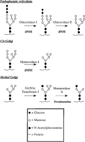
Fig. 2. A cartoon representation of N-linked oligosaccharide processing through the endoplasmic reticulum and the Golgi apparatus.
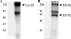
Fig. 3. Exposing [3H]choline-labelled Acanthocheilonema viteae PBS extracts to PAGE/fluorography under native (A) and reducing/denaturing (B) conditions illustrates that PC is transferred to tetrameric ES-62.
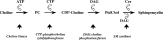
Fig. 4. The Kennedy Pathway of phosphatidylcholine biosynthesis.
 You have
Access
- 35
- Cited by