INTRODUCTION
Control of gastrointestinal (GI) nematodes presents an enormous challenge because such parasites are extremely important pathogens of domestic livestock (Nieuwhof and Bishop, Reference Nieuwhof and Bishop2005) and humans (Molyneux et al. Reference Molyneux, Bradley, Hoerauf, Kyelem and Taylor2003; Bethony et al. Reference Bethony, Brooker, Albonico, Geiger, Loukas, Diemert and Hotez2006; Hotez et al. Reference Hotez, Brindley, Bethony, King, Pearce and Jacobson2008, Reference Hotez, Fenwick, Savioli and Molyneux2009; Hotez and Kamath, Reference Hotez and Kamath2009). They are responsible for severe economic losses experienced by the livestock industry worldwide (Waller et al. Reference Waller, Dash, Barger, Le Jambre and Plant1995; Maciel et al. Reference Maciel, Giménez, Gaona, Waller and Hansen1996; Waller, Reference Waller1997; van Wyk et al. Reference van Wyk, Coles and Tammi Krecek2002; Bartley et al. Reference Bartley, Jackson, Jackson and Sargison2004; Leathwick et al. Reference Leathwick, Hosking, Bisset and McKay2009; Cezar et al. Reference Cezar, Toscan, Camillo, Sangioni, Ribas and Vogel2010; Miller et al. Reference Miller, Waghorn, Leathwick, Candy, Oliver and Watson2012; Veríssimo et al. Reference Veríssimo, Niciura, Alberti, Rodrigues, Barbosa, Chiebao, Cardoso, da Silva, Pereira, Margatho, da Costa, Nardon, Ueno, Curci and Molento2012) and cause extensive suffering and debilitation to human populations living in endemic regions (Yamamoto et al. Reference Yamamoto, Nagai, Kawabata, Leon, Ninomiya and Koizumi2000; Crompton and Nesheim, Reference Crompton and Nesheim2002; Bethony et al. Reference Bethony, Brooker, Albonico, Geiger, Loukas, Diemert and Hotez2006; Elliott et al. Reference Elliott, Kizza, Quigley, Ndibazza, Nampijja, Muhangi, Morison, Namujju, Muwanga, Kabatereine and Whitworth2007; Hotez et al. Reference Hotez, Brindley, Bethony, King, Pearce and Jacobson2008, Reference Hotez, Fenwick, Savioli and Molyneux2009; Hotez and Kamath, Reference Hotez and Kamath2009).
Although currently the main line of treatment and control of GI nematode infections relies heavily on the use of modern synthetic anthelmintics, for centuries in the past medicinal fruits and plants have been used by indigenous peoples worldwide to treat these infections (Caldwell and Caldwell, Reference Caldwell and Caldwell1929; Jonxis and Bekius, Reference Jonxis and Bekius1953; Stransky and Reyes, Reference Stransky and Reyes1955; Behnke et al. Reference Behnke, Buttle, Stepek, Lowe and Duce2008; Keiser and Utzinger, Reference Keiser and Utzinger2008). When fully explored and analysed, plant products could broaden the repertoire of existing anthelmintics, at a time when resistance to conventional synthetic anthelmintics has become widespread (Prichard, Reference Prichard1990; Geerts and Gryseels, Reference Geerts and Gryseels2000; Wolstenholme et al. Reference Wolstenholme, Fairweather, Prichard, von Samson-Himmelstjerna and Sangster2004; Behnke et al. Reference Behnke, Buttle, Stepek, Lowe and Duce2008).
Efficacy of ethnoveterinary therapies in treatment of both helminth and protozoan infections has been claimed in various publications (Hammond et al. Reference Hammond, Fielding and Bishop1997; Waller et al. Reference Waller, Bernes, Thamsborg, Sukura, Richter, Ingebrigtsen and Hoglund2001; Githiori et al. Reference Githiori, Athanasiadou and Thamsborg2006; Lans et al. Reference Lans, Turner, Khan and Brauer2007; Mwale and Masika, Reference Mwale and Masika2009) and a variety of different parts of plants (e.g. flowers, fruits, seeds and leaves) have been used for this purpose. Examples of effective drugs derived from plant sources are quinine and artemisinin. They are widely used for the treatment of malaria (Tagboto and Townson, Reference Tagboto and Townson2001; Taylor and Berridge, Reference Taylor and Berridge2006; Reyburn et al. Reference Reyburn, Mtove, Hendriksen and Seidlein2009). However, despite claims of efficacy, scientific validation of many of these therapies has either lacked conviction or simply not been conducted at all. In the present age, scientific validation of phytomedicine and nutraceuticals requires the same rigorous criteria as those used for chemically synthesized anthelmintics if they are to gain licences for use in animals and humans, and gain popularity as medicines for both (Geary et al. Reference Geary, Woo, McCarthy, Mackenzie, Horton, Prichard, de Silva, Olliaro, Lazdins-Helds, Engels and Bundy2010; Keiser and Utzinger, Reference Keiser, Utzinger, Xiao-Nong Zhou and Jürg2010; Woods and Knauer, Reference Woods and Knauer2010; Caffrey and Secor, Reference Caffrey and Secor2011; Kimura, Reference Kimura2011; Utzinger et al. Reference Utzinger, N'Goran, Caffrey and Keiser2011; Woods et al. Reference Woods, Vaillancourt, Wendt and Meeus2011). These criteria include definition, quality, efficacy, detailed information on the mode of action of the active compounds and identification of their potential side-effects.
Plant cysteine proteinases (CPs) from papaya (Carica papaya), pineapple (Ananas comosus), and fig (Ficus spp.) are known to kill parasitic nematode worms in vitro by a mechanism that is quite different to that of all commercially available synthetic anthelmintics and have also been shown to possess anthelmintic effects in vivo against a range of different GI nematodes (Satrija et al. Reference Satrija, Nansen, Bjørn, Murtini and He1994; Stepek et al. Reference Stepek, Buttle, Duce, Lowe and Behnke2005, Reference Stepek, Buttle, Duce and Behnke2006a , Reference Stepek, Lowe, Buttle, Duce and Behnke b , Reference Stepek, Lowe, Buttle, Duce and Behnke2007b , Reference Stepek, Lowe, Buttle, Duce and Behnke c ; Buttle et al. Reference Buttle, Behnke, Bartley, Elsheikha, Bartley, Garnett, Donnan, Jackson, Lowe and Duce2011; Luoga et al. Reference Luoga, Mansur, Buttle, Duce, Garnett and Behnke2012), including moderate effects on cestodes. These wide-spectrum qualities endow them with the potential to become novel treatments against human and livestock GI nematodes (Githiori et al. Reference Githiori, Athanasiadou and Thamsborg2006; Stepek et al. Reference Stepek, Lowe, Buttle, Duce and Behnke2007b ) and offer an alternative low-cost control strategy for these parasites (Tagboto and Townson, Reference Tagboto and Townson2001; Stepek et al. Reference Stepek, Lowe, Buttle, Duce and Behnke2007b ; Burke et al. Reference Burke, Wells, Casey and Miller2009; Nchu et al. Reference Nchu, Githiori, McGaw and Eloff2011; Maphosa and Masika, Reference Maphosa and Masika2012).
Current models used for testing the anthelmintic activities of CPs require the use of parasitic nematodes in laboratory animals such as mice, rats or hamsters (Stepek et al. Reference Stepek, Behnke, Buttle and Ducel2004, Reference Stepek, Buttle, Duce, Lowe and Behnke2005, Reference Stepek, Buttle, Duce and Behnke2006a , Reference Stepek, Lowe, Buttle, Duce and Behnke b , Reference Stepek, Lowe, Buttle, Duce and Behnke2007a , Reference Stepek, Lowe, Buttle, Duce and Behnke b ). Large-scale screening by these methods is not feasible because laboratory animals can only yield limited numbers of worms and moreover the costs associated with in vivo animal-based trials are becoming increasingly prohibitive. In order to test a wide range of CPs, a better solution would be to employ a free-living nematode which can be reared easily and cheaply in large numbers. An obvious candidate is Caenorhabditis elegans, which meets both of these criteria and is extremely well characterized as a model organism (Riddle et al. Reference Riddle, Blumenthal, Meyer and Priess1997).
Caenorhabditis elegans has already been used in high throughput in vitro screening assays for anthelmintic activity (Simpkin and Coles, Reference Simpkin and Coles1981) involving benzimidazoles and non-benzimidazoles (Boyd et al. Reference Boyd, McBride, Rice, Snyder and Freedman2010) and this has led to the discovery of several new drug compounds, has helped to elucidate the mechanisms of action of some anthelmintics (Sluder and Baumeister, Reference Sluder and Baumeister2004) and has provided useful information on mechanisms of anthelmintic resistance (Geary and Thompson, Reference Geary and Thompson2001; Sluder and Baumeister, Reference Sluder and Baumeister2004). It was therefore of interest to determine whether plant-derived CPs would also affect motility or cause evident cuticular damage to free-living nematodes. Here the hypothesis was tested that CPs are effective against free-living as well as GI nematodes. Our aim was to develop a rapid throughput assay to screen as yet untested plant sources for useful CP-based anthelmintic candidates. In this study, we assessed the efficacy of purified CPs from papaya (C. papaya) on wild C. elegans and cystatin null mutants at various temperatures and concentrations. Cystatin null mutants (cpi-1−/− and cpi-2−/−) were used to investigate whether C. elegans deploys cystatins CPI-1 and CPI-2 to resist CP attack and transgenic C. elegans strains with cystatin reporter gene fusions were used to monitor the patterns of expression of cystatin gene products.
MATERIALS AND METHODS
Source and maintenance of C. elegans strains
The wild-type C. elegans Bristol strain N2, and cystatin null mutants [cpi-1−/− (ok1213) and cpi-2−/− (ok1256)] were supplied by the Caenorhabditis Genetics Centre (University of Minnesota, Minneapolis, USA). The cpi-2::lacZ transgenic strain was kindly supplied by Dr S. Hashmi (Hashmi et al. Reference Hashmi, Zhang, Oksov, Ji and Lustigman2006). This comprises a translational fusion of the CPI-2 protein-coding sequence with the large β-galactosidase reporter gene expressed under cpi-2 promoter control (Hashmi et al. Reference Hashmi, Zhang, Oksov, Ji and Lustigman2006). The cpi-1::GFP strain (BC9219) was also a translational fusion of the CPI-1 coding sequence with the GFP reporter gene, and was supplied by the Baillie Genome GFP Project in Vancouver, Canada. In all cases, transgenes were introduced into the syncytial gonad by microinjection, and multiple extrachromosomal copies of the transgene were integrated into the genome by X irradiation, with any mutations removed by outcrossing (4–5×). All strains were maintained at 15 or 20 °C on Nematode Growth Medium (NGM) agar plates with a lawn of Escherichia coli strain P90C as food bacteria, as described previously (Guven et al. Reference Guven, Duce and de Pomerai1994).
Papaya latex supernatant (PLS) preparation
Preparation and active-site titration of micromolar concentrations of CPs was carried out as previously described (Buttle et al. Reference Buttle, Behnke, Bartley, Elsheikha, Bartley, Garnett, Donnan, Jackson, Lowe and Duce2011; Luoga et al. Reference Luoga, Mansur, Buttle, Duce, Garnett and Behnke2012). The active principles are now known to be CPs that occur naturally in various parts of the plant. Carica papaya spray-dried latex (Enzymase P1, Enzymase International S.A. Brussels, Belgium) was dissolved in water and centrifuged. The resultant supernatant was concentrated and freeze-dried. CPs are abundant in latex prepared in this way (Buttle et al. Reference Buttle, Dando, Coe, Sharp, Shepherd and Barrett1990).
Enzymatic activity (the molar concentration of active cysteine proteinase) of the latex was determined and standardized by active site titration against the cysteine proteinase-specific inhibitor L-trans-epoxysuccinyl-leucylamido-(4-guanidino) butane (E-64, Sigma-Aldrich Ltd, Dorset, UK) at each step of the purification with 1 mm L-cysteine as reducing agent. E-64 binds irreversibly to the active site of CP enzymes on a 1 : 1 molar basis (Zucker et al. Reference Zucker, Buttle, Nicklin and Barrett1985; Varughese et al. Reference Varughese, Ahmed, Carey, Hasnain, Huber and Storer1989). Aliquots of the concentrated material were stored in individual vials at −80 °C until use. All preparations of PLS were made from thawed aliquots and 1 mm of L-cysteine. Once the preparations were made, aliquoted and frozen, there was no deterioration of the enzyme activity.
Worm exposure to PLS
Depending on the assay, worm populations were either mixed (all life stages present) or were synchronized by L1 filtration and grown to the L4 stage (Mutwakil et al. Reference Mutwakil, Steele, Lowe and de Pomerai1997). Worms were washed off the NGM agar plates with approximately 5–10 mL of ice-cold K medium (53 mm NaCl, 32 mm KCl; Williams and Dusenbery, Reference Williams and Dusenbery1990) and transferred to a 50 mL centrifuge tube and allowed to settle on ice for 15 min. Most of the supernatant containing food bacteria was removed and discarded. The remaining suspension of worms was re-suspended in fresh cold K medium, and then the settling process and removal of supernatant was repeated 2–3 times, leaving a washed preparation of worms with few food bacteria present.
The worm suspension was transferred to a 10 mL beaker and stirred; constant gentle agitation was used so that equal numbers of worms from bulk preparations could be dispensed into individual wells of a flat-bottom Costar® 24-well culture plate (Corning Inc., USA). For each test well, 50 μL of worm suspension were added followed by 250 μL of each PLS concentration. Unless stated otherwise, four biological replicates including four K medium controls were set up. Closed plates were placed in a sealed humidified plastic box to minimize evaporation in an incubator set to the appropriate temperature.
Effects of PLS on motility
Motility scores were assessed using light microscopy (Leitz Wetzlar, Germany) based on a method by Stepek et al. (Reference Stepek, Buttle, Duce, Lowe and Behnke2005). Motility was assessed briefly and quickly (1–2 min per 24-well plate) and the assays returned to their original incubation temperatures. For this study, a single-blind method was used where the investigator designed, prepared the different PLS concentrations and set up the assays that were used. Another individual who was familiar with motility assessments but not privy to the assay setup verified the scores while a third individual recorded the outcome. The wells were assessed at low power (×4) for general behaviour of worms and then closely monitored at higher magnification (×40). An average score for the replicates was recorded. All scores reflect the behaviour of the majority of worms according to the adopted key: 5 for highly motile and thrashing vigorously; 4 for less motile but still thrashing; 3 for a marked reduction in both motility and thrashing; 2 for very sluggish movement, or motility confined to the ends of the worm; 1 for occasional very slow movement; and 0 for no visible movement whatsoever.
Blocking effects of specific CP inhibitor E-64 on activity of PLS
Synchronous cultures of fourth-stage (L4) larvae, prepared as described by Mutwakil et al. (Reference Mutwakil, Steele, Lowe and de Pomerai1997), were incubated in the presence or absence of PLS, and in the presence or absence of the specific CP inhibitor E-64 (Sigma-Aldrich Ltd, Dorset, UK). The concentration of E-64 was always the same or greater than the CP molar concentration, as it inhibits PLS by binding on a 1 : 1 molar basis, except in the 3000 μ m PLS where the highest concentration of 2800 μ m E-64 was used. Duplicate assays were conducted at double the concentrations explored in the experiment on motility assessments above, plus equal concentrations of E-64 were used. Controls included E-64 (Sigma-Aldrich Ltd, Dorset, UK) in the absence of PLS. Diluted aliquots of worms (10 μL containing 8–12 worms) were mixed with 50 μL of the PLS concentration under test and 60 μL of E-64 (thus, 10, 48, 240, 1200 and 2800 μ m of E64). Control wells without E-64 received 60 μL of K-medium in place of E-64. Worm motility was observed as described previously after 0 min, 2, 4, 24 and 48 h.
Temperature and concentration effects of PLS on C. elegans
Wild-type (Bristol N2) C. elegans were synchronized as above to obtain L4 larvae. Worm aliquots were diluted using K medium until 5–10 worms were present in 5 μL. The worm numbers were confirmed by checking several 5 μL aliquots under a low-power inverted microscope. Assays were incubated at temperatures differing by 4–5 °C i.e. 0, 4, 10, 15, 20 and 25 °C. Monitoring of cuticular effects of CPs was conducted at 0 min and then hourly up to 12 h of exposure to PLS.
Scanning electron microscopy (SEM) of wild-type C. elegans exposed to PLS
Since damage to worm cuticle in the wild-type was observable by light microscopy after overnight incubation, PLS at 45, 135, 240, 400 and 1200 μ m was used to examine the damage in more detail by SEM. Assays were set up at 0 and 20 °C and incubated overnight (24 h). Worms were fixed for 1 h in 2·5% (v/v) glutaraldehyde, which was replaced with 0·15 m sodium phosphate buffer (pH 7·2) for a further hour. The phosphate buffer was changed and samples left overnight at 4 °C. After removal of phosphate buffer, worms were further fixed in 1% (w/v) osmium tetroxide (Agar Scientific Ltd, UK) for 1 h. Worms were washed three times with distilled water and then dehydrated for 1 h in ascending concentrations of acetone (30–100%). Samples were dried with a Polaron E3000 critical point drying apparatus using CO2 as the transition fluid and then mounted on stubs using carbon paper. These were then coated with gold to a thickness of 50 nm using a Polaron freeze dryer sputter-coating unit (Polaron SC515). Images were observed and examined in a scanning electron microscope (JEOL JSM-840).
Susceptibility of dead C. elegans to PLS
In order to investigate the hypothesis that cystatins may cause active inhibition of CP activity, we tested whether CPs could attack dead worms with intact cuticles. 1 mL of 10 mm sodium azide was added to a washed pellet of mixed-stage wild-type N2 C. elegans. The mixture was vortexed and left at ambient temperature (20–25 °C) for 25 min. To ensure death of worms and intactness of cuticles, aliquots of the azide-treated worms were checked microscopically, then the remainder were washed twice with K medium before use. Four replicates each of 48 and 1200 μ m PLS were tested alongside PLS plus E-64, and E64 controls.
The susceptibility of wild-type C. elegans, cpi-null mutants and L2 Heligmosomoides bakeri to PLS
Caenorhabditis elegans strains (wild-type N2 Bristol, cpi-1−/− and cpi-2−/− null mutants) were synchronized to obtain L4 larvae. L2 H. bakeri harvested from feces of experimentally infected MKW (B&K Ltd, UK) mice were kindly provided by Wenceslaus Luoga. Worm aliquots were diluted using K medium until about 5 worms were present in 5 μL. The worm numbers were confirmed by checking several 5 μL aliquots by light microscopy. Monitoring of cuticular damage was conducted at 25 °C for 0, 30 and 60 min then hourly up to 4 h post-exposure to PLS.
Effect of PLS on expression of cpi-2
Determination of cpi-2 expression was carried out through detection of β-galactosidase activity in the cpi-2::lacZ transgenic strain of C. elegans. β-galactosidase activity was assessed using both in situ staining with X-gal (5-bromo-4-chloro-3-indoly-β-D-galactopyranoside) and enzyme activity determination with a 4-methylumbelliferyl-β-D-galactopyranoside (MUG) substrate. Using the cpi-2::lacZ transgenic strain, whenever cpi-2 gene expression is activated, lacZ expression will be detectable as β-galactosidase activity, which can be monitored readily by X-gal staining (Fire, Reference Fire1992).
Equal volumes of mixed populations of cpi-2::lacZ worms were exposed to various PLS concentrations for 30, 60, 90 and 120 min. Samples from each test condition were removed at each time point; the worms were then washed four times in ice-cold K-medium, with centrifugation (1931 g for 1 min; MiniSpin®plus, Eppendorf AG, Hamburg, Germany) between washes. To the worm pellet 100 μL of acetone were added to make the worms permeable to the X-gal substrate. This was left for 5 min before re-centrifuging for 1 min as above. Most of the acetone was removed with a pipette followed by drying in a sterile airstream for at least 1 h. 100 μL of X-gal staining mix were added to the worm pellets. It was made up of 0·2 m sodium phosphate buffer pH 7·5, 1 mm MgCl2, 5 mm K4[Fe(CN)6]·3H2O, 5 mm K3[Fe(CN)6], 0·004% (v/v) sodium dodecyl sulphate (SDS), 75 μg mL−1 kanamycin and 0·04% w/v of X-gal substrate in dimethylformamide. Staining reactions were incubated at 37 °C in a humidified chamber for between 2 and 24 h (usually 18 h). The tubes were checked regularly until the exposed worms (but not controls) turned blue.
After the colour had developed, the tubes were re-centrifuged at 1931 g for 1 min and the supernatant was removed. The remaining worm pellet was washed once with ice-cold K-medium, and finally resuspended in a small volume of 10% glycerol and placed either on a standard glass slide or Petri dish. The extent of β-galactosidase activity was determined by scoring all worms in ⩾10 randomly chosen microscope fields as either stained or unstained. Worms were observed under light microscopy and images captured on a Hiro High Resolution Microscope camera (American Lab and Science, Minnesota, USA) with TSView digital imaging software.
β-galactosidase hydrolyses the substrate MUG to produce 4-methylumbelliferone (MU) which is highly fluorescent in alkaline solution. The rate of MU formation after worms were treated with PLS was quantified fluorometrically in the supernatant as described previously (Daniells et al. Reference Daniells, Duce, Thomas, Sewell, Tattersall and de Pomerai1998; Power et al. Reference Power, David, Mutwakil, Fletcher, Daniells, Nowell, Dennis, Martinelli, Wiseman, Wharf and de Pomerai1998). Handling and initial processing were similar to the X-gal staining method. Samples from each test condition were removed at each time point, from 30 min and thereafter at 30 min intervals up to 120 min. Worms were washed four times in ice-cold K-medium, with centrifugation between washes as above. Samples were frozen overnight and thawed the following day. 100 μL of acetone were added to each worm pellet, then removed and the worm pellet was left to dry in a sterile airstream as above.
Once dry, 200 μL of reaction cocktail mix (25 mm Tris-HCl, pH 7·5, 125 mm NaCl, 2 mm MgCl2, 12 mm 2-mercaptoethanol, 0·3 mm MUG) were added to each tube, and the assays were incubated at 37 °C for 30 min. The reaction was stopped at each time point by adding 50 μL of trichloroacetic acid (TCA; 25% w/v). All tubes were centrifuged at 11 586 g for 1 min, and MU production estimated in the supernatant. MU fluorescence in the supernatant was measured in non-fluorescent black 96-well microtitre plates on a Perkin-Elmer™ 1420 Victor2 Multilabel Counter plate reader with narrow bandpass filters for excitation (355 nm) and emission (460 nm). The assay was calibrated against known concentrations of MU (0–300 pmol MU mL−1) on the same plate.
Assessment of interaction between exogenous PLS and β-galactosidase
Purified β-galactosidase (Sigma, UK) was diluted in 0·1 m sodium phosphate buffer (pH 7·0). Assays contained different concentrations of β-galactosidase only, or of β-galactosidase together with 22·5 μ m PLS final concentration in equal volumes. Extra controls included buffer only, buffer with PLS, E-64 only, PLS plus E-64, as well as 1·85 units β-galactosidase with 30 μ m E-64 and 22·5 μ m PLS. Duplicate assays were incubated for 30 or 60 min at 37 °C. After incubation, standard MUG assays were performed as described above, but omitting the permeation with acetone.
Effect of PLS on cpi-1 expression
Mixed stage cpi-1::GFP C. elegans (BC 9219) were exposed to various PLS concentrations at 0 and 20 °C in 24-well plates. Triplicate GFP fluorescence readings were taken at 4 and 24 h, using a Wallac Victor 1420 Multilabel counter reader with narrow bandpass filters for excitation (485 nm) and emission (535 nm) (Anbalagan et al. Reference Anbalagan, Lafayette, Antoniou-Kourounioti, Haque, King, Johnsen, Baillie, Gutierrez, Rodriguez Martin and de Pomerai2012). The wild-type Bristol N2 was used as a control strain.
Statistical analysis
All motility and temperature profile assessments of cuticular damage to worms were analysed using repeated measures General Linear Models (rmGLM) in SPSS (version 19.0). For analysis of changes in motility with time, TIME was fitted as the within-subject factor. For analysis of temperature profiles (TEMPERATURE), inhibition by E-64 (E-64 STATUS), and worm strain/species (WORM TYPE) we fitted these as between-subject factors. In experiments where PLS was either present or absent, TREATMENT was fitted as a between subject factor. The concentration of active enzyme or β-galactosidase (CONCENTRATION) was fitted as a covariate and where relevant we give gradients (β) and tests of departure from zero. When the data did not meet the requirements of sphericity (Mauchly's Test of Sphericity), we used the Huynh–Feldt adjustment to the degrees of freedom to interpret significance on the side of caution. Post hoc multiple comparison analysis was carried out using the Bonferroni test. Similarly, data on cpi-1 and cpi-2 expression, as well as β-galactosidase activities, were analysed using rmGLM in SPSS. Data on X-gal in situ staining as a result of cpi-2 expression were analysed using the χ 2 test.
RESULTS
The motility of wild-type C. elegans was not affected in the short-term at 20 °C when exposed to concentrations of CP that destroy parasitic stages of nematodes
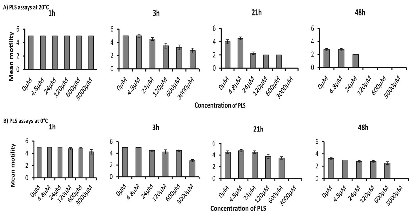
Generally, there were few visible changes to the thrashing behaviour or the cuticle of N2 worms in all treatments during the first 3 h after treatment (Fig. 1). Variable changes were noted thereafter, with 3000 μ m PLS showing the largest effect. In this group, most of the worms were no longer moving by 21 h (Fig. 1A).
The effect of incubation temperature on the motility of Caenorhabditis elegans exposed to different concentrations of PLS. Wild-type (N2) C. elegans were exposed to PLS at (A) 20 °C and (B) 0 °C.

At 48 h, all treatments showed drastic changes in worm motility with mortality considerably greater than 50% in treatments at and above 120 μ m PLS. Worms were largely non-motile at 0 °C whether or not there was any PLS present. Therefore, any thrashing that was seen was likely due to worms warming up as they were examined under the microscope. This effect was minimized by briefly and quickly assessing motility (1–2 min per 24-well plate) and returning the assays to their original temperature. The worms’ motility was less affected by PLS except at the very highest concentration (Fig. 1B).
The motility of worms declined in all treatments with time from 3 h onwards (main effect of TIME = F 3·7,162·8 = 58·4; P<0·001), and the rate of decline over time was dependent on concentration (2-way interaction TIME*CONCENTRATION = F 3·7,162·8 = 23·2; P<0·001), and on temperature (2-way interaction TIME*TEMPERATURE = F 3·7,162·8 = 8·9; P<0·001) but there was no 3-way interaction (P>0·05). There were significant overall effects of temperature (main effect of TEMPERATURE, F 1,44 = 8·3, P = 0·006) and of PLS concentration (main effect of CONCENTRATION, F 1,44 = 221·9; P<0·001) with negative gradients at all time-points examined (β at 1, 4, 24, 44 and 48 h = −0·32, −0·68, −0·72, −0·94 and −0·45, respectively, all significantly different from zero with t values ranging from −5·13 at 1 h to −9·2 at 48 h, all P values <0·001). There was no 2-way interaction.
PLS showed substantial nematicidal activity at 0 °C and attacked the cuticle of C. elegans
Wild-type C. elegans were not susceptible to PLS at room temperature when exposed for 24 h to concentrations of CP that are known to destroy parasitic stages of nematodes such as Trichuris muris, Heligmosomoides polygyrus and Haemonchus contortus. However, at low temperatures (0–4 °C), despite there being very little effect on thrashing activity, wild-type worms were more susceptible to CP attack based on observable damage to their cuticles. Major structural defects to the cuticle were seen in the assay at 0 °C as compared with relatively little in worms at 20 °C after overnight incubation (Fig. 2I). The defects mainly consisted of blistering and perforations of the cuticle leading to release of the worm's internal organs, and eventual death. This damage increased in intensity with time and PLS concentration. After 48 h, affected worms in higher PLS concentrations were destroyed and appeared as debris or were broken up into several indistinct sections. At lower concentrations, the numbers of worms affected were fewer and those with evident changes to their surface had irregular cuticles and indistinct anatomy. Worms in the 0 °C control group without CP were motile (once rewarmed to room temperature), intact and unaffected, showing no signs of structural changes or weakening.
Effects of PLS on cuticle of wild-type (N2) Caenorhabditis elegans. (I) Concentration-dependent effects of PLS on C. elegans structure exposed to PLS after 24 h exposure; panel (A) 0 °C and (B) 20 °C (×40 magnification). (II) Scanning electron microscopy of wild-type C. elegans incubated for 24 h with or without PLS at (A) 0 °C and (B) 20 °C. Scale bars, 10 μm. (III) E-64 added in equimolar amounts with PLS blocks the effects of PLS on motility and cuticular damage of C. elegans as shown by microscopy and motility scores; (A) PLS with E-64; (B) PLS without E-64. Duplicate tests per concentration were carried out at 0 °C.

The nematicidal effect of PLS was dependent upon the activity of CPs
Worms incubated with PLS in the absence of E-64 and maintained at 0 and 20 °C exhibited some cuticular damage. Those incubated with PLS plus E-64 did not have cuticular damage. Similarly, there was no cuticular damage seen in worms incubated with only E-64. This established that CPs were the major active molecules in PLS responsible for inflicting such damage.
There were significant concentration-related differences in motility scores among worms treated with PLS in the absence of E-64 (Fig. 2IIIb), which decreased with increasing time of incubation and PLS concentration. Treatment with PLS+E-64 had no effect on motility (Fig. 2IIIa). Overall, motility declined with time (main effect of TIME = F 2·3,99·4 = 6·8; P = 0·001), but the rate of decline differed depending on the concentration of enzyme activity (2-way interaction between TIME*CONCENTRATION; F 2·3,99·4 = 22·2; P<0·001). The 3-way interaction was also significant (for TIME*E-64 STATUS*PLS CONCENTRATION, F 2·3,99·4, = 20·6; P<0·001). There was an overall significant effect of enzyme concentration (for CONCENTRATION, F 1,44 = 51·2; P<0·001), with negative gradients at all time-points examined except at 2 h (β at 4, 24 and 48 h = −0·286, −0·386, −0·361, respectively, all significantly different from zero with t values −4·413, −4·738 and 4·257 at 4, 24 and 48 h, all P<0·001). There was also a 2-way interaction with presence/absence of E-64 (for E-64 STATUS*CONCENTRATION, F 1,44 = 47·7; P<0·001) since motility declined in a PLS concentration-dependent way without E-64, but not when E-64 was present.
SEM of worms incubated with various concentrations of PLS
The cuticle of wild-type C. elegans was observed and imaged as far as possible at equivalent points along the body of the worm. Following overnight incubation in PLS, the digestion of the cuticle to expose the internal structures was evident irrespective of concentration of treatment but was more severe at the lower temperature where shedding of the cuticle was visible (Fig. 2II). Digestion and shedding of the cuticle were clearly visible and more severe at lower than at higher temperatures. The damage was characterized by sloughing of the outer cuticle and obliteration of the smooth longitudinal and vertical ridges of the cuticle of worms. In contrast, control worms did not show any noticeable morphological change to the structure of the cuticle after 24 h of incubation in K medium.
The nematicidal effect of PLS was concentration, time, and temperature dependent
Figure 3 shows the percentages of worms that underwent cuticular damage when exposed to 0, 15, 45 and 1200 μ m PLS and incubated at different temperatures. Generally, with each increase in temperature, the time of onset of cuticular damage also increased, i.e. damage was observed later as temperature increased. Similarly, with each increase in concentration, the severity of damage increased. Strikingly, the severity of cuticular damage and proportion of worms affected was greater at lower temperatures (0, 4 or 10 °C) than at higher temperatures (15, 20 or 25 °C). At low temperatures wild-type C. elegans were highly susceptible to PLS attack and took less time to incur damage.
The effect of incubation temperature on C. elegans were exposed to 15, 45 and 1200 μ m PLS at 0, 4, 10, 15, 20 and 25 °C and their responses monitored over 12 and 24 h periods. Temperatures were grouped into lower (0, 4 and 10 °C) and higher (15, 20 and 25 °C) panels. Within each panel, percentages of worms whose cuticles were damaged are shown depicting concentration-, time- and temperature-dependent effects.

Overall, the number of worms damaged by PLS increased with time of incubation (for main effect of TIME, F 5·2,295·7 = 15·7; P<0·001), the rate of damage was dependent on temperature (for 2-way interaction TIME*TEMPERATURE = F 25·9,295·7 = 3·3; P<0·001) and there was also a highly significant 3-way interaction (for TIME*TEMPERATURE*CONCENTRATION, F 25·9,295·7 = 3·3; P<0·001). There were significant overall effects of temperature on the number of worms showing cuticular damage (main effect of TEMPERATURE = F 5,57 = 10·7; P<0·001), of CONCENTRATION (F 1,57 = 13·5; P = 0·001) as well as a 2-way interaction TEMPERATURE* CONCENTRATION (F 5,57 = 9·5; P<0·001). There were no significant differences among the lower temperature groups (0, 4 or 10 °C) or among the higher temperature groups (15, 20 or 25 °C). In post hoc tests, damage caused at 0 °C was significantly different from that seen at 15, 20 and 25 °C (all P<0·001), but not from that seen at 4 °C (P = 0·123) or 10 °C (P = 0·174).
Dead wild-type C. elegans pre-treated with sodium azide were highly susceptible to cuticular digestion by PLS
Dead wild-type worms, which had been pre-treated with sodium azide before being exposed to PLS, were highly susceptible to PLS attack and showed severe damage to the cuticle (Fig. 4B). Control dead worms incubated in K-medium were intact (Fig. 4A). PLS-induced damage was obliterated in the presence of E-64 (Fig. 4C) and E-64 alone did not damage dead worms (Fig. 4D).
Cuticular damage and digestion of dead wild-type Caenorhabditis elegans by PLS at room temperature after 30 min. Worms were pre-treated with 10 mm sodium azide for 30 min prior to exposure. (A) Control worms; (B) 48 μ m PLS only; (C) 48 μ m PLS+100 μ m E-64; (D) 100 μ m E-64 only. Scale bars show 50 μm.

C. elegans cystatin null mutants were highly susceptible to PLS
The nematicidal effect of PLS in cystatin null mutants (cpi-1−/− or cpi-2−/−) was greater and considerably more severe than that experienced by the wild-type N2 worms. Cuticular damage was noted between 30 min and 1 h irrespective of temperature of incubation (0 and 25 °C), and digestion of the cuticle was evident from 2 h onwards.
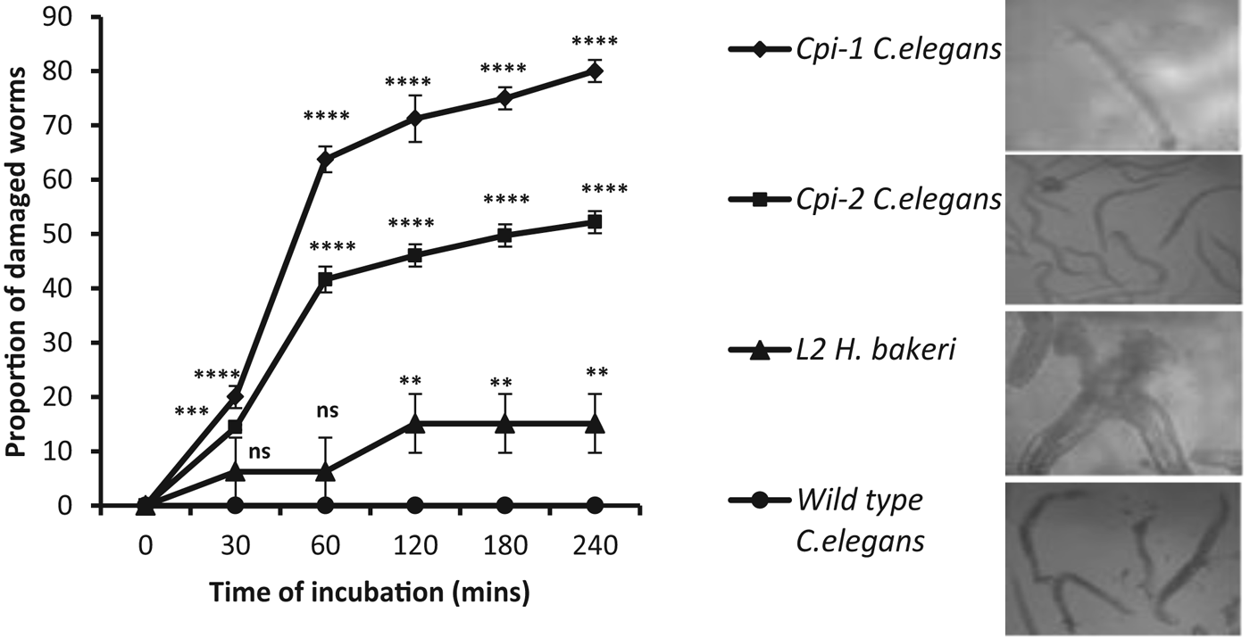
Figure 5 shows the response of different strains of C. elegans and L2 H. bakeri to 45 μ m PLS treatments at 25 °C. The number of damaged worms increased with time (main effect of TIME = F 4·8,57·4 = 332; P<0·001) and the rate of damage was dependent on the type of worm incubated (2-way interaction TIME*WORM TYPE = F 14·4,57·4 = 87·3; P<0·001). There was a significant overall effect of worm type (the main effect of WORM TYPE = F 3,12 = 1096·6; P<0·001). In contrast to wild-type C. elegans, cystatin-null mutants exhibited progressive damage to cuticles with increasing time of exposure from 30 min of treatment onwards. L2 H. bakeri showed only minimal damage levelling off after 120 min. In this experiment, wild-type C. elegans did not show damage after PLS attack even after 240 min of exposure.
The effect of incubation of L2 Heligmosomoides bakeri and wild-type C. elegans, cpi−1 and cpi-2 null mutants of C. elegans in 45 μ m PLS at 25 °C over a period of 30–240 min; the percentage of worms exhibiting cuticular damage is plotted against time. Means and SEM were calculated from 5 worms per well and involving four biological replicates. The statistical significance of differences from the solvent control is indicated for each group by asterisks: *P<0·05, **P<0·01, ***P<0·001, ****P<0·0001 or in their absence meaning not significant (ns). The pictorial inserts show examples of the type of damage that was seen for each strain.

PLS had nematicidal activity on cpi-2::lacZ transgenic C. elegans
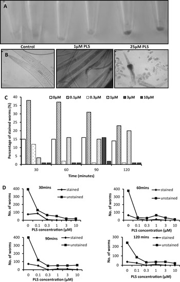
Incubation with PLS caused cuticular damage and death to the cpi-2::lacZ transgenic C. elegans (Fig. 6A). During microscopic observation, the overall number of worms in each count over time decreased with increasing PLS concentration. In addition there was debris from damaged worms, thus making scoring of staining of such worms difficult. We observed that this damage was progressive, initially forming blisters in the lower concentrations and eruption of the cuticular surface at higher concentrations, with subsequent release of the worms’ internal structures. The worms incubated in 3 and 10 μ m PLS for longer time periods (e.g. 120 min), exhibited complete cuticular digestion.
Tissue-specific expression of β-galactosidase in cpi-2::lacZ transgenic C. elegans. In situ staining assay with X-gal after exposure to PLS for 30 min at room temperature as seen in (A) bulk worm pellets in micro-tubes and (B) fixed worm preparations. Even low concentrations of PLS induced β-galactosidase activity, but also caused cuticular damage and subsequent digestion of cpi-2::lacZ C. elegans; (C) Proportion of worms expressing β-galactosidase (i.e. stained) after exposure to PLS for 30, 60, 90 and 120 min; (D) Damage and subsequent digestion of worms by PLS reduced the numbers of both stained and unstained worms.

Low PLS concentration induced cpi-2::lacZ expression and minimally damaged worms
Inducible cpi−2 expression was monitored as positive β-galactosidase staining of cpi-2::lacZ worms (Fig. 6A). In situ staining following exposure to PLS, revealed reporter gene expression exclusively in the pharyngeal and intestinal cells of larval and adult stages. Since 25 μ m PLS caused severe cuticular damage and death in transgenic C. elegans worms (Fig. 6B), further PLS concentrations were tested in the 0–10 μ m PLS concentration range. The time-course of transgene expression was monitored using both X-gal staining and MUG assays between 20 and 120 min. 0·1 μ m PLS induced the greatest cpi-2 expression in worms at each time point (Fig. 6C). Staining in worms incubated with higher PLS concentrations was variable, since there were few worms as a result of damage and digestion (Fig. 6C and D). However, there were no significant differences among the PLS concentrations (χ 2 = 84·0; P = 0·121) or exposure times (χ 2 = 42·22; P = 0·461) on the number of stained worms (Fig. 6D).
Expression of cpi-2::lacZ was time, temperature and concentration dependent (MUG assay)
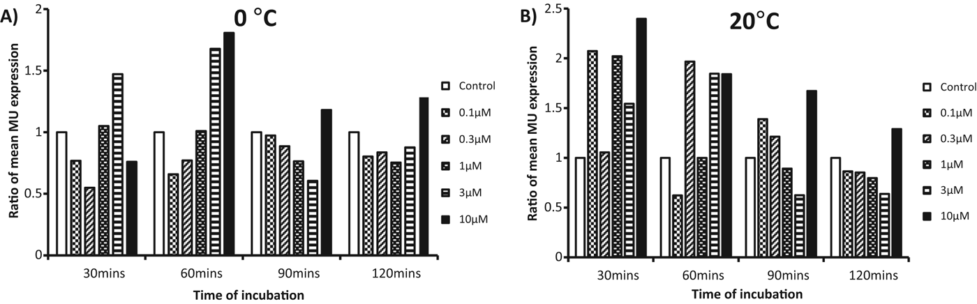
In the MUG assay, cpi-2 expression was elicited by PLS at both 0 and 20 °C (Fig. 7) and although expression appeared to be earlier (30 min), higher and lasted longer at some concentrations of PLS in worms incubated at 20 °C than those at 0 °C, this proved not to be significant (see below). At 0 °C, cpi-2 expression initially increased at 30 min only in 3 μ m PLS, peaked after 60 min in both 3 and 10 μ m PLS, but decreased thereafter except in the highest concentration. There was hardly any expression after 90 and 120 min in most PLS concentrations. At 20 °C, expression was noted after 30 and 60 min but decreased after 90 and 120 min (with most concentrations eliciting lower activities than the control).
PLS induces cpi-2 expression in cpi-2::lacZ C. elegans. Quantitative assay for β-galactosidase in cpi-2::lacZ transgenic C. elegans using the MUG assay. Mixed stage worms were exposed to varying concentrations of PLS from 0–120 min at (A) 0 °C and (B) 20 °C and the rate of MU formation in the supernatant was quantified fluorometrically. Data plotted as normalized fluorescence ratios (mean MU fluorescence in each test condition divided by that in the appropriate control with no PLS).

The expression of cpi-2 changed over time of incubation in PLS (main effect TIME = F 3,132 = 6·8; P<0·001), and the rate of change was dependent on concentration of PLS (2-way interaction TIME*CONCENTRATION = F 3,132 = 5·6; P = 0·001), but not on temperature (2-way interaction TIME*TEMPERATURE = F 3,44 = 0·429; P = 0·732). There was a significant overall effect of PLS concentration on cpi-2 expression (main effect of CONCENTRATION = F 1,44 = 12·4; P = 0·001), however, there was no overall effect of temperature on expression (for main effect of TEMPERATURE F 1,44 = 0·019; P = 0·890).
PLS digested β-galactosidase produced in cpi-2::lacZ C. elegans
Since the transgenic C. elegans (cpi-2::lacZ) strain was highly susceptible to PLS, we conducted assays using β-galactosidase from E. coli (Sigma, UK) to investigate whether PLS was responsible for the diminished β-galactosidase activity seen in the MUG assay above. Our hypothesis was that the low and insignificant activity reported above arose because PLS digested the cuticle and subsequently destroyed the transgene-encoded β-galactosidase, limiting its apparent expression.
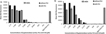
In vitro studies confirmed that purified β-galactosidase activity produced very high MU fluorescence at both 30 and 60 min of incubation (Fig. 8A and B). However, this was reduced when the β-galactosidase was incubated with PLS. For example, on incubation with 2·77 units of beta-galactosidase, fluorescence was reduced by 23·40 and 31·9% at 30 and 60 min, respectively. Thus, PLS was able to digest and inhibit the activity of β-galactosidase to form MU. All controls showed minimal activity; notably, incubation of 30 μ m E-64 with 22·5 μ m PLS and 1·85 units of β-galactosidase neutralized the digestion effect of PLS on β-galactosidase, resulting in high levels of MU fluorescence (Fig. 8A and B).
Effect of PLS on β-galactosidase activity in vitro at 37 °C at 30 and 60 min. Varying concentrations of β-galactosidase were incubated with 22·5 μ m PLS at (A) 30 min and (B) 60 min. Importance of cysteine proteinase and the effect of the inhibitor were tested with addition to PLS equimolar concentrations of E-64. Incubation of 30 μ m E-64, 22·5 μ m PLS plus 1·85 units of β-galactosidase neutralized the digestion effect of PLS on β-galactosidase, resulting in high levels of MU fluorescence.

Overall fluorescence (reflecting digestion of β-galactosidase) did not change significantly with time (main effect of TIME = F 1,28 = 0·002; P = 0·96), but varied significantly over time depending on presence/absence of PLS (2-way interaction TIME*TREATMENT = F 1,28 = 7·1; P = 0·013) and at different concentrations of β-galactosidase (3-way interaction TIME* TREATMENT*CONCENTRATION = F 1,28 = 20·4; P<0·001). There was a significant overall effect of concentration of β-galactosidase (main effect CONCENTRATION = F 1,28 = 21·1; P<0·001), with positive gradients at both time-points examined (β at 30, and 60 min = 12149·8, 8305·1, respectively, both significantly different from zero with t values of 4·351 at 30 min and 4·021 at 60 min, P = 0·003, P = 0·028, respectively), but not of presence/absence of 22·5 μ m PLS (TREATMENT) per se (F 1,28 = 0·05; P = 0·828) and there was no significant 2-way interaction (TREATMENT*CONCENTRATION = F 1,28 = 0·346; P = 0·561).
PLS induced cpi-1 expression but did not damage cpi-1::GFP C. elegans in the short term
The use of a GFP tagged transgene (cpi-1::GFP) as compared with lacZ tagged transgene (cpi-2::lacZ) might help to overcome some of the obstacles encountered with lacZ earlier. Furthermore, it should be much easier to monitor GFP fluorescence in living worms.
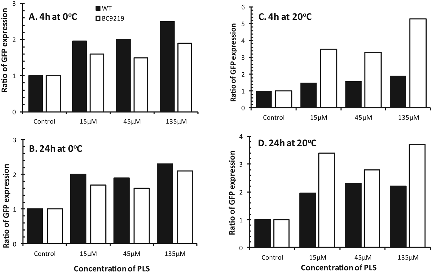
Unlike the cpi-2::lacZ worms, the survival of cpi-1::GFP C. elegans transgenic worms, when exposed to PLS, was comparable to the wild-type N2. Expression of cpi-1 in the presence of PLS was observed at both 4 and 24 h of incubation at 20 °C but not at 0 °C. Upregulation of cpi-1 was marked (net induction of 2·3–4·3-fold at 4 h and up to 2·8 fold at 24 h) in the BC9219 worms relative to the control worms (Fig. 9).
Effects of PLS on cpi-1::GFP and wild-type N2 C. elegans showing the downregulation at 0 °C (A and B) and upregulation at 20 °C (C and D) of cpi-1 as a response to PLS at 4 (A and C) and 24 h (B and D); as in Fig. 7 all data are shown as mean fluorescence ratios (each PLS-treated fluorescence reading divided by the appropriate control reading).

Expression of cpi-1 changed overall with time of incubation (main effect of TIME = F 1,121=7·1; P = 0·009), and the extent of the rise over time varied with concentration of active enzyme but differed significantly at different temperatures (for TIME*CONCENTRATION*TEMPERATURE, F 1,121 = 8·8; P = 0·004). There was a highly significant overall effect of concentration of PLS (main effect of CONCENTRATION = F 1,121 = 373·6; P<0·001), with significant positive gradients at both time-points examined (β at 4, and 24 h = 210·4, 190·4, respectively, both significantly different from zero with t values 5·471 at 4 h and 5·765 at 24 h, all P<0·001). However, the relationship with concentration of active enzyme differed between the nematodes (2-way interaction STRAIN*CONCENTRATION = F 1,121 = 49·5; P<0·001) and the concentration effect varied between the temperatures tested (2-way interaction CONCENTRATION*TEMPERATURE = F 1,121 = 5·9; P = 0·016).
DISCUSSION
Our study has shown that the effect of PLS on motility and cuticular damage of wild-type (Bristol N2) C. elegans was concentration, temperature and time dependent. The plant-derived CPs adversely affected motility and caused severe cuticular damage to, and consequent mortality of, the free-living nematodes. To the best of our knowledge, this is the first time that the nematicidal effect of plant CPs has been demonstrated against C. elegans. Furthermore, our evidence suggests that C. elegans can actively resist externally applied CPs by inhibiting their activities with cystatin gene products (Ce-CPI-1 and Ce-CPI-2) encoded by the cpi-1 and cpi-2 genes. The functions of these two gene products have previously been postulated in degradation of the old cuticle and development of the new cuticle during moulting as well as oogenesis (Murray et al. Reference Murray, Manoury, Balic, Watts and Maizels2005; Hashmi et al. Reference Hashmi, Zhang, Oksov, Ji and Lustigman2006). The data presented here suggest that they also serve an important role in protecting worms against exogenous CPs which in nature may be derived from soil bacteria or fungi, or the rotting fruits and vegetation within which these nematodes reside (Kiontke et al. Reference Kiontke, Felix, Ailion, Braendle, Penigault and Fitch2011).
Generally, there were few visible changes to the thrashing behaviour or the cuticle of C. elegans exposed to PLS during the first 3 h after treatment. These became more evident after overnight incubation. In C. elegans assays, normal worm movement (thrashing) is observed in a toxicant-free environment and is indicative of uninhibited activity (Cronin et al. Reference Cronin, Mendel, Mukhtar, Kim, Stirbl, Bruck and Sternberg2005). However, in the presence of drugs or toxicants, worm movement is altered (Anderson et al. Reference Anderson, Boyd and Williams2001; Boyd et al. Reference Boyd, Cole, Anderson and Williams2003). Major loss of worm motility was observed at higher concentrations of PLS. Unfortunately, our visual inspection method is qualitative and somewhat subjective, and may therefore fail to adequately deal with subtle differences in motility, especially for hyperactive worms in higher motility score categories. Notwithstanding this drawback, such behavioural endpoints measured by scoring are deemed useful in resolving mechanisms of toxicity (Anderson et al. Reference Anderson, Boyd and Williams2001). The use of sophisticated automated systems has been introduced recently (Feng et al. Reference Feng, Cronin, Wittig, Sternberg and Schafer2004; Cronin et al. Reference Cronin, Mendel, Mukhtar, Kim, Stirbl, Bruck and Sternberg2005; Buckingham and Sattelle, Reference Buckingham and Sattelle2009), but, in our experience, the way these systems capture data does not cope well with CP assays on C. elegans due to an inability to account for worms lost or destroyed as a result of cuticular damage by CPs, or confusion caused by debris or worm fragments following on from this.
Cuticular damage to wild-type C. elegans was more noticeable at 0 °C than at ambient room temperature, suggesting that at higher temperatures and with increased metabolic activity the worms were able to block the efficacy of externally applied CPs, but were less able to do so at lower temperatures. Notably, C. elegans are active and routinely cultured at temperatures of 15–25 °C. Higher concentrations of PLS, lower temperatures of incubation and/or longer exposure times were required to show any clear effect on wild-type C. elegans. It may be relevant that the epicuticle of these worms is covered with a negatively charged glycoprotein surface coat which might actually serve to bind and concentrate the papaya proteinases, most of which are highly basic (pIs between 8·75 and 11·5) (Barrett et al. Reference Barrett, Rawlings and Woessner1998). In all the assays, we strove to strike a balance between exposures of worms to high (up to 3000 μ m) vs low PLS concentrations (less than 25 μ m). This is because other researchers have found that a small proportion of pharmacologically active compounds (2%; 308 out of 14 100) produce a clearly defined phenotype involving slow growth, lethality, uncoordinated movement and morphological defects, when the 25 μ m screening concentration was used in C. elegans (Burns et al. Reference Burns, Kwok, Howard, Houston, Johanson, Chan, Cutler, McCourt and Roy2006; Kwok et al. Reference Kwok, Ricker, Fraser, Chan, Burns, Stanley, McCourt, Cutler and Roy2006).
Caenorhabditis elegans were not affected as acutely or as severely as the parasitic stages of nematodes such as H. bakeri. Findings from previous studies show that the CP effect on loss of motility and damage to the cuticle of parasitic stages of GI nematodes and cestodes is much quicker and occurs at lower PLS concentrations than those observed here with C. elegans. Motility is severely reduced within 15–40 min and cuticles are usually totally degraded within 2 h (Stepek et al. Reference Stepek, Behnke, Buttle and Ducel2004, Reference Stepek, Buttle, Duce, Lowe and Behnke2005, Reference Stepek, Buttle, Duce and Behnke2006a , Reference Stepek, Lowe, Buttle, Duce and Behnke2007b , Reference Stepek, Lowe, Buttle, Duce and Behnke d ; Luoga et al. Reference Luoga, Mansur, Buttle, Duce, Garnett and Behnke2012). The nematicidal effect of PLS has been shown to be dependent upon the activity of CPs. In order to establish that the effect was mediated solely by CPs, we exploited the cysteine proteinase-specific blocking properties of E-64. Pre-incubation of PLS with E-64 in the worm assay blocked and abolished the activity of PLS and hence prevented worm damage. This demonstrated that CPs were indeed responsible for the observed effects, in agreement with findings on CPs tested against parasitic nematodes and cestodes (Stepek et al. Reference Stepek, Lowe, Buttle, Duce and Behnke2007b ; Buttle et al. Reference Buttle, Behnke, Bartley, Elsheikha, Bartley, Garnett, Donnan, Jackson, Lowe and Duce2011; Luoga et al. Reference Luoga, Mansur, Buttle, Duce, Garnett and Behnke2012). The mechanism by which natural plant CPs act against parasitic and free-living nematodes as well as cestodes is by hydrolysis of the worms’ surface (i.e. cuticle and tegument, respectively). In so doing, they weaken the surface, and the effect seen is the release of the worm's internal structures driven by the high internal hydrostatic pressure in the nematodes’ pseudocoelomic cavity, which subsequently leads to death (Stepek et al. Reference Stepek, Lowe, Buttle, Duce and Behnke2007b ). High turgor pressure inside nematodes has been suggested to be a survival strategy, as it regulates and limits the rate of water transport across cuticular surfaces (Kotze, Reference Kotze2012). However, once the cuticle is breached, there is rapid evisceration and subsequent mortality.
The thick cuticle of free-living nematodes has been reported to act as a formidable barrier to many molecules (Ségalat, Reference Ségalat2006); as a result, many anthelmintic candidates may therefore not show any effects because they fail to penetrate across the cuticle into the animals. These worms have been known to possess a wide range of physical and xenobiotic defence systems which when fully deployed make many pharmacological compounds ineffective (Lindblom and Dodd, Reference Lindblom and Dodd2006; Burns et al. Reference Burns, Wallace, Wildenhain, Tyers, Giaever, Bader, Nislow, Cutler and Roy2010), and this has discouraged the use of C. elegans as a chemical–genetic model system. Wild-type C. elegans express cystatin-like proteins which apparently can block the activity of externally applied CPs; these are encoded by the Ce-cpi-1 (K08B4.6) and Ce-cpi-2a (R01B10.1) genes described by Hashmi et al. (Reference Hashmi, Zhang, Oksov, Ji and Lustigman2006). At lower temperatures these CP inhibitors are not sufficiently expressed by the worms or are present in smaller quantities enabling PLS to effectively damage the cuticle. This difference was seen as early as 4 h after exposure to PLS. While this timescale may be too rapid for the de novo synthesis of cystatins that would be required, two complementary explanations for this anomaly can be advanced. A mammalian cystatin (cystatin F) is known to be stored in vesicles (Langerholc et al. Reference Langerholc, Zavašnik-Bergant, Turk, Turk, Abrahamson and Kos2005). Therefore, as an immediate response to PLS attack, it is plausible that a ‘quick release’ of cystatins that are stored in lysosomal-like vesicles could take place and the cystatin-containing vesicles are then trafficked to the cuticle surface when they are needed. Low temperatures have been reported to prevent membrane fluidity resulting in major constraints in vesicle trafficking (Dunn et al. Reference Dunn, Hubbard and Aronson1980), which in this study would have precluded the cystatin trafficking from happening and facilitated damage to the cuticle by PLS. The response data for cpi-1::GFP fluorescence or of β-galactosidase staining for cpi-2::lacZ also suggest that after the early release of small quantities of stored cystatins, there is a rapid induction of both the cpi-1 and cpi-2 genes. Hence, active cystatin production and mobilization, rather than a thick cuticle, may be largely responsible for the failure of CPs to successfully attack these worms.
Similarly, when the secretion of cystatins is impaired in C. elegans (as in cpi-null mutants) or when the worms are killed before testing, the worms become incapable of mounting any effective resistance against CP attack; dead wild-type worms, unlike live ones, cannot produce any protective response (such as cystatins), thereby rendering their cuticles more sensitive to CP attack. The results obtained with azide-killed worms support this hypothesis that the defence against CPs is dependent on the worms being alive and therefore capable of secreting endogenous cystatins. For both cpi-1 and cpi-2 null mutants, there was cuticular damage between 30 min and 1 h after PLS exposure irrespective of the temperature of testing, suggesting that the products of these genes were indeed the blocking agents preventing CPs from damaging wild-type worms. The manner and severity of this cuticular damage is consistent with that reported in parasitic nematodes (Stepek et al. Reference Stepek, Behnke, Buttle and Ducel2004, Reference Stepek, Buttle, Duce, Lowe and Behnke2005; Behnke et al. Reference Behnke, Buttle, Stepek, Lowe and Duce2008).
There are no reports of cysteine proteinase inhibitors in the cuticle of the parasitic stages of gastrointestinal nematodes (Stepek et al. Reference Stepek, Behnke, Buttle and Ducel2004). This may partly explain why CPs have a quicker and more severe effect on parasitic nematodes than on wild-type C. elegans. Apart from low levels from eating some fruits, the mammalian intestine does not contain CPs and therefore worms have no need for a defence against these enzymes. Furthermore, several potential disadvantages of using C. elegans as a model for parasitic species have been advanced (Geary et al. Reference Geary, Sangster and Thompson1999; Geary and Thompson, Reference Geary and Thompson2001). For example, free-living nematodes live naturally in soil or aqueous environments (though C. elegans is now adapted to culture conditions), while parasitic worms live in animal or plant hosts and therefore have to cope with the hostile environment they provide. Subtle drug-induced changes in parasitic nematodes may be enough to expel the worms from the host (Geary et al. Reference Geary, Sangster and Thompson1999), but comparable effects on worms in culture may not be detectable by existing assays.
Papaya latex supernatant induced expression of both cystatin genes (cpi-1 and cpi-2). Caenorhabditis elegans strains carrying either a reporter green fluorescent protein transgene (cpi-1::GFP) or a β-galactosidase reporter construct (cpi-2::lacZ) were used to explore the expression and inducibility of these two cystatin genes. Although CPs were shown to induce β-galactosidase activity in cpi-2::lacZ worms by both X-gal staining and quantitative MUG assays, the cuticles of these transgenic C. elegans were adversely affected by the PLS. It was difficult, therefore, to measure and establish a clear concentration–response effect of PLS on gene expression with either method. Even very low concentrations of PLS caused visible damage to these worms and therefore any X-gal staining in such worms would have been removed rapidly by degradation of the worm tissues. This scenario is compounded by the fact that CPs in PLS also attack β-galactosidase itself (Fig. 8), abolishing its enzymatic activity. We had anticipated that the transgenic cpi-2::lacZ strain should be relatively insensitive to PLS as it is a non-mutant worm with a fully functional cpi-2 gene of its own, although the reverse proved to be the case. One possible explanation is that the design of this transgene may have blocked the secretion of cystatin fusion proteins, and perhaps CPI-2 itself, across the endoplasmic reticulum (ER) membrane due to the large size of the attached β-galactosidase moiety. The retention of secreted cystatins within the secretory cells may have hindered their activity on the worm surface and thus made the worms more prone to PLS attack. This was apparently not the case for a cpi-1::GFP translational fusion, perhaps because GFP is a much smaller protein than β-galactosidase; this strain was seemingly more resistant to CP attack than that carrying the β-galactosidase reporter. However, the sensitive detection of GFP can be hampered by auto-fluorescence, an intrinsic property arising from endogenous molecules within gut cells (Monici, Reference Monici and El-Gewely2005; Igarashi et al. Reference Igarashi, Yoshinari, Yokota, Sugi, Sugihara, Ikeda, Sumiya, Tsuji, Mori, Tochio, Harada and Shirakawa2012).
In this paper we have shown that free-living C. elegans worms, across their optimal temperature range (15–25 °C), were able to resist CP attack, at least in part through the action of secreted cystatins CPI-1 and CPI-2; both of the available cystatin null mutants were far more sensitive than wild-type N2 C. elegans, and both of these genes were inducible by PLS in transgenic worms. In conclusion, we now have a better understanding of C. elegans’ extensive physical and enzymatic response to CPs and using this information attempts to circumvent its defences can be made. When taken together, the results for wild-type and cpi-null mutants point towards a cheap and effective rapid throughput C. elegans-based assay for screening the nematicidal activities of plant-derived CPs that have yet to be tested. A double cpi-1−/−/cpi-2−/− null mutant – unless it proves to be non-viable – might provide an even more sensitive tool for such screening, and we are currently attempting to breed such a double mutant strain by conventional crossing.
ACKNOWLEDGEMENTS
We sincerely thank Fadlul Mansur, late Wenceslaus Luoga, Tim Smith, Ann Lowe, Declan Brady and Darren Hepworth for technical support and advice throughout this work and Ian Duce for advice with electron microscopy. Some nematode strains used in this work were provided by the Caenorhabditis Genetics Center (which is funded by the NIH National Center for Research Resources), the Baillie Lab (Simon Fraser University, Burnaby, Canada) and Dr S. Hashmi (formerly of the New York Blood Centre).
FINANCIAL SUPPORT
AMP was funded and supported by the Commonwealth Scholarship, University of Nottingham and University of Zambia.