Computed tomographic (CT) angiography is increasingly being performed for the evaluation of congenital heart disease (CHD). Reference Burchill, Huang and Tretter1,Reference Suranyi, Varga-Szemes and Hlavacek2 However, despite acquiring a volumetric dataset, the analysis of the CT dataset is generally based on a multi-planar interpretation of two-dimensional images. Reference Gupta, Spicer and Anderson3 Patient-specific three-dimensional printed models of the heart permit excellent three-dimensional visualisation of intracardiac anatomy. Reference Vukicevic, Mosadegh, Min and Little4–Reference Illman, Hosking and Harris7 The printing of surface-rendered three-dimensional models, however, need specialised software, printing material, and trained personnel limiting its routine use. Reference Valverde, Gomez-Ciriza and Hussain6,Reference Illman, Hosking and Harris7 Some cardiologists and cardiac surgeons now use these surface-rendered virtual models without printing. Reference Garekar, Bharati and Kothari8 Virtual dissection and cinematic rendering, modifications of volume rendering, are other techniques of creating three-dimensional virtual models to show intracardiac anatomy. Reference Gupta, Spicer and Anderson3,Reference Mori, Fukuzawa and Takaya9–Reference Anderson and Gupta14
However, when viewed on two-dimensional screens, the third dimension of these three-dimensional virtual models is relatively obscured. Reference Goo, Park and Yoo10–Reference Anderson and Gupta14 Extended reality techniques, such as virtual reality and augmented reality, augment the third dimension, but only with expensive equipment. Reference Goo, Park and Yoo10,Reference Rowe, Chu, Recht, Lin and Fishman11 In this technical report, we describe a simple technique of anaglyph stereoscopic visualisation of virtual models. The production of anaglyph images using open-source software and viewing with inexpensive anaglyph three-dimensional glasses makes this technique affordable and easy to use.
Methods
Rationale
Human eyes do not have the three-dimensional vision. Instead, it is the binocular vision that highlights the third dimension during natural vision. A pair of spatially separated eyes generate different perspectives of an object which are merged by the brain to create stereovision. Stereoscopic visualisation is the norm in human vision, but it is challenging to replicate the same on a computer. Reference Banks, Read and Allison15
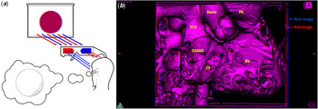
Extended reality techniques such as augmented and virtual reality simulate stereovision by sending two different viewpoints of an object to each eye. These techniques provide high-quality stereoscopic visualisation but require specialised equipment. Reference Goo, Park and Yoo10,Reference Banks, Read and Allison15,Reference Ong, Krishnan and Huang16 A similar stereoscopic visualisation is possible using anaglyph stereovision. In this technique, a pair of closely overlapping red and blue images called stereo pair is created. When viewed using red–cyan anaglyph glasses, only the red-coloured image of the stereo pair reaches the right eye. In contrast, the red filter allows only the blue-coloured image to reach the left eye. These dissimilar colour-coded inputs to the left and the right eye are equivalent to different perspectives viewed during natural vision and thus help in stereoscopic visualisation (Fig 1a). 17,18
Principle of anaglyph stereovision (a) and a representative stereo virtual dissection image obtained from Horos (b) coded in red and blue. When viewed using red–cyan anaglyph glasses, the blue glass on the right eye filters out the blue image while the red glass on the left eye prevents the red image from reaching the left eye. This colour-separated dissimilar inputs from each eye are merged by the brain to produce stereovision. Panel A is adapted from https://www.techeblog.com/3d-technology/ (last accessed on 15 July 2020). CS=coronary sinus; OSASD=ostium secundum atrial septal defect; PA=pulmonary artery; RV=right ventricle; SCV=superior caval vein.

Equipment for anaglyph stereovision
Horos is an open-source version of OsiriX (Pixmeo, Switzerland), a Mac-based software. It is freely available under GNU lesser general public license, version 3 (LGPL-3.0). The software permits a full range of post-processing of DICOM datasets, including virtual dissection of CT angiograms. 19,20
Horos-based virtual dissection and stereovision are possible on a Macintosh computer with modest technical specifications. The authors use a MacBook Pro (Retina, 13-inch, early 2015 model) laptop with a 2.7 GHz Intel Core i5 processor and 8GB RAM for virtual dissection and stereovision. The aforementioned personal computer has an Intel Iris Graphics 6100 card (Apple Inc, Cupertino, California, USA) and is powered by macOS High Sierra version 10.13.1 to run the Horos software. Reference Gupta, Spicer and Anderson3
Anaglyph stereovision – creation and interactive editing
The CT dataset is first imported to Horos. A virtual model of the heart is created using the three-dimensional volume rendering function of the software. Virtual dissection is then achieved by adjusting the 16-bit colour look-up table editor embedded in the programme. The virtual volume is further edited using cropping, scissoring, and the clip editing function of the programme to focus on the area of interest. Reference Gupta, Spicer and Anderson3 The details of the steps for obtaining virtual dissection have been described in an earlier publication. Reference Gupta, Spicer and Anderson3
Once the virtual dissection is achieved, the “stereovision” function of the Horos software duplicates the virtual model to produce two colour-coded copies in less than 30 seconds. The two colour-coded copies of the virtual model, one coded in red and the other in blue, are slightly separated spatially and maintain their relative position during interaction with the virtual model (Fig 1b). Reference Gupta, Spicer and Anderson3,19,20 The stereo pair of the virtual model, when viewed using red–cyan anaglyph glasses, permits stereoscopic visualisation. The quality of three-dimensional visualisation, however, depends upon the quality of virtual dissection model of the heart.
Sharing anaglyph stereo virtual dissection
The stereoscopic visualisation is best achieved within the Horos software. It is not possible to export a fully functional virtual model for interaction outside the Horos software. The representative frames in any desired projection, nonetheless, can be exported as an image. Using “Fly Through” mode or by rotating the virtual cardiac model, it is also possible to create a video of multiple sequential anaglyphs to provide different perspective of the cardiac structure being visualised. Supplementary video 1 obtained using “Fly Through” function provides sequential anaglyph stereoscopic visualisation of stenosed mitral valve secondary to rheumatic heart disease. Supplementary video 2, on the other hand, demonstrates different perspective of the virtual cardiac model of a patient with double outlet right ventricle with a large interventricular communication in subaortic region.
These images and videos, when viewed using red–cyan anaglyph glasses in any standard photo viewing software, retain stereoscopic visualisation. The stereo images and videos, just like any other digital image, can be easily shared via Bluetooth, email, WhatsApp, or any other forms of electronic file sharing.
Moreover, the images can also be shared in printed format. The stereo image printed on a photo paper permits stereoscopic visualisation. The quality of visualisation in printed format, however, depends upon the quality of photo paper and colour printing. Despite good quality printing, the print version is not as bright as its electronic equivalent. This is because the computer monitor uses additive colour mixing with red, green, and blue (RGB) as the primary colours. Photo printing, on the other hand, is based on a subtractive colour model using cyan, magenta, yellow, and black (CMYK) colours. Besides, the light emitted from the monitor augments the depth perception while the brightness of the printed version relies heavily on the ambient light and therefore, stereoscopic visualisation of the printed format is much better in a well-illuminated environment. 19
Clinical application
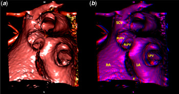
Anaglyph-based stereoscopic visualisation can be applied to any CT dataset once optimal volume rendering and virtual dissection is achieved. Reference Gupta, Spicer and Anderson3 We demonstrate application of anaglyph stereoscopic visualisation of CT datasets from patients with structural heart disease (Figs 1–5). Viewing of anaglyph stereo images using red–cyan anaglyph glasses provides an unequivocal sense of depth, thus making the appreciation of the third dimension much better. A comparison of an anaglyph image with a corresponding image without stereovision (Fig 2) highlights the importance of the depth perception achieved by application of this technique.
Virtual dissection image in LAO cranial projection without (panel A) and with (panel B) stereovision from a patient with sinus venosus interatrial communication. The relative position of connection of right upper and lower pulmonary vein is easily appreciable in panel B when seen using red–cyan anaglyph glasses. The patient also has a secundum atrial septal defect (#). LA=left atrium; LPV=left pulmonary vein; RA=right atrium; RLPV=right lower pulmonary vein; RUPV=right upper pulmonary vein; SCV=superior caval vein.

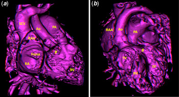
Stereo virtual dissection in anteroposterior (a) and left anterior oblique (b) projection from CT dataset of a 20-month-old child with double outlet right ventricle having interventricular communication (#) in subaortic region (see Supplementary video 2). An enhanced depth perception when seen using red–cyan anaglyph glasses amplifies appreciation of the posterior location of fossa ovalis and superior caval vein (SCV) relative to right atrial appendage (RAA). It also helps in ascertaining the relationship of interventricular communication (#) with the aorta and the subvalvular apparatus of the tricuspid valve. PA=pulmonary artery; RA=right atrium; RAA=right atrial appendage; RV=right ventricle; SCV=superior caval vein.

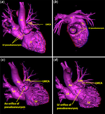
Stereo virtual dissection showing external appearance (panels A and B), aortic (panel C), and LV orifice (panel D) of a complex LV pseudoaneurysm in an adult following infective endocarditis of aortic valve. Anaglyph stereoscopic visualisation using red–cyan anaglyph glasses provides an unequivocal sense of depth highlighting the relative position of various cardiac structures. Ao=aorta; LA=left atrium; LAA=left atrial appendage; LMCA=left main coronary artery; LV=left ventricle.

Stereo virtual dissection image from systolic phase of CT dataset from a patient with paravalvular leak at mitral position as seen from the head end in surgical view. The leak (#) is located at 4 o’clock position. The stereoscopic visualisation from the head end of the patient using red–cyan anaglyph glasses enables perception of location of the leak relative to the left atrial appendage, aorta, and atrial septum. This enhanced 3D visualisation is helpful in planning interventional closure of paravalvular leaks. Ao=aorta; LAA=left atrial appendage; MV=mitral valve; PA=pulmonary artery; RA=right atrium.

Discussion
Stereovision brings viewing of virtual models closer to natural vision. There are obvious advantages of such a visualisation of 3D virtual models of the heart. Unlike other techniques for extended reality, Horos-based anaglyph stereovision is easy and inexpensive and requires just a click of the “stereovision” button in the software after virtual dissection is achieved. Further, unlike virtual reality, anaglyph-based stereoscopic visualisation is possible using easily available and inexpensive red–cyan anaglyph glasses. Although stereo pairs can be produced in other colour combinations, the red–cyan is the most commonly used anaglyph stereo pair. Also, red–cyan anaglyph glasses are most readily available making this combination the most preferred. Furthermore, almost instantaneous production of anaglyph stereo pair makes it suitable for real-time editing and viewing of 3D virtual models.
A similar anaglyph-based stereoscopic visualisation can also be produced using photo editing software but is time-consuming and has a learning curve. Reference Lechanoine, Smirnov and Armani-Franceschi22 Although being reported in the setting of cardiac CT angiograms, a similar approach can be used for other regions of the body. Further, like volume rendering, anaglyph-based stereoscopic visualisation is also possible for surface-rendered virtual models of the heart.
Ease of sharing in electronic and printed format makes it a perfect tool for education, training, and discussion among team members. Stereoscopic visualisation during interventional and surgical procedures is known to improve the operator’s confidence and reduce procedure time. Reference Lechanoine, Smirnov and Armani-Franceschi22–Reference Jourdan, Dutson and Garcia24 A similar approach using True3D software has been shown to improve surgeon’s understanding of the anatomy while planning surgery for CHDs. Reference Lu, Ensing and Ohye25 A fusion of stereoscopic visualisation of CT virtual model at the time of interventional procedure is also likely to improve outcomes and potentially reduce procedure-related complications. Reference Kliger, Jelnin and Sharma26
Nevertheless, unlike other techniques of stereoscopic visualisation, being based on colour separation, anaglyph stereovision does not provide a true sense of colour. The perception of depth may require few seconds before it can be optimally appreciated. In fact, the depth perception using anaglyph-based stereoscopic visualisation varies from person to person. Besides, like any other form of stereoscopic visualisation, prolonged viewing can produce discomfort in some viewers.
Conclusion
We report the first anaglyph stereoscopic visualisation of intracardiac anatomy from CT angiograms. This stereoscopic display may benefit medical students, cardiologists, radiologists, and cardiac surgeons by providing an easy, customisable, and low-cost alternative technique for stereoscopic display.
Acknowledgements
The authors acknowledge the help received from the faculty members of the Department of Cardiovascular Imaging and Endovascular Interventions, AIIMS, New Delhi for making the CT datasets available. The authors also thank the faculty members of the Department of Cardiology, AIIMS, New Delhi and Prof Robert H Anderson from University of Newcastle, Newcastle Upon Tyne, UK for providing critical comments regarding the stereoscopic visualisation technique.
Financial support
This research received no specific grant from any funding agency, commercial, or not-for-profit sectors.
Conflicts of interest
None.
Supplementary material
To view supplementary material for this article, please visit https://doi.org/10.1017/S1047951121001323