Introduction
Otological surgery is conventionally performed using an operating microscope. Identification of complex anatomical structures, illumination of the operative field and three-dimensional (3D) vision are most valuable for an otolaryngologist. A 3D exoscope allows both hands to work over the surgical field while viewing the monitor, similar to endoscopic surgery.
This paper reports the first case of simultaneous bilateral cochlear implant surgery performed exclusively with a 3D exoscope. It also discusses the optimum operative set-up as well as the feasibility of the 3D exoscopic system as an alternative to the microscope.
Materials and methods
A three-year-old boy with bilateral profound sensorineural hearing loss was selected for cochlear implant surgery. The patient positioning, surgical instruments and surgical technique conducted using the 3D exoscope were similar to that of the microscopic procedure.
Bilateral cochlear implant surgery was performed in the same sitting via a canal wall-up mastoidectomy with a facial recess approach. A cochleostomy method was chosen because the round window membrane was completely covered by bone, as demonstrated on the computed tomography scan.
We obtained written permission from our institution to use the Vitom 3D exoscope system in our surgery. Institutional review board approval and written consent were not required.
Equipment
The Vitom 3D exoscope system provided by the Karl Storz company (Tuttlingen, Germany) was used to perform the cochlear implant surgery. It is a video system that merges a 4000 pixel (‘4K’) display high-definition resolution view with 3D technology. The images are presented on a flat screen and the 3D vision is attained with polarised passive glasses.
There are two types of holding system: the Versacrane™ Light, which is mounted on a mobile stand, and the ARTip Cruise™, which we used, that is clamped to the operating table.
The 3D camera has a working distance of 20–50 cm and offers a magnification power of 8–30 times. The Image1 Pilot (Karl Storz) is a control unit with a 3D wheel for intuitive control of the camera system.
Operating theatre set-up
In our setting, the camera was positioned approximately 30 cm from the target. The screen was positioned at eye level and placed directly across the operating table in front of the surgeon, to achieve an optimal head-up surgical view.
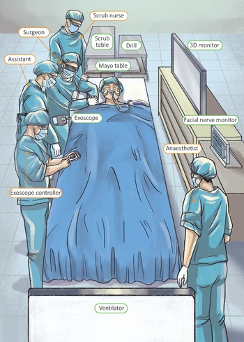
We began our procedure with the right-side cochlear implantation. In our attempt to achieve optimum visualisation, we set up the camera in two different positions. Initially, the camera was mounted on the opposite side to where the surgeon was sitting. Subsequently, we placed the camera on the same side next to the right side of the surgeon. For the left-side cochlear implant surgery, the camera was placed on the left side of the first surgeon, in between the surgeon and the assistant. In both cases, the scrub nurse stood next to the head of the patient.
Surgeons, anaesthetists, trainees and medical students wore 3D passive polarising eyeglasses. A designated team member operated the Image1 Pilot control unit. Intra-operatively, the surgeon instructed the team member to adjust the magnification, focus and light intensity, and to take recordings and images of the surgery.
The operating theatre setting for the right-side cochlear implant surgery is illustrated in Figure 1.
Operating theatre set-up for the right-side cochlear implant surgery. The exoscope is positioned on the surgeon's right side, in between the surgeon and the assistant. The scrub nurse is positioned on the left side of the surgeon. A separate team member operates the Image1 Pilot control unit. The three-dimensional (3D) monitor is in front of the surgeon and the assistant.

Results
Bilateral simultaneous cochlear implant surgery was performed exclusively with the Vitom 3D exoscope. The operation time for both sides totalled 2.5 hours in duration. The recorded time for the change and set-up of the camera system to the opposite side for the second cochlear implant surgery was approximately 10 minutes.
The surgery was successfully completed with no peri-operative complications. The patient was discharged home the following day. There were no complications reported during subsequent clinic follow up.
Both the operation time and the surgical outcome for the patient were comparable with previous cochlear implant surgical procedures performed in our centre using the operating microscope.
Discussion
To the best of our knowledge, this is the first paper to illustrate bilateral simultaneous cochlear implant surgery performed exclusively with the 3D exoscope system. Only six cases have been described in the literature regarding the application of the 3D imaging system in cochlear implant surgery and not all used the exoscope exlcusively.Reference Chen, Hsieh and Hsu1–Reference Colombo, Ferreli, Di Bari, Cugini, Miceli and De Virgilio4 In our case, both left- and right-side cochlear implant procedures were performed entirely with the 3D exoscope system.
This paper demonstrates that the 3D exoscope is a feasible alternative to the microscope in certain cases. Smith et al.Reference Smith, Kozin, Kanumuri, Barber, Backous and Nogueira2 reported seven cases of post-auricular or transmastoid approaches made using the exoscope exclusively. Garneau et al.Reference Garneau, Laitman, Cosetti, Hadjipanayis and Wanna3 and Rubini et al.Reference Rubini, Di Gioia and Marchioni5 described their experiences utilising the 3D exoscope during lateral skull base surgery. In our experience with the 3D exoscope, standard drilling during the mastoidectomy and posterior tympanotomy ensued safely, with excellent identification of all key landmarks. At no point was the exoscope switched to the operating microscope. While some authors have reported that 3D exoscopes are better suited for procedures performed through wider surgical fields, such as mastoidectomies,Reference Smith, Kozin, Kanumuri, Barber, Backous and Nogueira2 we did not encounter problems when visualising through a posterior tympanotomy for the electrode insertion, as this system offers a magnification power of 8–30 times.
We would like to emphasise that proper set-up in terms of the position and location of the camera is crucial for optimal visualisation. We believe that mounting the camera on the same side as the surgeon was more suitable compared to the opposite side, as the camera is nearer to the operative field. It is also easier for the assistant to manually adjust the angle of the stand and camera if placed on the same side. During the right-side cochlear implantation procedure, we mounted the camera to the left of the surgeon (Figure 1). It is imperative to position the camera as directly on top of the operative field as possible, to prevent the surgeon's operating hand from blocking the camera. We chose the ARTip Cruise holding system, in which the exoscope is clamped to the operating table, because of its compact nature, easy manoeuvrability and precise positioning.
The operating microscope, with its large cumbersome design, restricts the surgeon and assistants to certain positions around the operative field. In contrast, the exoscopic camera unit is compact. This allows for increased workspace, easier manoeuvring of instruments and less constraint for the surgical assistants, which improves their ability to assist.
The monitor-based display and ease of exoscope positioning also allow for superior ergonomics for the surgeon. Babar-Craig et al.Reference Babar-Craig, Banfield and Knight6 reported that otology surgeons had the highest prevalence of back and neck pain. This is likely related to microscope work, which involves the surgeons bending their neck or back into ergonomically challenging positions and sitting for extended durations. Pre-operatively, we set up the camera systems in different positions, because we wanted to determine which would give the surgeon the best visualisation, while allowing freedom of movement.
The 3D exoscope is ideal for teaching and training, as the trainees’ view is similar to that of the surgeon's, with the added benefit of a 3D and high-definition view. The conventional operative microscope screen allows the residents to observe the surgical procedure two-dimensionally, as only the surgeon has a stereoscopic vision. As our hospital is a teaching centre, it is imperative that the residents are able to follow the surgery with the same high-quality view as the surgeon.
Nevertheless, there are some shortcomings of the Vitom 3D exoscope. The screen must be positioned precisely at a 90-degree angle in front of the first surgeon to achieve clear visualisation of the surgical field. If the screen is viewed more laterally, a gradual loss of 3D depth and resolution is noted. The assistant and the scrub nurse need to position themselves as near as possible to the first surgeon's line of sight to obtain the best view.
Other limitations of the exoscope include reduced illumination in narrow surgical corridors and loss of resolution at higher magnifications.Reference Smith, Kozin, Kanumuri, Barber, Backous and Nogueira2 Smith and colleaguesReference Smith, Kozin, Kanumuri, Barber, Backous and Nogueira2 reported low lighting, with a reduced subjective image quality, when visualising through the transcanal approach and a posterior tympanotomy for round window exposure. In our case, we did not encounter this problem and were able to perform the operation smoothly.
As the exoscope is a rather novel operative tool, there was reservation that adaptation to a new system would involve a steep learning curve. Rubini et al.Reference Rubini, Di Gioia and Marchioni5 reported that this method requires previous training on endoscopic and microscopic surgery, because the skills arising from both techniques are essential to perform surgery using the exoscope. However, we found no difference in the operating times when the exoscope surgery technique was compared with previous surgical procedures performed using the microscope. We also found that insertion of the electrode with the 3D system was comparable, if not easier, to that with the operating microscope.
Conclusion
This paper demonstrates that exclusive use of the 3D exoscope is a viable alternative to the operating microscope for selected otological cases. Larger scale studies should be conducted to assess the effectiveness of this 3D imaging system. It is clear that the 3D exoscopic technique is potentially very promising for future surgical procedures provided that cases are selected carefully, to prevent compromising exposure, efficiency or patient safety.
Competing interests
None declared