The anatomy of the ventricular septum in patients with complex ventricular septal defects can be difficult to characterise, even with the aid of computed tomography or cardiac MRI. Rapid prototyping has proven invaluable in accurately depicting the complex three-dimensional anatomy present in various complex CHDs, including complex ventricular septal defect, allowing visualisation from multiple points of view.Reference Riesenkampff, Rietdorf and Wolf 1 We describe here an instance where such printing clarified the morphology of an unusual interventricular communication.
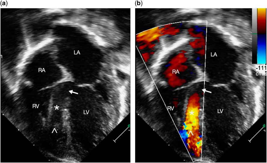
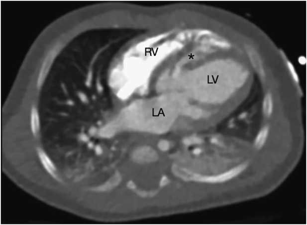
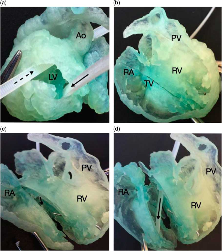
A 1-month-old infant with signs of heart failure was referred for surgical repair of a ventricular septal defect. Echocardiographic interrogation had demonstrated an extremely unusual and large tunnel-like defect, which commenced within the muscular ventricular septum from the posterior aspect of the basal left ventricle, and then coursed inferiorly, terminating in the anterior and apical part of the right ventricle, with an apparent large and unrestrictive entrance and exit (Fig 1). Given the unusual and complex nature of this tunnel-like defect, the surgeon requested computed tomographic imaging, which confirmed the described anatomy (Fig 2). A three-dimensional model was subsequently created utilising a Stratasys J750 3D printer (Stratasys, Eden Prairie, Minnesota, United States of America) with Polyjet technology, with the specific aim of better demonstrating the complex anatomy, with the additional benefit of the surgeon being able to manoeuvre the anatomy in hand in order to elicit the optimal approach for surgical closure. The specific printer and printing technology were chosen given the advantage over Fused Deposition Modeling technology for printing multiple materials, multicoloured and high-resolution models enabling the printing of flexible material models for better physical manoeuvrability, and allowing for procedural simulation. The model demonstrated that the exit of the defect in the right ventricle was not only apical but also very anterior. It was narrowed significantly by excessive right ventricular trabeculations, being bordered by the septomarginal trabeculation and the attachment of the tensor apparatus of the tricuspid valve to the ventricular septum, these latter features creating the seemingly “tunnel-like” course of a muscular inlet defect (Fig 3, Video 1).
Transthoracic echocardiogram showing apical four-chamber, ( a ) two-dimensional, and ( b ) colour comparison image, demonstrating the apparently large and unrestrictive tunnel-like ventricular septal defect (white asterisk). The entrance extends from the posterior aspect of the basal muscular septum of the left ventricle (LV) (white arrow), coursing inferiorly, and exiting the apical septum (white caret) anteriorly into the right ventricle (RV). The tensor apparatus of the tricuspid valve attaches near the superior border of the defect, adjacent to its apparent exit into the RV. LA=left atrium; RA=right atrium.

Computed tomography showing an axial plane image demonstrating the apparently large tunnel-like ventricular septal defect (black asterisk) extending from the posterior aspect of the basal septum of the left ventricle (LV), coursing inferiorly, and exiting anteriorly into the apex of the right ventricle (RV), adjacent to the attachment of the moderator band to the base of the septomarginal trabeculation with additional adjacent heavy septoparietal trabeculations. LA=left atrium.

( a ) Three-dimensional printed model displayed in a left anterior oblique orientation, with a portion of the left ventricle (LV) free wall dissected to demonstrate the large entrance to the complex, large tunnel-like defect (zip tie notated with solid black arrow), located in the posterior aspect of the basal septum on the LV aspect, inferior and posterior to the LV outflow tract (zip tie notated with dashed black arrow). ( b ) The same model is demonstrated in a right anterior oblique view with the anterior surface of the right atrium (RA), right ventricle (RV), and pulmonary artery removed. A metal pin is demonstrated in the small exit of the tunnel-like ventricular septal defect into the anterior apical region of the RV, bordered superiorly and anteriorly by the moderator band attachment to the base of the septomarginal trabeculation, and posteriorly and inferiorly by adjacent heavy trabeculations. The tricuspid valve and tensor apparatus have been removed. ( c ) An oblique sagittal dissection through the model is partially opened to demonstrate the tunnel-like ventricular septal defect (zip tie notated with solid black arrow) coursing inferiorly from the LV entrance, to the small, restrictive anterior exit into the RV (metal pin). ( d ) The oblique sagittal dissection through the model is now completely opened, and the model is rotated to better demonstrate the body of the large tunnel-like ventricular septal defect (zip tie notated with solid black arrow) with the small, restrictive exit into the RV (metal pin). AO=ascending aorta; PV=pulmonary valve; TV=tricuspid valve.

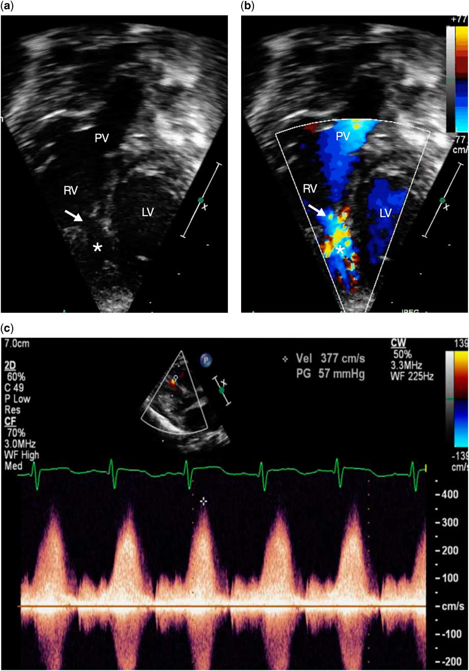
A repeat focussed echocardiogram was then obtained, assessing the demonstrated exit of the defect by sweeping more anteriorly than usual, in this way confirming its small and restrictive right ventricular orifice (Fig 4, Video 2). The understanding provided by the three-dimensional model also helped the sonographer to optimise the angle of the spectral Doppler interrogation, utilising unusual imaging planes, and hence improving the accuracy and identification of the high-pressure restriction across the defect. On the basis of this new information, we reconsidered our initial decision to proceed with surgical repair, and instead opted to continue conservative medical management. At the most recent follow-up, the child was thriving, with improvement in the symptoms reflecting heart failure, secondary to the defect becoming more restrictive at its site of exit into the right ventricle.
A follow-up transthoracic echocardiogram showing apical, ( a ) two-dimensional, and ( b ) colour comparison image positioned anteriorly at the level of the pulmonary valve (PV), demonstrating the smaller, restrictive, and anteriorly directed exit (white arrow) from the tunnel-like ventricular septal defect (white asterisk) into the right ventricle (RV). This anteriorly positioned exit was previously not demonstrated on the echocardiograms obtained before obtaining the computed tomography images with subsequent construction of the three-dimensional model. ( c ) Continuous wave Doppler interrogation of the anteriorly positioned exit from the tunnel-like ventricular septal defect confirming its restrictive nature. LV=left ventricle.

Acknowledgements
The authors are indebted to Mac Cameron from Stratasys for his involvement in printing these three-dimensional heart models. They also thank Dr Robert Anderson for his guidance in understanding the complexity of the septal anatomy.
Financial Support
This research received no specific grant from any funding agency, commercial, or not-for-profit sectors.
Conflicts of Interest
None.
Supplementary material
To view supplementary material for this article, please visit https://doi.org/10.1017/S104795111600202X