




No CrossRef data available.
Article contents
The molecular diagnosis of Marteilia refringens and differentiation between Marteilia strains infecting oysters and mussels based on the rDNA IGS sequence
Published online by Cambridge University Press: 14 September 2004
Abstract
Marteilia refringens is a paramyxean parasite which infects the flat oyster Ostrea edulis and mussels (Mytilus galloprovincialis), where it has been attributed to a separate species, Marteilia maurini, by several authors. Doubts persist though as to the existence or not of two species of Marteilia in Europe. We have devised a molecular method for the diagnosis of M. refringens based on 358 bp nested-PCR of the rDNA intergene spacer (rDNA IGS) which is capable of detecting 0·5 fg of M. refringens DNA. Molecular characterization of this spacer indicates that the Marteilia parasites which infect oysters and mussels are two different strains of the same species.
Keywords
Information
- Type
- Research Article
- Information
- Copyright
- 2004 Cambridge University Press
INTRODUCTION
Marteilia refringens, the aetiological agent of Abers disease (Grizel et al. 1974), is a protozoan parasite found in different commercially exploited molluscs such as the European flat oyster Ostrea edulis (Grizel et al. 1974), the most appreciated of all the world's edible oysters, and the mussel Mytilus galloprovincialis, another widely consumed species of shellfish (Villalba et al. 1993). M. refringens is widely present along the European Atlantic coast from western Brittany in the north to the northern Mediterranean in the south (Audemard et al. 2001). The parasite is included in the International Aquatic Animal Health Code of the World Organization for Animal Health.
Another species within the genus (M. sydneyi), the aetiological agent of ‘QX disease’ in the Australian Sydney rock oyster, Saccostrea commercialis (Perkins & Wolf, 1976), can be distinguished by the number of secondary and tertiary cells in their characteristic cell-within-cell structure. In 1982 another species of the same genus, M. maurini, was described in M. galloprovincialis in the Adriatic (Comps, Pichot & Papagianni, 1982). Although the criteria used in the recognition of two species of Marteilia in Europe, ultrastructural characteristics and host specificity, have since been put into doubt (Villalba et al. 1993; Longshaw et al. 2001), epizootiological data and molecular data based on their ITS-1 sequence suggest that distinct types of Marteilia do infect bivalve molluscs in Europe (Le Roux et al. 2001).
Currently used methods for the diagnosis of M. refringens are based on histological examination, which requires a high level of expertise. Doubts also persist concerning the effectiveness and reliability of these techniques during the first stages of infection or at very low infection rates, suggesting the need to develop a more sensitive method that can be used routinely in the diagnosis of this parasitic disease. To this end Pernas and co-workers (Pernas et al. 2001) described a method for the molecular detection of M. refringens based on the amplification by PCR of an anonymous repetitive DNA sequence of the parasite. This method proved to be very sensitive but the location in the genome of the amplified sequence is unknown and may always belong to a highly preserved region amongst different species. In this study we have devised a molecular diagnostic method based on a specific, located DNA sequence of the parasite, the intergenic spacer (IGS) between ribosomal genes (rDNA). In contrast to other primers designed for M. refringens detection in 18S or ITS-1 sequences (Le Roux et al. 1999, 2001) we have used the rDNA IGS as a useful marker because of its high variability and faster sequence evolution than ribosomal genes and ITS regions. Thus we also used the rDNA IGS as a taxonomic marker to differentiate between Marteilia species present in oysters and mussels from different sites in Europe.
MATERIALS AND METHODS
Purification of parasite cells and DNA isolation
M. refringens cells were obtained from naturally infected flat oysters (O. edulis) from Huelva (SW Spain) and infected mussels (M. galloprovincialis) from Huelva, Vigo (NW Spain), Trieste and Venice (NW Italy) following the protocol described by Robledo, Mialhe & Figueras (1995) with minor modifications. Parasitized oysters and mussels were identified by microscopical examination of digestive-gland smears stained with VOE dye (Gutierrez, 1977). From 20 to 40 heavily infected digestive glands were homogenized in conservation buffer (0·25 M EDTA, 30% w/v NaCl, 20% v/v DMSO) using an Ultra-Turrax. Before homogenization the superficial gonad was removed to improve purification efficiency. The homogenate was sieved progressively through 300, 200 and 75 μm nylon meshes and the filtrate centrifuged at 2500 g for 30 min at 4 °C. The pellet was diluted in PBS and placed on a 35% (w/v) sucrose gradient to be centrifuged at 2500 g for 30 min at 4 °C. The resulting pellet was resuspended in PBS and placed in a 40%/20% (v/v) Percoll gradient to be centrifuged again at 2500 g for 30 min at 4 °C. The brown interface containing the parasite cells was recovered using a Pasteur pipette. We thus obtained between 1·7 and 1·8×106 sporonts/ml.
Genomic M. refringens DNA was purified according to Winnepenninckx, Backeljau & Wachter (1993). Parasite cells were transferred to a centrifugation tube containing 10 ml of preheated (60 °C) CTAB buffer (2% w/v CTAB, 1·4 M NaCl, 0·2% (v/v) β-mercaptoethanol, 20 mM EDTA, 100 mM TrisHCl pH 8·0, 0·1 mg/ml proteinase K). After incubation at 60 °C for 3–6 h, DNA was extracted using a standard phenol/chloroform protocol and precipitated with ethanol. Genomic DNA to be used as negative controls for PCR amplifications was obtained in the same way from the abductor muscles of non-infected flat oysters and mussels.
Isolation and characterization of the IGS of the nuclear ribosomal genes
We amplified the M. refringens rDNA IGS obtained from infected flat oysters using sense primer 26S designed in the 28S DNA ribosomal gene for the amplification of rDNA IGS in plants (Bena et al. 1998). This primer is preserved in the large ribosomal gene sequence of distant evolutive taxa ranging from microorganisms to mammals (personal observation). The specific antisense primer 18S was designed in our laboratory from the 18S DNA ribosomal sequence of M. refringens obtained by Berthe et al. (2000) (Fig. 1). Amplification was carried out from 750 ng of M. refringens DNA as template in 50 μl total volume containing 150 ng of each primer, 20 mM dNTPs, 2·5 mM MgCl2 and 3 units of Taq polymerase (Amersham Biosciences) in Taq buffer. The PCR was conducted with an initial denaturation at 94 °C for 5 min, 30 cycles of denaturation at 92 °C for 15 sec and annealing-extension at 68 °C for 5 min plus a final step a 72 °C for 10 min in a GeneAmp 2700 Applied Biosystems Thermocycle. The amplified product was cloned into pGEM-T Easy Vector System (Promega) and the nucleotide sequences of rDNA IGS were determined by Sanger's method (Sanger, Nicklen & Coulson, 1977) using an ABI Prism Dye Terminator Cycle Sequencing kit (Perkin-Elmer). For the complete sequencing of the rDNA IGS, 4 primers IN-1, IN-2, IN-3 and IN-4, were designed from the rDNA IGS sequence obtained beforehand (Fig. 1). The sequences were analysed and compared using the Basic Local Alignment Search Tool (BLAST). The complete rDNA IGS sequence of M. refringens isolated from oysters has been entered into the EMBL database with Accession number AJ604561.

Fig. 1. Schematic representation of the nuclear ribosomal DNA repeating unit and location of the primers used in the amplification and sequencing of ribosomal intergenic region. (A) IGS, intergenic spacer; ETS, external-transcribed spacer; NTS, non-transcribed spacer; ITS, internal-transcribed spacer. Arrows indicate the region from which the primers were designed for the amplification and sequencing of the rDNA IGS from Marteilia refringens. (B) Description of primers used.
Development of a molecular diagnostic test for Marteilia refringens
For the diagnostic PCR we designed primers located in the rDNA IGS sequence of M. refringens. The first round of PCR was made using the primers MT-1 (sense) and MT-2 (antisense) and nested-PCR with the primers MT-1B (sense) and MT-2B (antisense) (Fig. 1). To determine the sensitivity of the PCR with the diagnostic primers we prepared 1 μg to 0·1 fg dilutions of DNA from M. refringens. To determine the detection limit of primers from infected oyster tissues we made an experimental approximation consisting of mixing oyster DNA with each of the M. refringens DNA dilutions.
We compared the effectiveness of our primers with the diagnostic primers described by other authors. The comparison between the sensitivity of primers MT-1 and MT-2 to that of other primers described in the 18S gene (SAS1 and SS2) (Le Roux et al. 1999) and in the ITS-1 spacer (Pr4 and Pr5) (Le Roux et al. 2001) of M. refringens was made using both primer pairs SAS1/SS2 and Pr4/Pr5 in PCR amplifications from M. refringens DNA dilutions according to Le Roux and co-workers (1999, 2001).
Comparison of Marteilia purified from oysters and mussels
The MT-1/MT-2 fragment of rDNA IGS and the Pr4/Pr5 fragment of ITS-1 from 1–3 infected oysters and mussels were amplified, cloned and sequenced. A comparative analysis of the 2–5 cloned sequences per individual allowed us to obtain the variability between Marteilia purified from oysters and that from mussels. Multiple sequence alignment was done using the MegAlin program of the DNAstar package (LASERGEN). Intraspecific variability and interspecific divergence were calculated using the Kimura 2-parametres model distance matrix (Kimura, 1980). Neighbour-joining phylogenetic analyses were made of the MT-1/MT-2 sequences using the MEGA package (Kumar et al. 2001). Genetic distances were calculated according to Kimura 2-parametres (Kimura, 1980). The partial ITS and IGS sequences have been entered into the EMBL database with Accession numbers AJ629334 to AJ629376.
RESULTS
Amplification of the ribosomal intergenic region of Marteilia refringens
M. refringens DNA purified from the digestive glands of infected oysters (O. edulis) was amplified with the preserved 26S primer and the M. refringens 18S specific primer, both of which anneal to ribosomal genes (Fig. 1). These primers amplified a PCR product of 3455 bp only when the DNA of the parasite was used as template. When DNA from non-infected oysters was used PCR consistently gave a negative result (Fig. 2A), indicating that the 26S and 18S primers did not cross-react with oyster DNA. The specificity of the PCR product was also confirmed by Southern blot hybridization. A hybridization signal appeared only in the presence of parasite DNA, no signal being detected for oyster DNA (data not shown).

Fig. 2. Amplification of Marteilia refringens rDNA IGS. (A) Amplified rDNA IGS using primers 26S and 18S. PCR was carried out from different templates: lane 2, genomic DNA from non-infected oyster; lane 3, genomic DNA from non-infected mussels; lane 4, genomic DNA from M. refringens purified from digestive glands of infected oysters; lane 5, negative control. The molecular weight markers used are 100 base-pair Ladder (Amersham Biosciences), lane 1, and DNA Molecular Weight Marker II (λ-phage digested with Hind III, Roche), lane 6. (B) PCR amplification in the rDNA IGS sequence with primers MT-1 and MT-2: lane 1, molecular weight marker (100 base-pair Ladder (Amersham Biosciences). PCR was carried out from different templates, always using 200 ng of DNA: lane 2, genomic DNA from lightly infected oyster; lane 3, genomic DNA from moderately infected oyster; lane 4, genomic DNA from highly infected oyster; lanes 5 and 6, genomic DNA from M. refringens purified from digestive glands of infected oysters; lane 7, genomic DNA of non-infected oysters; lane 8, negative control of PCR. The intensity of the amplified product correlated with the level of infection of the host. Highly infected oysters displayed an intense DNA band on the gel whilst lightly infected ones were not so bright.
A comparison of our M. refringens rDNA IGS cloned sequences with other sequences deposited in the GenBank and EMBL databases showed homology only in the 3′ and 5′ ends with the 18S and 28S ribosomal genes of M. refringens and different organisms respectively. The 205 bp of the 3′ end of the cloned fragments matched the 5′ end of the 18S ribosomal gene from M. refringens and the 5′ end matched the 3′ end of the 28S ribosomal gene of different organisms. The end of the rDNA IGS and beginning of the 28S ribosomal gene was established in the light of these comparisons. Excluding these homologous regions, the rDNA IGS separating the genes for small and large rRNA's was 2822 bp.
Development of a molecular test for the diagnosis of Marteilia refringens infection
We designed the primers MT-1 and MT-2 for the amplification of a DNA fragment located within the rDNA IGS of M. refringens (Fig. 1). These primers amplified the expected 525 bp product when either the DNA of the parasite or the DNA of infected oysters was used as template (Fig. 2B).
To determine the sensitivity of PCR with the primers we prepared MT-1 and MT-2 dilutions from the DNA of M. refringens. The effectiveness of the primers was tested by using them in PCR amplification of Marteilia DNA taken from parasite cells purified from the digestive glands of infected oysters. The PCR was capable of repeatedly detecting 5 pg of DNA (Fig. 3A).

Fig. 3. Efficiency and sensitivity of the diagnostic test devised for the detection of Marteilia refringens using primers MT-1 and MT-2. (A) Amplification from M. refringens DNA purified from digestive glands of infected oysters. The 525 bp fragment was obtained from different quantities of parasite DNA: lane 2, 100 pg; lane 3, 50 pg; lane 4, 25 pg; lane 5, 10 pg; lane 6, 5 pg; lane 7, 2·5 pg. Lane 8 is the negative control. (B) Amplification from M. refringens DNA mixed with oyster DNA. The product was obtained by mixing 1 μg of oyster DNA with different quantities of parasite DNA: lane 2, 1000 pg; lane 3, 750 pg; lane 4, 500 pg; lane 5, 200 pg; lane 6, 100 pg; lane 7, 50 pg; lane 8, 25 pg of DNA. Lane 9 is the negative control. At the bottom, nested-PCR gels of pure Marteilia DNA in (C) and Marteilia DNA mixed with oyster DNA (D). The 358 fragment was obtained from: lane 2, 50 pg; lane 3, 10 pg; lane 4, 5 pg; lane 5, 1 pg; lane 6, 0·5 pg; lane 7, 50 fg; lane 8, 0·5 fg; lane 9, 0·1 fg. Lane 1 corresponds to 100 base-pair Ladder (Amersham Biosciences) in gels.
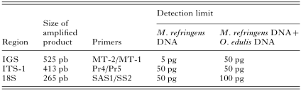
To determine the detection limit of primers from infected oyster tissue we experimented by mixing oyster DNA with each M. refringens dilution. Figure 3B shows that the addition of host DNA in excess of the DNA dilution slightly compromised the sensitivity of the test, up to 50 pg of DNA of Marteilia. The sensitivity of the MT-1 and MT-2 primers was compared to the sensitivity of the SAS1/SS2 and Pr4/Pr5 primers described in the 18S gene and the ITS-1 spacer respectively (Le Roux et al. 1999, 2001) (see Table 1). In amplifying pure Marteilia DNA both pairs of primers proved to be less sensitive than the MT-1/MT-2 primers and in amplifying the DNA of Marteilia mixed with that of oysters the SAS1/SS2 primers once more proved to be less sensitive than primers MT-1/MT-2. Primers Pr4/Pr5 showed the same detection limit as MT-1/MT-2.
The detection limit of the MT-1 and MT-2 primers increased substantially on using nested-PCR. The nested-PCR designed in our laboratory was carried out with primers MT-1A and MT-1B located within the 525 bp fragment amplified in the first round of PCR. These primers yielded a 358 bp fragment, which was the size we expected. This second PCR increased the limit of detection to 0·5 fg of pure M. refringens DNA (Fig. 3C) and to 50 fg of M. refringens DNA mixed with oyster DNA (Fig. 3D).
The specific amplifications were never observed with oyster DNA either in the first or second round of PCR.
rDNA IGS as taxonomic marker for Marteilia purified from oysters and mussels
We obtained a total of 25 sequences of the MT-1/MT-2 fragment from M. refringens cells purified from the digestive glands of infected oysters from Huelva and from infected mussels from Huelva, Vigo, Trieste and Venice. There was no significant polymorphism between sequences obtained from oysters and mussels and neither were there any mutations at nucleotide positions to allow us to differentiate between the sequences obtained from either host. An analysis of variability between sequences was made using the data set based on the aligned MT-1/MT-2 sequences of the rDNA IGS. The variable positions corresponded to shared polymorphisms amongst all of them, although some of these variable nucleotide positions appeared mainly in the parasite sequences from just one of the hosts (Fig. 4A).

Fig. 4. Sequences of Marteilia purified from infected oysters and mussels. (A) Partial sequence of the MT-1/MT-2 rDNA IGS fragment (positions 181–210 and 401–420 of 525 bp fragment) in which the polymorphic positions (boxed) are present mainly in the sequence of Marteilia from oysters and mussels. They are the only 2 of these positions that coincide with enzyme restriction sites, Hpa II (CCGG, 195–198) and Hae III (GGCC, 412–415) (in bold). Only the variable parasite sequences from each population are represented. (B) Partial sequence of the Pr4/Pr5 ITS-1 fragment (positions 301–360 of 413 bp fragment) compared with consensus sequences of Marteilia from mussels (M. maurini, Mm) and from oysters (M. refringens, Mr) described by Le Roux et al. (2001). Four diagnostic positions shared by the 2 species (boxed) are located in this fragment. Only the variable sequences are represented. The clones were obtained from only one infected oyster or mussel. The rDNA IGS and ITS-1 fragments were obtained from the same infected oyster or mussel.
Intraspecific sequence analysis showed that the percentage of differences (p) was low both between sequences from oysters (0·0<p<2·3) and from mussels (0·6<p<1·8).
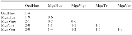
Interspecific sequence analysis was carried out on the premise that the MT-1/MT-2 parasite sequences obtained from oysters and mussels were sequences belonging to different species and also taking into account the different populations from which they were sampled. This analysis revealed that the interspecific distances between sequences from oysters and the different mussel populations were of the same order as the intraspecific distances (Table 2).
Nevertheless, when a phylogenetic tree was constructed the sequences separated into 2 distinct groups supported by a bootstrap value of 99%, one being composed of the majority of the mussel sequences and the other of the majority of the oyster sequences (Fig. 5).

Fig. 5. Phylogenetic tree deduced from 25 partial rDNA IGS sequences of Marteilia refringens obtained by PCR amplification from 5 samples. The names of the sequences correspond to the host species, Oed: Ostrea edulis and Mga: Mytilus galloprovincialis, and their geographical location, Hue: Huelva, Vigo: Vigo, Tri: Trieste and Ven: Venice. The numeric values indicate the clone. Numbers to the left correspond to bootstrap values.
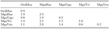
Finally, we amplified the Pr4/Pr5 ITS-1 fragment in the same samples in which we had previously amplified the MT-1/MT-2 rDNA IGS fragment. All the sequences obtained from Marteilia purified from oysters and the majority of those obtained from mussels were type O, corresponding to M. refringens. Only 2 sequences from one infected mussel from Huelva (MgaHue-18 and MgaHue-26) were type M, assigned to M. maurini. Nevertheless, another 2 sequences (MgaHue-11 and MgaHue-50) from the same infected mussel were type O (Fig. 4B; Table 3).
DISCUSSION
We have amplified the ribosomal intergene spacer (rDNA IGS) of M. refringens isolated from O. edulis using a universal primer located at the 3′ end of the 28S rDNA (Bena et al. 1998), taking advantage of the high level of evolutionary rDNA-sequence preservation, and a specific primer designed in our laboratory from the only gene sequence of the parasite known until now, 18S rDNA (Berthe et al. 2000).
The rDNA IGS sequence information was used to design primers for the molecular diagnosis of this parasite: MT-1 and MT-2 for the first round of PCR and MT-1A and MT-1B for nested-PCR. The nested-PCR designed was capable of repeatedly detecting 0·5 fg of M. refringens DNA compared to the 150 fg detected by Pernas and co-workers' nested-PCR molecular diagnostic method (Pernas et al. 2001). Furthermore, with the Pernas' technique the site of the amplified sequence in the genome is not known and thus the amplified fragment might be a gene region preserved in other Paramyxea, which makes it considerably less specific than the IGS-sequence method. Other pairs of primers have also been described for PCR and the in situ detection of M. refringens. Primers SAS1 and SS2, were designed within the 18S rRNA gene of the parasite (Le Roux et al. 1999) to be specific for its detection. These primers were less sensitive than primers MT-1/MT-2 under our experimental conditions and when the amplified fragment was used as a probe it cross-reacted with M. sidneyi and Marteilioides sp. in tissue sections of their respective hosts (Kleeman et al. 2002). Another primer pair, Pr4 and Pr5, described within the ITS-1 sequence of M. refringens (Le Roux et al. 2001), were also less sensitive than the MT primers. These primers are located in a variable region in one of the internal transcribed spacers of ribosomal genes. Nevertheless, some authors have calculated that the more preserved fragments of intergenic spacer evolve 1·5 times more quickly than internal ones (Bena et al. 1998). We conclude therefore that: (a) the IGS-primers should be even more specific than primers described in ITSs; and (b) on the other hand, our method is more sensitive than those devised hitherto, detecting as it does 0·5 fg of DNA.
With regard to differentiating between the 2 European species of Marteilia, M. refringens and M. maurini, neither the SAS1/SS2 primers nor Pr4/Pr5 were able to make any clear distinction. The 18S gene sequence amplified by the SAS1/SS2 primers was identical for Marteilia purified from both oysters and mussels (Berthe et al. 2000). In the case of the ITS-1 fragment amplified by primers Pr4/Pr5 there were 2 haplotypes, which the authors identified as belonging to M. refringens (type O) and M. maurini species (type M). This differentiation was based on 4 diagnostic positions between the sequences of 413 bp and on 16 nucleotic positions fixed within the sequences of Marteilia purified from oysters and variable in Marteilia from mussels and 20 nucleotic positions variable in the former and fixed in the latter. An analysis of the less-preserved rDNA IGS sequence should support this idea.
Thus we analysed the MT-1/MT-2 fragment in infected mussels from Trieste and Venice, where M. maurini was first defined as a different species, Vigo where the species of Marteilia in mussels was type M in the ITS-1 sequence, and Huelva, where the presence of Marteilia in mussels had not been described until now. We compared the sequences obtained from these mussels with the M. refringens isolated from O. edulis from Huelva. The result of the phylogenetic analysis shows that the majority of rDNA IGS sequences are separated into 2 well-defined clusters depending upon the host. Nevertheless we can find sequences from only 1 infected oyster present in the 2 clusters and the same occurs with sequences obtained from mussels. If 2 species of the parasite exist then these 2 species should be present in the same host at the same time and this situation should not be at all scarce because it occurs throughout the population. Co-infection by 2 or more parasites in the same host is habitual and should be the situation in these molluscs, but in this case the intergenic distances between the sequences obtained from infected oysters and mussels are far too close, bearing in mind the high variability of a sequence such as rDNA IGS. The intergenic distances between other close species of mollusc parasites are higher: 20% between Perkinsus atlanticus and P. marinus; 35% between P. atlanticus and P. andrewsi and 42% between P. marinus and P. andrewsi (Navas et al. 2003). Altogether, our results show that if some differentiation does exist between the parasite sequences from the oyster and mussel hosts in question it is not sufficient to consider them as being 2 separate species.
Nevertheless, it could be that the MT-1 and MT-2 primers designed in the rDNA IGS are exclusive to the spacer sequence from M. refringens and do not amplify the homologous fragment of a second species, M. maurini, due to divergences between the sequences. If this were true, we would amplify the MT-1/MT-2 fragment of M. refringens in the different samples studied and the distances would correspond to the intraspecific variability between them. To determine whether the parasite which infects mussels is, in fact, a different species of Marteilia from that which parasitizes flat oysters and that the results were not due to the marker used (the rDNA IGS), we amplified the ITS-1 in the same samples using primers Pr4/Pr5. This fragment has been used in the past to differentiate between 2 species of Marteilia (Le Roux et al. 2001). We compared the sequences obtained with the consensus sequences described for M. refringens and M. maurini by Le Roux and co-workers. The results obtained from this analysis turned out to be the same as those obtained from rDNA IGS: the interspecific distances were in the same range as the intraspecific ones and sequences from only one infected mussel presented both the nucleotide specific to M. refringens and that specific to M. maurini.
We conclude therefore that in the samples we have studied the sequences do not belong to a different species of the parasite, although some differentiation does exist, which may be due to 2 incompletely differentiated species or, more probably, to 2 different strains.
In summary, in this work we show a quick, easy, highly sensitive and specific technique to diagnose the presence of M. refringens using nested-PCR of a 358 bp fragment located in the intergene spacer. Furthermore, the variability of this sequence suggests very strongly that, in the samples analysed, the Marteilia protozoan parasites purified both from oysters and from mussels constitute 2 different strains of the same species.
This research was supported by a grant from the Plan Andaluz de Investigación (Group No. CVI0200), a grant from the Junta Nacional Asesora de Cultivos Marinos (JACUMAR), and by a co-operation agreement between the University of Granada and the Andalusian Ministry for Agriculture and Fisheries. We thank Dr Pablo Presa (University of Vigo, Spain) for his generous donation of samples and Dr Frank Berthe (IFREMER, France) for his valuable suggestions. We also thank our colleague A. L. Tate for revising our English text.
References
REFERENCES
AUDEMARD, C.,
BARNAUD, A.,
COLLINS, C. M.,
LE ROUX, F.,
SAURIAU, P.,
COUSTAU, C.,
BLACHIER, P. &
BERTHE, F. C. (2001).
Claire ponds as an experimental model for Marteilia refringens life-cycle studies: new perspectives.
Journal of Experimental Marine Biology and Ecology 257,
87–108.CrossRefGoogle Scholar
BENA, G.,
JUBIER, M. F.,
OLIVIERI, I. &
LEJEUNE, B. (1998).
Ribosomal external and internal transcribed spacers: combined use in the phylogenetic analysis of Medicago (Leguminosae).
Journal of Molecular Evolution 46,
299–306.CrossRefGoogle Scholar
BERTHE, F.,
LE ROUX, F.,
PEYRETAILLADE, E.,
PEYRET, P.,
RODRIGUEZ, D.,
GOUY, M. &
VIVARES, P. (2000).
Phylogenetic analysis of the small subunit ribosomal RNA of Marteilia refringens validates the existence of phylum Paramyxea (Desportes and Perkins, 1990).
Journal of Eukaryotic Microbiology 47,
288–293.CrossRefGoogle Scholar
COMPS, M.,
PICHOT, Y. &
PAPAGIANNI, P. (1982).
Recherche sur Marteilia maurini n. sp. parasite de la moule Mytilus galloprovincialis Lmk.
Revue des Travaux de l'Institut des Pechês Maritimes 45,
211–214.Google Scholar
GRIZEL, H.,
COMPS, M.,
BONAMI, J.,
COUSSERANS, F.,
DUTHOIT, J. &
LE PENNEC, M. (1974).
Recherches sur l'agent de la maladie de la glande digestive de Ostrea edulis Linnè.
Bulletin de l'Institut des Peches Maritimes 240,
7–30.Google Scholar
GUTIERREZ, M. (1977).
Técnicas de coloración del agente de la enfermedad de la ostra plana, Ostrea edulis L.
Investigación Pesquera 41,
643–645.Google Scholar
KIMURA, M. (1980).
A simple method for estimating evolutionary rate of base substitutions through comparative studies of nucleotide sequences.
Journal of Molecular Evolution 16,
111–120.CrossRefGoogle Scholar
KLEEMAN, S. N.,
LE ROUX, F.,
BERTHE, F. &
ADLARD, R. D. (2002).
Specificity of PCR and in situ hybridization assays designed for detection of Marteilia sydneyi and M. refringens.
Parasitology 125,
131–141.CrossRefGoogle Scholar
KUMAR, S.,
TAMURA, K.,
JAKOBSER, I. B. &
NEI, M. (2001).
MEGA2: Molecular Evolutionary Genetics.
Analysis software, Arizona State University,
Tempe, Arizona, USA.
LE ROUX, F.,
AUDEMARD, C.,
BARNAUD, A. &
BERTHE, F. (1999).
DNA probes as potential tools for the detection of Marteilia refringens.
Marine Biotechnology 1,
588–597.CrossRefGoogle Scholar
LE ROUX, F.,
LORENZO, G.,
PEYRET, P.,
AUDEMARD, C.,
FIGUERAS, A.,
VIVARES, C.,
GOUY, M. &
BERTHE, F. (2001).
Molecular evidence for the existence of two species of Marteilia in Europe.
Journal of Eukaryotic Microbiology 48,
449–454.CrossRefGoogle Scholar
LONGSHAW, M.,
FEIST, S. W.,
MATTHEWS, A. &
FIGUERAS, A. (2001).
Ultrastructural characterisation of Marteilia species (Paramyxea) from Ostrea edulis, Mytilus edulis and Mytilus galloprovincialis in Europe.
Diseases of Aquatic Organisms 44,
137–142.CrossRefGoogle Scholar
NAVAS, J. I.,
DE LA HERRÁN, R.,
GARRIDO-RAMOS, M. A.,
LÓPEZ, I.,
QUINTERO, M. C.,
GARCÍA-PRADO, I. M.,
RUIZ-REJÓN, C. &
RUIZ-REJÓN, M. (2003).
Aplicación del diagnóstico molecular de Perkinsus atlanticus para la detección del parásito en diferentes especies de almejas del litoral suratlántico y el estudio de la variabilidad intraespecífica. IX Congreso Nacional de Acuicultura, Cádiz. pp.
321–322.
PERKINS, F. &
WOLF, P. (1976).
Fine structure of Marteilia sydneyi sp. n.-haplosporidian pathogen of Australian oysters.
Journal of Parasitology 62,
528–538.Google Scholar
PERNAS, M.,
NOVOA, B.,
BERTHE, F.,
TAFALLA, C. &
FIGUERAS, A. (2001).
Molecular methods for the diagnosis of Marteilia refringens.
Bulletin of the European Association of Fish Pathologists 21,
200–208.Google Scholar
ROBLEDO, J. A. F.,
MIALHE, E. &
FIGUERAS, A. J. (1995).
Purification of several phases of the parasite Marteilia (Protozoa: Ascetospora) from Mussels (Mytilus galloprovincialis). In
Techniques in Fish Inmunology-4, Inmunology and Pathology of Aquatic Invertebrates (ed. Stolen, J. S., Fletcher, T. C., Smith, S. A., Zelikoff, J. T., Kaattari, S. L., Anderson, R. S., Södernäll, K. & Weeks-Perkins, B. A.), pp.
117–121.
SOS Publications,
Fair Haven, NJ, USA.
SANGER, F.,
NICKLEN, S. &
COULSON, A. R. (1977).
DNA sequencing with chain-terminating inhibitors.
Proceedings of the National Academy of Sciences, USA 74,
5463–5467.CrossRefGoogle Scholar
VILLALBA, A.,
MOURELLE, S. G.,
LÓPEZ, M. C.,
CARBALLAL, M. J. &
AZEVEDO, C. (1993).
Marteiliasis affecting cultured mussels Mytilus galloprovincialis of Galicia (NW Spain). I. Etiology, phases of the infection and temporal and spatial variability in prevalence.
Diseases of Aquatic Organisms 16,
61–72.Google Scholar
WINNEPENNINCKX, B.,
BACKELJAU, T. &
DE WACHTER, R. (1993).
Extraction of high molecular weight DNA from molluscs.
Trends in Genetics 9,
407.Google Scholar
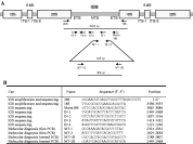
Fig. 1. Schematic representation of the nuclear ribosomal DNA repeating unit and location of the primers used in the amplification and sequencing of ribosomal intergenic region. (A) IGS, intergenic spacer; ETS, external-transcribed spacer; NTS, non-transcribed spacer; ITS, internal-transcribed spacer. Arrows indicate the region from which the primers were designed for the amplification and sequencing of the rDNA IGS from Marteilia refringens. (B) Description of primers used.
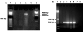
Fig. 2. Amplification of Marteilia refringens rDNA IGS. (A) Amplified rDNA IGS using primers 26S and 18S. PCR was carried out from different templates: lane 2, genomic DNA from non-infected oyster; lane 3, genomic DNA from non-infected mussels; lane 4, genomic DNA from M. refringens purified from digestive glands of infected oysters; lane 5, negative control. The molecular weight markers used are 100 base-pair Ladder (Amersham Biosciences), lane 1, and DNA Molecular Weight Marker II (λ-phage digested with Hind III, Roche), lane 6. (B) PCR amplification in the rDNA IGS sequence with primers MT-1 and MT-2: lane 1, molecular weight marker (100 base-pair Ladder (Amersham Biosciences). PCR was carried out from different templates, always using 200 ng of DNA: lane 2, genomic DNA from lightly infected oyster; lane 3, genomic DNA from moderately infected oyster; lane 4, genomic DNA from highly infected oyster; lanes 5 and 6, genomic DNA from M. refringens purified from digestive glands of infected oysters; lane 7, genomic DNA of non-infected oysters; lane 8, negative control of PCR. The intensity of the amplified product correlated with the level of infection of the host. Highly infected oysters displayed an intense DNA band on the gel whilst lightly infected ones were not so bright.

Fig. 3. Efficiency and sensitivity of the diagnostic test devised for the detection of Marteilia refringens using primers MT-1 and MT-2. (A) Amplification from M. refringens DNA purified from digestive glands of infected oysters. The 525 bp fragment was obtained from different quantities of parasite DNA: lane 2, 100 pg; lane 3, 50 pg; lane 4, 25 pg; lane 5, 10 pg; lane 6, 5 pg; lane 7, 2·5 pg. Lane 8 is the negative control. (B) Amplification from M. refringens DNA mixed with oyster DNA. The product was obtained by mixing 1 μg of oyster DNA with different quantities of parasite DNA: lane 2, 1000 pg; lane 3, 750 pg; lane 4, 500 pg; lane 5, 200 pg; lane 6, 100 pg; lane 7, 50 pg; lane 8, 25 pg of DNA. Lane 9 is the negative control. At the bottom, nested-PCR gels of pure Marteilia DNA in (C) and Marteilia DNA mixed with oyster DNA (D). The 358 fragment was obtained from: lane 2, 50 pg; lane 3, 10 pg; lane 4, 5 pg; lane 5, 1 pg; lane 6, 0·5 pg; lane 7, 50 fg; lane 8, 0·5 fg; lane 9, 0·1 fg. Lane 1 corresponds to 100 base-pair Ladder (Amersham Biosciences) in gels.
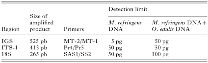
Table 1. Detection limit of pure Marteilia refringens DNA and M. refringens DNA plus oyster DNA by PCR and nested PCR with different pairs of primers
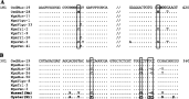
Fig. 4. Sequences of Marteilia purified from infected oysters and mussels. (A) Partial sequence of the MT-1/MT-2 rDNA IGS fragment (positions 181–210 and 401–420 of 525 bp fragment) in which the polymorphic positions (boxed) are present mainly in the sequence of Marteilia from oysters and mussels. They are the only 2 of these positions that coincide with enzyme restriction sites, Hpa II (CCGG, 195–198) and Hae III (GGCC, 412–415) (in bold). Only the variable parasite sequences from each population are represented. (B) Partial sequence of the Pr4/Pr5 ITS-1 fragment (positions 301–360 of 413 bp fragment) compared with consensus sequences of Marteilia from mussels (M. maurini, Mm) and from oysters (M. refringens, Mr) described by Le Roux et al. (2001). Four diagnostic positions shared by the 2 species (boxed) are located in this fragment. Only the variable sequences are represented. The clones were obtained from only one infected oyster or mussel. The rDNA IGS and ITS-1 fragments were obtained from the same infected oyster or mussel.
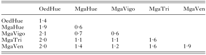
Table 2. Divergence (percentage) among MT-1/MT-2 fragments from Marteilia purified from oysters (Oed) and mussels from Huelva (MgaHue), Vigo (MgaVigo), Trieste (MgaTri) and Venice (MgaVen)
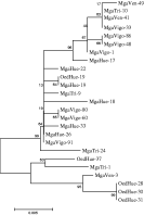
Fig. 5. Phylogenetic tree deduced from 25 partial rDNA IGS sequences of Marteilia refringens obtained by PCR amplification from 5 samples. The names of the sequences correspond to the host species, Oed: Ostrea edulis and Mga: Mytilus galloprovincialis, and their geographical location, Hue: Huelva, Vigo: Vigo, Tri: Trieste and Ven: Venice. The numeric values indicate the clone. Numbers to the left correspond to bootstrap values.
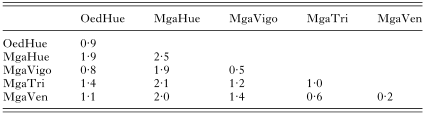
Table 3. Divergence (percentage) among Pr4/Pr5 fragments from Marteilia purified from oysters (Oed) and mussels from Huelva (MgaHue), Vigo (MgaVigo), Trieste (MgaTri) and Venice (MgaVen)
 You have
Access
- 52
- Cited by