Introduction
Akopovaite, Li2Al4(OH)12(CO3)(H2O)3, was discovered by V.Yu.K., L.A.P. and A.A.A. during field work in 1993 at the Karasu–Karavshinskoye tin deposit, in a pegmatite vein located in the upper reaches of the Asan–Usan glacier (area #2, 39.5893°N, 70.2489°E, northern slope of the Turkestan Range, Kyrgyzstan). The small size of the crystals and admixture with gibbsite, Al(OH)3, and, probably, amorphous Al(OH)3, resulted in a long period of study for this mineral species. The new mineral was named after Anna Vartanovna Akopova (Cyrillic: Анна Вартановна Акопова) (b. 1952), a chemistry teacher in the gymnasium #14, Rostov-on-Don, Russia. The remarkable style of her teaching, including experimental work, has promoted many of her students to choose a career in the field of Natural Sciences, including the first author of the present paper.
The mineral and its name have been approved by the International Mineralogical Association Commission on New Minerals, Nomenclature and Classification (IMA2018-095 Karpenko et al., Reference Karpenko, Pautov, Zhitova, Agakhanov, Krzhizhanovskaya, Siidra and Rassulov2018). The type material (holotype fragment) has been deposited in the Fersman Mineralogical Museum, Russian Academy of Sciences, Leninskiy prospect, 18-2, Moscow, 119071, Russia, Catalogue # 96260.
The hydrotalcite supergroup, which includes akopovaite, consists of more than 40 minerals (Mills et al., Reference Mills, Christy, Génin, Kameda and Colombo2012a) known as natural layered double hydroxides (LDHs). The majority of hydrotalcite-supergroup mineral species are represented by hydroxides with positively-charged layers built by divalent (Mg, Fe2+, Mn, Ni, Cu, Ca and Zn) and trivalent (Al, Fe, Mn, Co and Cr) cations alternating with negatively charged interlayers [e.g. (CO3)2–, (OH)–, Cl–, (SO4)2– and (Sb(OH)6)–] and water molecules. Natural [dritsite (Zhitova et al., Reference Zhitova, Pekov, Chaikovskiy, Chirkova, Yapaskurt, Bychkova, Belakovskiy, Chukanov, Zubkova, Krivovichev and Bocharov2019a) and akopovaite (reference therein)] and synthetic LiAl2-members (see references for synthetic analogue of akopovaite and related LDHs in Table 1) are structurally based on gibbsite layers, whereas all the other LDHs are composed of divalent and trivalent cations and brucite-type layers. The characterisation of akopovaite was facilitated by the previously described synthetic analogue of akopovaite and related LDHs (Devyatkina et al., Reference Devyatkina, Kotsupalo, Tomilov and Berger1983; Drewien et al., Reference Drewien, Eatough, Tallant, Hills and Buchheit1996; Isupov, Reference Isupov1999; Isupov et al., Reference Isupov, Chupakina, Tarasov and Shestakova2007; Serna et al., Reference Serna, White and Hem1977, Reference Serna, Rendon and Iglesias1982; Table 1). Theу have [LiA12(OH)6]q+(Xm –)q/m⋅nH2O stoichiometry and gibbsite-type metal–hydroxide layers with octahedral voids occupied by Li+. The presence of Li+ results in a positive charge compensated by the (CO3)2– groups in the interlayer.
Some synthetic structurally characterised LiAl2-LDH with carbonate anions.

n.d. – not determined
Occurrence
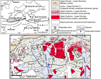
The mineral was found at the Karasu–Karavshinskoye Sn deposit Batken region, Kyrgyzstan located in the headwaters of Karasu River, Turkestan Range (Fig. 1). It has long been known for its pegmatite veins (Weber, Reference Weber1913). However, a systematic study of the geology and mineralogy of pegmatites began only after the famous Tajik–Pamir expedition in the first half of the 1930s (Gavrusevich, Reference Gavrusevitch1932; Ionin, Reference Ionin1934; Arapov, Reference Arapov1935) and resulted in the discovery of several pegmatite fields with rich Sn mineralisation on the northern slope of the Turkestan Range, including Tamyngen, Dzhaupaya, Dukenek, Karasu–Karavshinskoye and Aksu–Karavshinskoye by the team headed by N.V. Ionin (Ionin, Reference Ionin1934). These pegmatites bear beryl and are rich in Fe–Mn–Li-phosphate mineralisation (Kogan et al., Reference Kogan, Beus, Deberin, Paltova and Eventov1950; Beus, Reference Beus1951; Ginzburg, Reference Ginzburg1952). They are related to the Karavshinsky intrusive complex of the late Carboniferous to early Permian age, which includes the Karavshinsky (25 km2) two-phase granitic massif (Fig. 1). Pegmatites are localised both within the massif and in the host Lower Silurian andalusite schists (Kogan et al., Reference Kogan, Beus, Deberin, Paltova and Eventov1950). Some of them bear cassiterite mineralisation and are enclosed within the boundaries of the Karasu–Karavshinskoye deposit. There are several types of pegmatite veins, namely biotite–microcline, muscovite–microcline, muscovite–albite and spodumene–albite. The latter includes the pegmatite vein of #2 area, located on the right side of the Asan–Usan glacial moraine, where akopovaite was found (Fig. 1b). The vein is ~110 m long and ~12 m wide, located relatively far from the contacts of the intrusion (2.5 km to the south), and associated with quartz-micaceous schists. The abundant minerals in the pegmatite are quartz, albite, microcline–‘perthite’, muscovite and spodumene. Less common are beryl, schorl, columbite-group minerals, cassiterite, various phosphates (triphylite, griphite, amblygonite–montebrasite series members, etc.) and ‘lepidolite’. Rare minerals include microlite-group members and secondary uranyl phases, such as autunite and torbernite (Kogan et al., Reference Kogan, Beus, Deberin, Paltova and Eventov1950). Andalusite, tourmaline, quartz and muscovite are abundant in the near-contact zone of the pegmatite with schists. Pegmatite veins with akopovaite can be subdivided into four zones (Kogan et al., Reference Kogan, Beus, Deberin, Paltova and Eventov1950): (1) quartz–feldspar zone; (2) quartz–muscovite zone; (3) albite zone; and (4) the central zone containing large spodumene crystals (up to 40 cm long). The samples containing akopovaite were only found in dumps of vein material, therefore we can only suppose that they originate from the albite zone or the quartz-spodumene zone.
Geographical location (upper left) of the Karasu-Karavshinskoye deposit (the arrow points to the enlarged area, shown below) (1 – Kazakhstan, 2 – Tajikistan, 3 – Uzbekistan, 4 – China) and geological map (lower) of the Karavshinskiy batholite; polygons mark boundaries of Dukenek (1) and Karasu-Karavshinskoye deposits (2) and Aksu-Karavshinskoye occurrence (3) (drawn after A. V. Berezanskiy).

Appearance, physical and optical properties
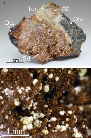
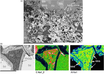
Pegmatite samples with akopovaite consist of light, pale yellow granular albite and translucent white quartz, sometimes dark because of fine mica inclusions (albite and quartz compose 90 vol.% of the rock). Muscovite plates (1.5–2 cm across) and black to dark blue and green schorl are subordinate minerals (Fig. 2). Macroscopically, pegmatite samples with akopovaite look multi-coloured due to the presence of films of Mn and Fe oxides (hydroxides) of brown and light-brown colours in cracks and between albite and quartz grains. Akopovaite aggregates occur in areas (1–4 cm across) of alteration and (or) leaching of primary pegmatite phosphates. These areas comprise fir-like cavities morphologically similar to skeletal crystals and aggregates of unaltered phosphates abundant in other parts of the deposit. These cavities are encrusted with brown crystals of Mn-rich siderite [(Fe0.80Mn0.20)(CO3) to (Fe0.53Mn0.47)(CO3)] sprinkled by akopovaite spherulitic aggregates (Fig. 2). The interstices between siderite crystals also contain akopovaite aggregates and are commonly contoured by a rim consisting of Fe and Mn oxides (hydroxides), unidentified Fe–Al phosphates and montebrasite relicts (Fig. 3). Occasionally akopovaite fills box-like pseudomorphs, which have a morphology outwardly similar to those of triphylite crystals (Fig. 3) (Beus, Reference Beus1951).
(a) General view of the pegmatite sample with akopovaite, containing albite (Ab) and quartz (Qtz); brown colour is due to the presence of thin films of Fe–Mn hydroxides. Tur – schorl. Sample size is 5.5 × 4.5 cm. (b) Akopovaite forms white and yellowish spherulites on siderite (Sd).

(a) Back-scatter electron (BSE) image of leached Fe–Mn phosphate areas in muscovite (Ms) – albite (Ab) aggregate, filled with Mn-rich siderite (Sd) and akopovaite (Akp), surrounded with montebrasite (Mnb). Zones with akopovaite are contoured with a complex polyphase rim (R) consisting of Fe–Mn hydroxides, Fe–Al phosphates and montebrasite relicts. (b) BSE image and X-ray elemental maps of the interstice between siderite crystals (Sd), filled with akopovaite (Akp), and contoured by a polycomponent rim, with various Fe–Al-phosphates (Phosp), montebrasite (Mnb) and Fe–Mn-oxides and/or hydroxides (Fe-Mn-ox). Note: black (in BSE) and red (for CKα) areas correspond to the epoxy resin. Scale bar (a) 600 μm and (b) 40 μm.

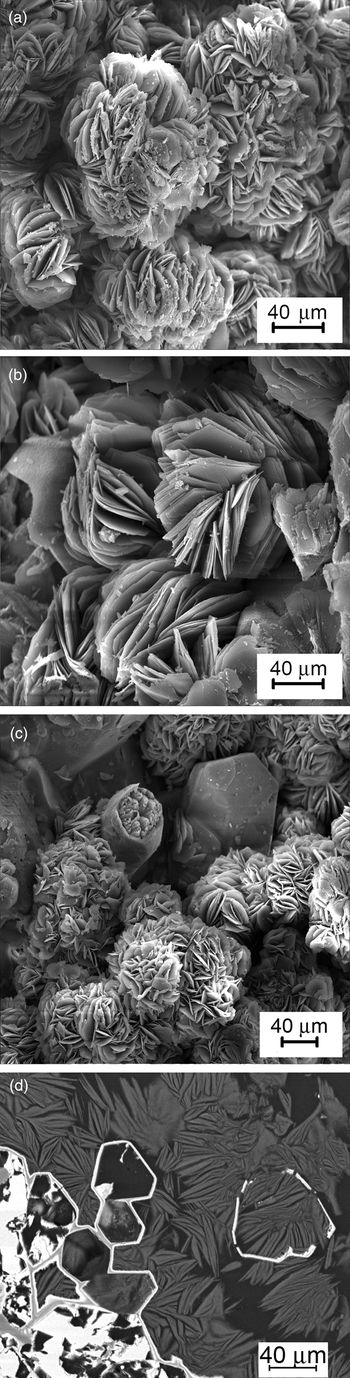
Rosette-like aggregates of akopovaite are composed of pseudohexagonal thin curved plates up to 15–30 μm wide and several μm thick (Fig. 4). Both small size and poor quality of the plates precluded single-crystal X-ray diffraction (SCXRD) investigations and optical methods. The considerable amount of gibbsite and X-ray amorphous Al(OH)3 admixtures also complicated the characterisation of the mineral.
Akopovaite rosettes: (a,b) aggregates of lamellar pseudohexagonal crystals forming rosettes; (c,d) hollow crystals (triphylite pseudomorph?), filled by akopovaite aggregates. (a–c) Scanning electron microscopy image; and (d) BSE image.

Akopovaite is white or yellowish with a silky lustre. The cleavage is perfect along {001}, which is characteristic for all hydrotalcite-supergroup minerals. Micro-indentation measurements, performed on a PMT-3 with a load of 0.5 g, are close both in basal and cross-sections and give a mean value VHN = 24 kg/mm2 (range 20–31) which corresponds to a Mohs hardness of ~1. The density of akopovaite aggregates measured in Clerici solution is 2.12(2) g/cm3. The value obtained is in a good agreement with the calculated density (2.14 g/cm3) taking into account the presence of the Al(OH)3 admixture estimated as ~10%.
Photoluminescence studies were undertaken on a 50 μm diameter section of akopovaite using a LOMO MSFU-312 microspectrophotometer (with spectral resolution ≥ 2 nm); N2-laser with λ = 337.1 nm (3.67 eV) LGI-505 (PLASMA JSC). The study of the luminescent properties of akopovaite was carried out according to the method developed by Rassulov (Reference Rassulov2005). Akopovaite is luminescent with a weak bluish–greenish colour after excitation with a mercury-quartz lamp with both shortwave (254 nm) and mediumwave (365 nm) filters. After excitation with a nitrogen laser (λex = 337.1 nm) it luminesces green. The latter gives the most intensive bands (495, 516 and 538 nm) due to the admixtures containing [UO2]2+ cations (Tarashchan, Reference Tarashchan1978) in amounts below the limits of the microprobe analysis. The [UO2]2+ cations can be absorbed by amorphous Al(OH)3 or by akopovaite, although the latter is less likely.
An optical study was carried out on thin sections; separate plates were studied in immersion liquids in transmitted light using Leitz Ortholux and Polam L-211 polarising microscopes. Akopovaite is optically anisotropic. Under the microscope akopovaite plates show low interference colours in crossed polars, with an undulatory mosaic extinction because of deformations, which prevented determination of whether the mineral is uniaxial or biaxial. Therefore, it was only possible to measure n´ = 1.535(3) (at 589 nm) in a cleavage plane.
The mineral is easily soluble, both in concentrated and diluted (up to 0.5 vol.%) HCl and HNO3 followed by the release of CO2. The dissolution of akopovaite aggregates in weak acids leads to the formation of fine-grained precipitate corresponding to gibbsite (identified by powder XRD).
Chemical composition
Electron-microprobe analysis (for Al and Fe) was done using a Jeol JXA-50A microprobe, equipped with an Oxford instruments energy-dispersive spectrometer (voltage = 20 kV, current = 2 nA and beam diameter = 5 μm, 4 points) and a Jeol Superprobe 733, equipped with a Si(Li)-detector with an ATW2 ultra-thin window and INCA control system (voltage = 20 kV, current = 1 nA with a rastered beam, 4 points). Aluminium and Fe were determined by electron microprobe (Table 2). The following standards were used: albite, microcline, corundum (AlKα) and hematite (FeKα). Contents of other elements with Z > 8 are below their detection limits. The mineral was analysed in polished and thin-polished sections of pegmatite samples, processed with a diamond suspension using the Texmet 1000 cloth of Buehler Ltd., USA.
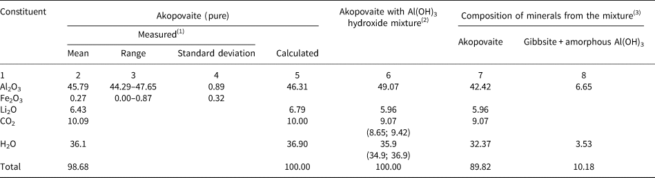
Compositional data (wt.%) for akopovaite and its mixture with gibbsite and amorphous Al(OH)3.

(1) Al2O3, Fe2O3 – microprobe (8 points), Li2O – from the ratio method (see text); CO2 and H2O – normalised data, derived from the columns (6–8). Formula calculated on the sum of cations = 6 atoms per formula unit is Li1.94(Al4.05Fe0.02)Σ4.07(OH)12(CO3)1.03(H2O)3.03.
(2) Al and Li determined from the Al/Li ratio, measured by ICP-OES (see text); CO2 and H2O measured from two weights by CHN-elemental analysis method (in brackets there are results of separate analyses).
(3) Values calculated from columns 4 and 6.
Initial qualitative determination of Li was done using an Opton FMD-4 atomic-absorbtion spectrometer (AAS). Later, inductively coupled plasma optical emission spectrometry (ICP-OES) (Varian ICP-OES 720-ES spectrometer) was used for direct Li determination using the Al:Li ratio. The following chemicals were used: HNO3; H2O with resistivity = 18.2 MΩ/cm and multi-element standard solutions produced by Merck (Germany).
The contents of carbon and hydrogen in akopovaite were measured using Perkin Elmer 2400 Series II and Carlo-Erba 1106 analysers equipped with a thermal conductivity detector and CHN analysis with chromatographic completion. The following standards were used: benzoic acid, acetanilide and hydrotalcite from Snarum, Norway.
The CHN analysis shows the absence of N, and ICP-OES the absence of Be and B in akopovaite.
Certain parts of the mineral aggregates (without weighing) were placed into polypropylene tubes and dissolved in 15 ml of 1% HNO3 solution. The solution was left for 30 min at room temperature and filtered with a 0.45 μm press filter (Varian) to remove undissolved particles, which may consist not only of gibbsite, but also of an amorphous alumina admixture, described previously for synthetic LiAl2-LDHs (Isupov et al., Reference Isupov, Chupakina, Tarasov and Shestakova2007). Afterwards HNO3 acid was added to the solution in order to increase its concentration to 4% (in both blank and reference solutions).
The Al:Li ratio, measured by ICP-OES is 8.11 (average of five measurements), and has been used for calculation of the Li2O content in akopovaite (Table 2). A test on the dissolution of fine-grained (<10 μm) pure gibbsite under the same conditions (temperature, time and HNO3 concentration) showed that <1 wt.% of it dissolves in 1% HNO3, i.e. the majority of the Al in the solution belongs to akopovaite. Sample # 92365 (gibbsite from Amarkantak, India from the systematic collection of the Fersman Mineralogical Museum; checked by powder XRD) was used for the test described above.
The direct determination of CO2 and H2O was carried out using two probes of 0.670 and 0.495 mg, by CHN analysis. The two probes were purified initially: siderite particles were removed using a Sochnev magnet. As the fraction contains admixtures of gibbsite and possible amorphous Al(OH)3, the obtained contents of CO2 and H2O correspond to that of the total proportion of both components in a mixture. In order to normalise the composition for 100% of akopovaite, the amount of total Al(OH)3 (gibbsite and possible amorphous phases) in the mixture was measured. Similar to the procedure described above, tiny parts of mineral aggregates were dissolved in HNO3 (1:1) until complete decomposition (T = 105°C, t ≈ 30 min). Deionised water (18 МΩ/сm) was added in order to reach 4% HNO3 concentration after which the Al/Li ratio was measured in the solutions using ICP-OES. Results of three measurements provide a ratio of Al/Li = 9.36, which was used to obtain an Al(OH)3 content of 10.2 wt.% (Table 2). It should be noted, that normalised values of CO2 and H2O are in a good agreement with calculated ones and correspond to the stoichiometry of the hydrotalcite-supergroup minerals (Mills et al., Reference Mills, Christy and Schmitt2016; Zhitova et al., Reference Zhitova, Krivovichev, Yakovenchuk, Ivanyuk, Pakhomovsky and Mikhailova2018, Reference Zhitova, Krivovichev, Pekov and Greenwell2019b). The empirical formula calculated on the basis of Li+Al+Fe = 6 apfu is Li1.94(Al4.05Fe0.02)Σ4.07(OH)12(CO3)1.03(H2O)3.03. The simplified formula is Li2Al4(OH)12(CO3)(H2O)3.
Raman spectroscopy
The Raman spectrum of akopovaite was obtained with a Horiba Jobin-Yvon LabRam HR800 spectrometer, equipped with an Ar+ laser (λ = 514 nm) at 50 mW output power and ~6 mW power at the sample surface for an area of 2 μm × 2 μm. The spectrum was recorded at room temperature from randomly oriented akopovaite rosette-like aggregates placed on a glass slide and was further processed using LabSpec (Horiba) and Origin (Originlab corp) software.
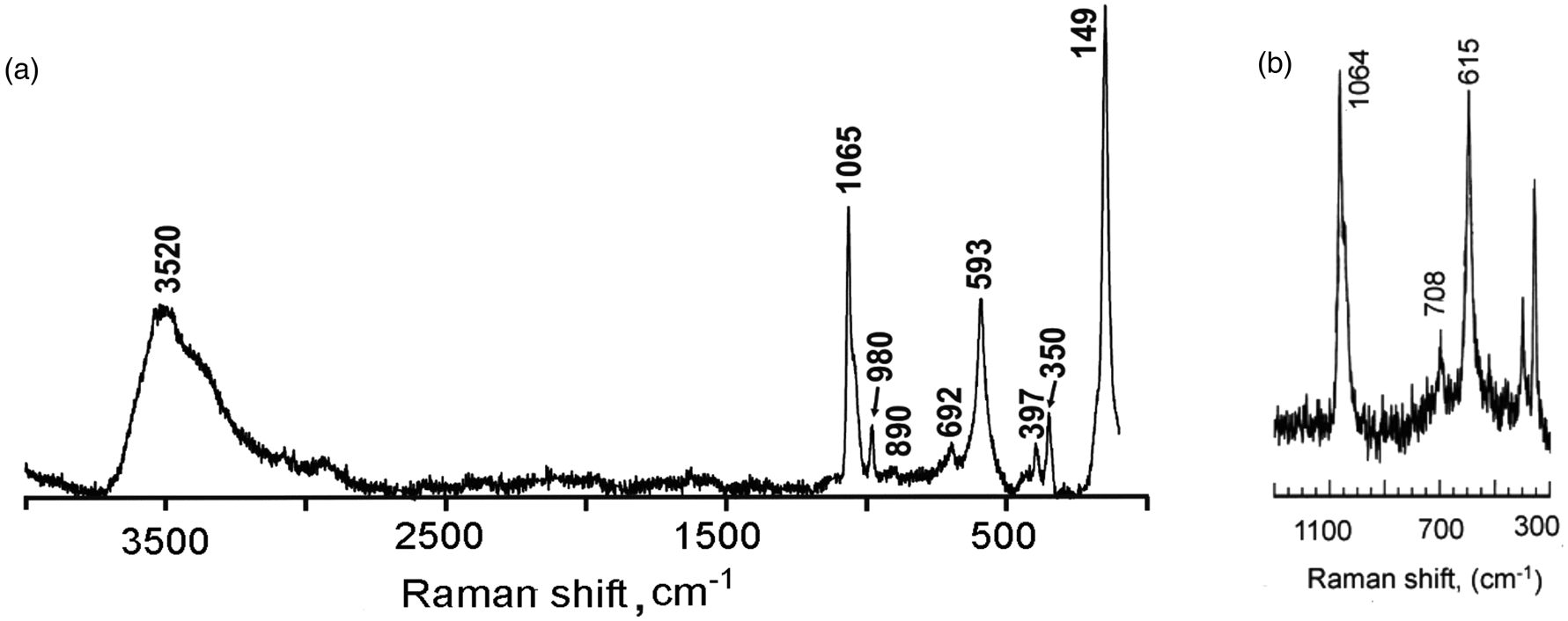
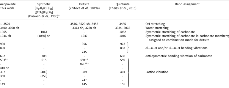
The Raman spectrum of akopovaite is shown in Fig. 5. The band assignment is provided in Table 3 and compared with its synthetic analogue (Drewien et al., Reference Drewien, Eatough, Tallant, Hills and Buchheit1996), dritsite (Zhitova et al., Reference Zhitova, Pekov, Chaikovskiy, Chirkova, Yapaskurt, Bychkova, Belakovskiy, Chukanov, Zubkova, Krivovichev and Bocharov2019a) and quintinite (Theiss et al., Reference Theiss, López, Frost and Scholz2015). The Raman spectrum reveals the presence of carbonate groups and different OH vibrations that correspond to hydroxyl and interlayer water (Table 3).
The Raman spectra of (a) akopovaite and (b) synthetic Al-Li LDH (Drewein et al., Reference Drewien, Eatough, Tallant, Hills and Buchheit1996).

Raman bands (in wavenumber, cm–1) of akopovaite compared to its synthetic analogue, dritsite and quintinite.

*Only bands in the range 1300–300 cm–1 were given. The values in the brackets were not described in the text, but estimated from the band position in the given spectra. Sh – shoulder. **Al⋅⋅⋅O stretching, ***Li⋅⋅⋅O stretching.
Powder X-ray diffraction
Preliminary X-ray identification of the mineral was performed with powder XRD data (Debye-Scherrer method using an RKD 57.3 mm camera, FeKα radiation and Mn filter) and shows it's similarity with synthetic LiAl2-LDH (Devyatkina et al., Reference Devyatkina, Kotsupalo, Tomilov and Berger1983; Drewien et al., Reference Drewien, Eatough, Tallant, Hills and Buchheit1996; Serna et al., Reference Serna, Rendon and Iglesias1982). Powder XRD data for the full-profile analysis were collected by means of a Rigaku R-Axis Rapid II single-crystal diffractometer using Debye-Scherrer geometry (d = 127.4 mm). The diffractometer is equipped with a rotating anode (CoKα, λ = 1.79026, voltage = 40 kV and current = 15 mA), microfocus optics and a cylindrical image plate detector. Five different rosette-like aggregates were investigated and gave almost identical powder patterns. The data were integrated using the software package Osc2Tab/SQRay (Britvin et al., Reference Britvin, Dolivo-Dobrovolsky and Krzhizhanovskaya2017). Topas 4.2 software (Bruker AXS, 2009) was used for the refinement of the unit-cell parameters and crystal structure by the Rietveld method (Table 4). Previously reported Rietveld refinements on synthetic LiAl2-LDHs indicated that Li–Al ordering combined with a rhombohedral stacking sequence of layers results in a reduction of the symmetry from trigonal to monoclinic (Thiel et al., Reference Thiel, Chiang and Poeppelmeier1993; Britto et al., Reference Britto, Thomas, Kamath and Kannan2008; Britto and Kamath, Reference Britto and Kamath2009 and Reference Britto and Kamath2011). These results were later confirmed by the SCXRD study and structure refinement of quintinite-1M, Mg4Al2(OH)12(CO)3(H2O)3 (Krivovichev et al., Reference Krivovichev, Yakovenchuk, Zhitova, Zolotarev, Pakhomovsky and Ivanyuk2010b). In the latter, it was suggested that quintinite-1M is isotypic with the monoclinic polytype of synthetic Li2Al4(OH)12(CO)3(H2O)3 (Krivovichev et al., Reference Krivovichev, Yakovenchuk, Zhitova, Zolotarev, Pakhomovsky and Ivanyuk2010b). It is worth noting that Li–Al ordering has been detected in the first (and to date only) single-crystal refinement of LiAl2-LDHs done on dritsite-2H (Zhitova et al., Reference Zhitova, Pekov, Chaikovskiy, Chirkova, Yapaskurt, Bychkova, Belakovskiy, Chukanov, Zubkova, Krivovichev and Bocharov2019a). Therefore the Rietveld refinement was performed starting from atom coordinates of quintinite-1M (monoclinic, C2/m, a = 5.266(2), b = 9.114(2), c = 7.766(3) Å, β = 103.17(3)° and V = 362.9(2) Å3) in which the Mg site (in quintinite) was assigned to Al (in akopovaite), whereas the Al site (in quintinite) was assigned to Li (in akopovaite).
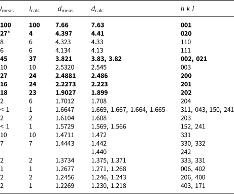
Powder X-ray diffraction data (d in Å) for akopovaite.

* Overlap with gibbsite (see Fig. 6)
The strongest lines are given in bold.
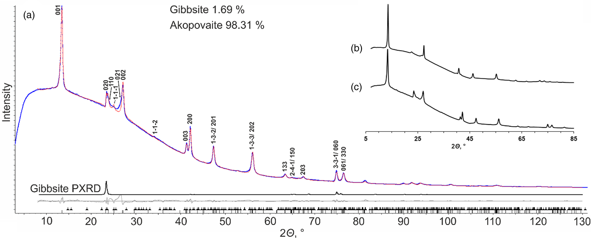
(a) Rietveld fit (red line) of the observed powder X-ray diffraction (blue line) profile of akopovaite. (b) Powder X-ray diffraction pattern obtained for quintinite-1M from Rudnogorskoe Fe-deposit (Siberia, Russia) and (c) akopovaite (CoKα).

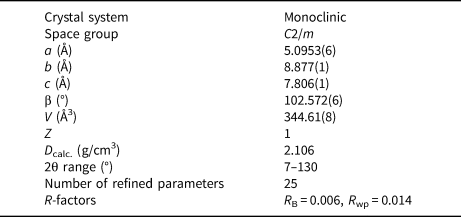
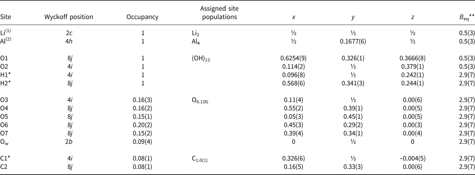
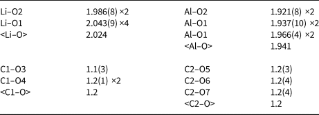
The indexed powder XRD pattern, obtained using a Gandolfi-like motion, is provided in Table 4. The Rietveld refinement confirmed that the mineral is monoclinic, space group C2/m, a = 5.0953(6), b = 8.877(1), c = 7.806(1) Å, β = 102.572(6)°, V = 344.61(8) Å3 and Z = 1 (Table 5). The final refinement consisted of 25 parameters (coordinates and isotropic displacement parameters) and final convergence was achieved at R B = 0.006 and R wp = 0.014 (Table 5). Crystal data and parameters of the crystal-structure refinement are given in Table 5. The atomic coordinates, site occupancies and isotropic displacement parameters are listed in Table 6 and interatomic distances are provided in Table 7.
Crystallographic data and refinement parameters for akopovaite.

Atomic coordinates, site occupancies and isotropic displacement parameters for akopovaite.

*Coordinates were fixed during the refinement.
(1) Corresponds to the Al site in quintinite; (2) corresponds to the Mg site in quintinite.
**B = 8π2U 2, where U 2 is the mean-square amplitude of the vibration.
Selected interatomic distances in the crystal structure of akopovaite.

The difficulty in the structure refinement of akopovaite is in the presence of light elements and statistic disorder of interlayer C and O atoms (also characteristic for single-crystal structure refinements of other hydrotalcite-supergroup minerals) (Krivovichev et al., Reference Krivovichev, Yakovenchuk, Zhitova, Zolotarev, Pakhomovsky and Ivanyuk2010a,Reference Krivovichev, Yakovenchuk, Zhitova, Zolotarev, Pakhomovsky and Ivanyukb; Zhitova et al., Reference Zhitova, Yakovenchuk, Krivovichev, Zolotarev, Pakhomovsky and Ivanyuk2010, Reference Zhitova, Popov, Krivovichev, Zaitsev and Vlasenko2017a,Reference Zhitova, Ivanyuk, Krivovichev, Yakovenchuk, Pakhomovsky and Mikhailovab, Reference Zhitova, Krivovichev, Yakovenchuk, Ivanyuk, Pakhomovsky and Mikhailova2018, Reference Zhitova, Krivovichev, Pekov and Greenwell2019b). Therefore, atom coordinates were refined with soft restraints applied for the interlayer atoms. The atomic coordinates were fixed for H1, H2 and C1. Two isotropic-displacement parameters were refined for: (1) Li, Mg, O1 and O2 atoms of the metal–hydroxide layer; and (2) interlayer C and O atoms and hydrogen H1 and H2 atoms. The occupancy of atoms was fixed in accordance with the chemical formula determined by the number of different methods. Neutral scattering factors were used for all atoms. The background was modelled using a Chebyshev polynomial approximation. The peak profile was described using the fundamental parameters approach. Refinement of preferred orientation parameters confirmed the presence of a significant preferred orientation along the [001] direction. The gibbsite model (Balan et al., Reference Balan, Lazzeri, Morin and Mauri2006) was added to the refinement because of the intimate intergrowth of akopovaite with gibbsite as indicated by the comparison of the calculated and observed intensity of the reflection at 4.397 Å (Fig. 6). The Rietveld refinement showed good agreement between experimental and calculated diffraction patterns (Fig. 6). Less smooth fitting is observed in the 2θ region 23–27°, due to stacking faults causing non-uniform broadening of reflections, as previously shown for the synthetic analogue of akopovaite by Britto and Kamath (Reference Britto and Kamath2011).
The crystallographic information files have been deposited with the Principal Editor of Mineralogical Magazine and are available as Supplementary material (see below).
Discussion
Crystal structure
The synthetic LiAl2 members of the LDH family have been well characterised structurally (Table 1). The position of Li+ cations is dictated by the localisation of voids in gibbsite layers and therefore obey $ \sqrt 3 $ × $ \sqrt 3 $
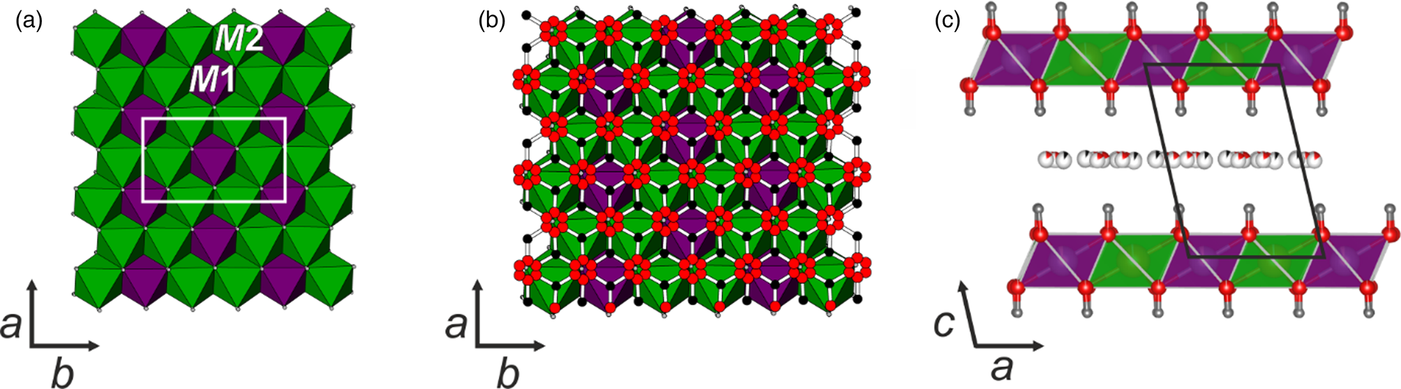
 superstructure within the xy plane. Both strict ordering of Li and Al cations and the significant difference in their scattering power result in the presence of an intensive additional (110) reflection at ~4.40 Å in the powder XRD pattern as observed for synthetic LiAl2-members (Britto et al., Reference Britto, Thomas, Kamath and Kannan2008 and Britto and Kamath, Reference Britto and Kamath2009 and Reference Britto and Kamath2011, Sissoko et al., Reference Sissoko, Iyagba, Sahai and Biloen1985) and recently in dritsite-2H (Zhitova et al., Reference Zhitova, Pekov, Chaikovskiy, Chirkova, Yapaskurt, Bychkova, Belakovskiy, Chukanov, Zubkova, Krivovichev and Bocharov2019a). The crystal structure of akopovaite consists of alternating gibbsite-like layers where Li fills octahedral voids in gibbsite layers, and interlayers represented by carbonate groups and water molecules (Fig. 7).
Projections of the crystal structure of akopovaite (M1 = Li and M2 = Al, oxygen – red, carbon – black, hydrogen – grey): (a) metal–hydroxide layer {[Al4(OH)12]Li2}2+, (b) metal–hydroxide layer and interlayer and (c) stacking of metal–hydroxide layers and interlayers along the c axis.

The interlayer is represented by disordered carbonate groups and water molecules, no peculiarities can be deduced from the Rietveld refinement, but we believe that all carbonate hydrotalcite-supergroup members possess a similar interlayer arrangement to that found for quintinite (Krivovichev et al., Reference Krivovichev, Yakovenchuk, Zhitova, Zolotarev, Pakhomovsky and Ivanyuk2010a,Reference Krivovichev, Yakovenchuk, Zhitova, Zolotarev, Pakhomovsky and Ivanyukb; Zhitova et al., Reference Zhitova, Yakovenchuk, Krivovichev, Zolotarev, Pakhomovsky and Ivanyuk2010, Reference Zhitova, Popov, Krivovichev, Zaitsev and Vlasenko2017a, Reference Zhitova, Krivovichev, Yakovenchuk, Ivanyuk, Pakhomovsky and Mikhailova2018); zaccagnaite (Lozano et al., Reference Lozano, Rossi, La Iglesia and Matesanz2012); takovite (Mills et al., Reference Mills, Whitfield, Kampf, Wilson, Dipple, Raudsepp and Favreau2012b); pyroaurite (Allmann, Reference Allmann1968; Ingram and Taylor, Reference Ingram and Taylor1967; Zhitova et al., Reference Zhitova, Ivanyuk, Krivovichev, Yakovenchuk, Pakhomovsky and Mikhailova2017b); and hydrotalcite (Mills et al., 2016; Zhitova et al., Reference Zhitova, Krivovichev, Pekov and Greenwell2019b). In the interlayer, low-occupied O sites form toroidal rings, while low-occupied C sites are located between three such rings forming triangles of CO3 groups (Fig. 7). It is worth noting that the structural model indicates that the refined occupancies of the two independent carbonate anions are similar to each other and also to that of the water molecule (Table 6). The carbonate groups and water contents are in a good agreement with the empirical formula (Table 6).
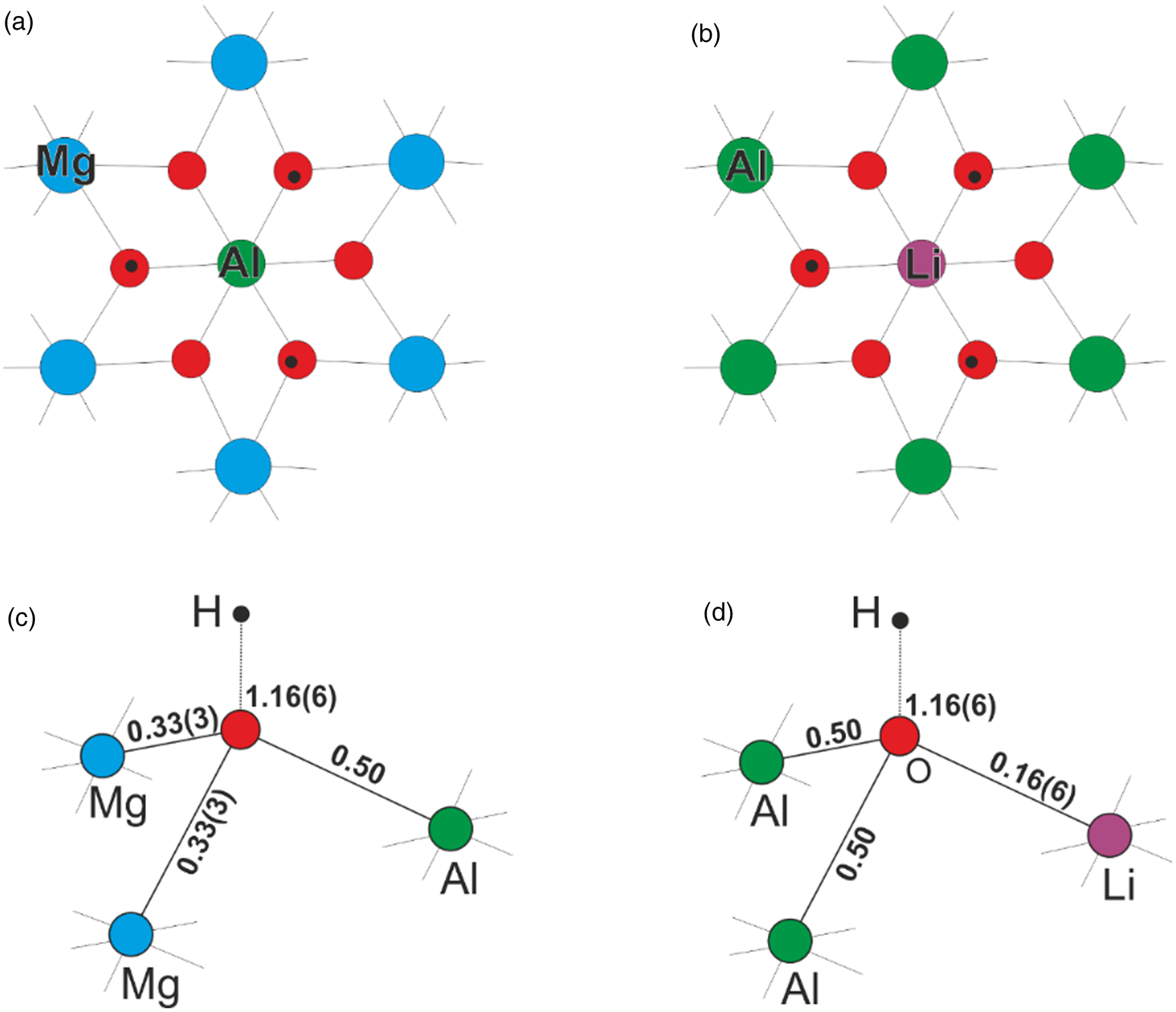
Despite heterovalent substitution of cations in the metal–hydroxide layer when comparing quintinite and akopovaite, i.e. Mg2Al → Al2Li, both minerals have the same (2+) charge of metal–hydroxide layer per 6 cations. In akopovaite (as well as in stoichiometric quintinite), all oxygen atoms in the metal–hydroxide layer are identical in terms of bond valences as each oxygen is coordinated by LiAl2 (or Mg2Al in quintinite) and gains 1.16(6) valence units (Fig. 8). The same is observed for recently described dritsite (Zhitova et al., Reference Zhitova, Pekov, Chaikovskiy, Chirkova, Yapaskurt, Bychkova, Belakovskiy, Chukanov, Zubkova, Krivovichev and Bocharov2019a) and chlormagaluminite (Zhitova et al., Reference Zhitova, Krivovichev, Pekov and Yapaskurt2019c) that are isotypic taking into account Mg2Al → Al2Li substitution and differ from akopovaite and quintinite by the dominance of Cl– in the interlayer instead of (CO3)2–.
Local arrangement of atoms in the metal–hydroxide layer: (a) quintinite and (b) akopovaite; schemes of bond-valence distribution (in valence units = vu) for O atoms in the metal–hydroxide layer: (c) quintinite and (d) akopovaite.

The gibbsite-type layers are stacked by pseudo-rhombohedral periodicity, i.e. cations in each first and fourth layer are located strictly under each other (Fig. 7), as indicated by the reflections in the 2θ region 40–60° (Evans and Slade, Reference Evans and Slade2006). The reduction of the symmetry from rhombohedral to monoclinic is due to Li‒Al ordering (the same symmetry reduction was reported for quintinite-1M due to Mg‒Al ordering by Krivovichev et al., Reference Krivovichev, Yakovenchuk, Zhitova, Zolotarev, Pakhomovsky and Ivanyuk2010b) indicated by the appearance of superstructure reflections in the 2θ region 20–30°.
As akopovaite is isotypic to quintinite, Mg4Al2(OH)12(CO3)(H2O)3, the structural formula of akopovaite can be written as [Al4Li2(OH)12][(CO3)(H2O)3] where the position of Li in akopovaite corresponds to the Al site in quintinite (Mg2Al → Al2Li). It is important to note that the akopovaite structure is composed of gibbsite-like layers (not brucite-like ones as in quintinite and other hydrotalcite-supergroup members) with Li filling octahedral voids. It is a characteristic structural feature of LiAl2-LDHs that should be reflected by the following structural formula, {[Al4(OH)12]Li2}[(CO3)(H2O)3], that points to the possibility of Li leaching (Sissoko et al., Reference Sissoko, Iyagba, Sahai and Biloen1985).
Relationship with other minerals
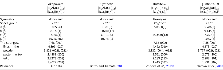
Akopovaite, Li2Al4(OH)12(CO3)(H2O)3, reported in this paper, is a new member of the hydrotalcite supergroup (Mills et al., Reference Mills, Christy, Génin, Kameda and Colombo2012a) and belongs to a large family of LDHs consisting of natural and synthetic species. The mineral is a carbonate analogue of recently reported dritsite, however akopovaite has been found exclusively as the 1M polytype, whereas dritsite has been found as the 2H polytype; akopovaite-1M can be considered as isotypic with quintinite-1M (Table 8). It is worth noting that the literature review shows that both 1M and 2H polytypes of synthetic LiAl2-LDHs were obtained independently via different routes (Britto and Kamath, Reference Britto and Kamath2011) suggesting the possibility of akopovaite-2H also being found in Nature.
Crystallographic data for akopovaite-1M, its synthetic analogue, dritsite-2H and quintinite-1M.

* The strongest lines in the powder pattern are calculated by the INDX program (LMPG suite, Laugier and Bochu, Reference Laugier and Bochu2004) using single-crystal X-ray diffraction data.
The mechanism of akopovaite formation in Nature may be similar to that of its synthetic analogues. We suggest two possible scenarios: (1) the hydrothermal interaction of components presented in akopovaite under alkaline conditions and temperature not higher than 110–140°C (Serna et al., Reference Serna, Rendon and Iglesias1982; Sissoko et al., Reference Sissoko, Iyagba, Sahai and Biloen1985; Isupov et al., Reference Isupov, Chupakina, Tarasov and Shestakova2007; Britto et al., Reference Britto and Kamath2011; Britto, Kamath, Reference Britto and Kamath2011); (2) the precipitation or intercalation of Li+ into the Al(OH)3 matrix (Isupov, Reference Isupov1999; Britto, Kamath, Reference Britto and Kamath2009; Reference Britto and Kamath2011). The possible sources of the chemical components for akopovaite are montebrasite (Li and Al) and siderite (Fe and C).
Acknowledgements
The authors would like to thank Pavel V. Khvorov and Timur K. Berkeliev for help in material collection; Karybek E. Ibraev and Anatoly V. Berezanskiy for help in field work organisation; Nikita V. Chukanov for the discussion of Raman spectra interpretation; Vasily D. Shcherbakov for the collection of preliminary Raman spectra not presented herein; Frank Hawthorne and Elena V. Sokolova for attempts to obtain single-crystal X-ray diffraction data; Pavel M. Kartashov and Igor V. Pekov for providing samples of hydrotalcite from Snarum (Norway) and quintinite from Kovdor (Russia) used for comparison. We thank members of the IMA CNMNC for their comments. The XRD studies and Raman spectroscopy were supported by the Russian Science Foundation (grant 17-77-10023) and carried out using facilities of XRD and Geomodel Resource Centers of St. Petersburg State University. Finally we thank Nikita V. Chukanov, the anonymous reviewer and Structures Editor Peter Leverett for their comments and corrections and Principal Editor Stuart Mills for the manuscript handling.
Supplementary material
To view supplementary material for this article, please visit https://doi.org/10.1180/mgm.2020.10.