

Introduction
Digeneans have complex life cycles that pass through several generations, alternating between free-living and parasitic stages, and almost always include multiple animal hosts. Despite their remarkable diversity, all these life cycles involve asexual parthenogenetic reproduction occurring in the first intermediate host, whose role is typically played by molluscs (Ataev Reference Ataev2017; Cribb et al. Reference Cribb, Bray, Olson, Timothy and Littlewood2003; Esch et al. Reference Esch, Barger and Fellis2002; Galaktionov and Dobrovolskij Reference Galaktionov and Dobrovolskij2003; Poulin and Cribb Reference Poulin and Cribb2002). The miracidium larva, which forms in the egg of the hermaphroditic adult, enters the mollusc using either passive or active mechanisms, undergoes regressive metamorphosis and in the overwhelming majority of digeneans develops into a mother sporocyst. The mother sporocyst reproduces parthenogenetically giving rise to the next generation of parthenitae represented by rediae or daughter sporocysts. These stages produce numerous cercariae, the transmissive larval stage that leaves the mollusc to ensure continuation of the life cycle. In many species, daughter parthenitae can also produce more of their kind to replenish the infrapopulation within the mollusc. The successful colonisation of the first intermediate host and its transformation into a potent source of infection are largely dependent on the successful development of mother sporocysts and their ability to counteract host defences and to overcome additional risks arising in those species in which parthenitae migrate through the host’s body during early development (reviewed in Ataev Reference Ataev2017; Galaktionov and Dobrovolskij Reference Galaktionov and Dobrovolskij2003).
In naturally infected molluscs, mother sporocysts are typically very few in number, and as a result, they rarely become the focus of attention in digenean studies. Most aspects of this important life cycle stage, including fine morphology of these parasites, remain poorly and unevenly studied. Fairly detailed information on the structure and development of mother sporocysts is available only for species of the genus Schistosoma Weinland, 1858 (see, for example, Bahia et al. Reference Bahia, Avelar, Vigorosi, Cioli, Oliveira and Mortara2006; Basch and DiConza Reference Basch and DiConza1974; Coustau et al. Reference Coustau, Ataev, Jourdane and Yoshino1997; Diaz Soria et al. Reference Diaz Soria, Attenborough, Lu, Fontenla, Graham, Hall, Thompson, Andrews, Rawlinson, Berriman and Rinaldi2024; Køie and Frandsen Reference Køie and Frandsen1976; Meuleman et al. Reference Meuleman, Holzmann and Peet1980; Meuleman et al. Reference Meuleman, Lyaruu, Khan, Holzmann and Sminia1978; Pan Reference Pan1996; Poteaux et al. Reference Poteaux, Gourbal and Duval2023), whose adult forms are major human parasites. These studies rely on the material obtained through experimental infection of molluscs or on cultures grown in vitro in specially designed media and employ a variety of methods including light, electron, and confocal microscopy, immunocytochemistry, and modern techniques of molecular genetic analysis.
The morpho-biological characteristics of mother sporocysts in many other digenean species remain largely unknown or inadequately described. While the germinal material of mother sporocysts has been examined in a few species at the light-microscopic level (Ataev Reference Ataev2017; Ataev et al. 1997; Cort et al. Reference Cort, Ameel and Van der Woude1954; Dobrovolskij et al. 2000; Galaktionov and Dobrovolskij Reference Galaktionov and Dobrovolskij2003), their somatic structures including those involved in reproduction have so far received very little attention. Outside of schistosomes, ultrastructural studies of mother sporocysts are rare and typically focus only on the tegument (Køie et al. Reference Køie, Christensen and Nansen1976; Matthews and Matthews Reference Matthews and Matthews1991; Pinheiro et al. Reference Pinheiro, Franco-Acuña, Oliveira-Menezes, Brandolini, de Souza and DaMatta2011; Southgate Reference Southgate1970; Threadgold Reference Threadgold, Bereiter-Hahn, Matoltsy and Richards1984). As with most digeneans, our knowledge of the first-generation parthenitae in hemiuroid digeneans is very limited, and therefore, the aim of this study was to examine one of the representatives of this superfamily, providing the first detailed description of the microanatomy and ultrastructure of mature mother sporocysts in Bunocotyle progenetica (Markowski, 1936) Chabaud et Buttner, 1959, a monoxenous parasite of the marine mud snails Peringia ulvae (Pennant, 1777) and Ecrobia ventrosa (Montagu, 1803). Following on our previous work on the highly unusual tegument of the mother sporocysts in this species (Podvyaznaya and Filimonova Reference Podvyaznaya and Filimonova2024), the present study uses transmission electron microscopy and confocal microscopy to examine the fine organisation of their muscle and nervous systems, as well as the structures related to reproduction.
Material and methods
Mature sporocysts of B. progenetica containing embryonic rediae were obtained from the mud snails Peringia ulvae collected during cold and warm seasons in the Sukhaya Salma Inlet of the Chupa Bay, White Sea. For transmission electron microscopy, the material was fixed in cold 3% glutaraldehyde in 0.1 M cacodylate buffer (pH = 7.4), followed after 8–10 days by postfixation in 1% osmium tetroxide in the same buffer at 4°C for 2 hours. Sucrose was added to the fixatives and rinsing buffer to adjust the osmolarity to 760 mOsm. After the final rinse in buffer, sporocysts were dehydrated in an ascending series of ethanol and acetone and embedded in an Epon-Araldite mixture following the standard protocol (Mollenhauer Reference Mollenhauer1964). Serial thin sections of sporocysts were cut on a Leica EM UC6rt ultramicrotome (Leica Microsystems GmbH, Wetzlar, Germany), stained with an aqueous solution of uranyl acetate and lead citrate according to Reynolds, and examined using a Morgagni 268 transmission electron microscope (FEI, Eindhoven, Netherlands) at the ‘Taxon’ Research Resource Centre (Zoological Institute of the Russian Academy of Sciences, St. Petersburg, Russia). Live sporocysts were studied at the light-microscopic level using a Leica DMLS microscope (Leica Microsystems GmbH, Wetzlar, Germany) and an Olympus CH-40 microscope (Olympus, Tokyo, Japan) equipped with an Olympus XC-30 digital camera.
For confocal microscopy, sporocysts were fixed in 4% paraformaldehyde in 0.01 M phosphate-buffered saline (PBS) for 6 h at ambient temperature and then transferred to 0.01 M PBS containing 0.05% sodium azide (Sigma-Aldrich, St. Louis, Missouri, USA) for storage at 4–6°C. All subsequent steps were performed on an orbital shaker at room temperature. The worms were permeabilised for 2–4 h in 0.01M PBS containing 0.25% Triton X-100 (Tr-PBS; Sigma-Aldrich, St. Louis, Missouri, USA) and blocked with Image-iT FX Signal Enhancer (Molecular Probes, Eugene, Oregon, USA) for 1 h. Blocked sporocysts were rinsed 4 × 15 min in Tr-PBS and incubated for 12–24 h in antibodies against serotonin (5HT) (Sigma-Aldrich, St. Louis, Missouri, USA; diluted 1:600) or against FMRFamide (Abcam, Cambridge, Massachusetts, USA; diluted 1:600) to stain for FMRFamide-related peptides (FaRPs). After incubation with primary antibodies, the animals were rinsed again 4 × 15 min in Tr-PBS and incubated for 5 h with Alexa Fluor 488-conjugated secondary antibodies (Life Technologies, Carlsbad, California, USA; diluted 1:300). After that, the sporocysts were rinsed for 15 min in Tr-PBS and stained for 2–3 h with TRITC-conjugated phalloidin (Sigma-Aldrich, St. Louis, Missouri, USA), then rinsed once in 0.01M PBS and mounted on slides with Vectashield containing DAPI (Vector Laboratories, Burlingame, California, USA). Staining specificity was verified by omitting primary antibodies.
Confocal images were acquired on a Leica TCS SP5 confocal laser scanning microscope (Leica Microsystems GmbH, Wetzlar, Germany) at the ‘Taxon’ Research Resource Centre (Zoological Institute of the Russian Academy of Sciences, St. Petersburg, Russia). All confocal images presented in this study are shown as maximum intensity projections of Z-stacks.
Results
General morphology
Mature mother sporocysts of B. progenetica reside in the lacunar system of the host; in naturally infected molluscs, they occur in very low numbers (usually 1–2 individuals per host). The elongated, vermiform sporocysts (1000–1700 μm x 150–250 μm) are quite mobile; they are capable of contraction, significant elongation, and can flex in various directions. Most of the sporocyst body is occupied by a well-developed brood cavity, which has its own lining and is surrounded by a thin body wall (Figure 1). The body wall consists of an outer anuclear layer of the tegument, an underlying basal lamina, subtegumental muscles, and 2–3 layers of various cells, including tegumental and parenchymal cells, as well as cellular elements of the muscle and nervous systems (Figures 7a–d, 8a, 9a, d, e) and sparsely distributed undifferentiated cells. A small cone-shaped region at the anterior end of the sporocyst is filled with densely packed cells (Figure 1). The composition of these cells does not differ from that in the other parts of the body, but there is a higher proportion of muscle tissue. This cellular mass is traversed by a birth canal that opens to the exterior via a terminal birth pore (Figures 1, 4a, b). The brood cavity contains simultaneously various embryonic stages of rediae, from small germinal balls to fully formed rediae (Figure 1), with observed counts varying from approximately one to 15 embryos.
General view of a live mother sporocyst of Bunocotyle progenetica. Scale bar: 200 μm. Abbreviations: bc, brood cavity; bp, birth pore; em, early embryo; re, embryonic redia; sbw, sporocyst body wall.

Brood cavity and birth canal
The structures involved in the reproduction of B. progenetica mother sporocysts include a brood cavity, its lining, germinal cells, a birth canal, and a birth pore (Figures 2–4). These parthenitae lack a germinal mass.
Ultrastructure of the brood cavity lining in mother sporocysts of Bunocotyle progenetica. (a) Perikaryon of the syncytial lining of the brood cavity. (b) Details of the syncytial lining. Note the gap junction (arrowhead) with a cytoplasmic extension of a parenchymal cell. (c) Squamous cellular epithelium lining the anterior region of the brood cavity. (d) Connection of the syncytial lining with the cellular epithelium in the anterior part of the brood cavity; note the septate junction. Scale bars: a – 2 μm; b – 0.5 μm; c, d – 1 μm. Abbreviations: bc, brood cavity; bl, basal lamina; cel, epithelial cell; hd, hemidesmosome; in, invaginations of plasma membrane; ip, irregular apical projections; la, lamellar projections; mf, myofibril; n, nucleus; nu, nucleolus; pa, parenchymal cells and their extensions; sj, septate junction; syl, syncytial brood cavity lining; tc, tegumental cell.

TEM micrographs showing the germinal material in mother sporocysts of Bunocotyle progenetica. (a, b) Developing germinal cells embedded in the syncytial lining of the brood cavity. (c) Micrograph showing an early embryo (germinal ball) and a group of germinal cells enclosed by the syncytial lining of the brood cavity. Scale bars: a, c – 2 μm; b – 1 μm. Abbreviations: bc, brood cavity; em, embryo; ge, germinal cell; n, nucleus; pa, parenchymal cells; syl, syncytial brood cavity lining.

TEM micrographs showing the birth canal of a mother sporocyst of Bunocotyle progenetica. (a) The distal-most part of the birth canal near the birth pore. (b) Longitudinal oblique section of the birth canal showing its three regions. Approximate boundaries of the distal and middle regions are indicated by white square brackets. (c) Details of the epithelial lining of the middle region of the birth canal. Note the septate junction connecting adjacent cells. (d) Details of the squamous epithelium of the proximal region of the birth canal. The inset shows an enlarged view of the septate junction. Scale bars: a, b – 2 μm; c – 0.5 μm; d – 1 μm. Abbreviations: bc, brood cavity; bin, basal invaginations with expanded tips; bl, basal lamina; bp, birth pore; dbi, distal birth canal region; ip, irregular apical projections; mbi, middle birth canal region; mep, epithelium of middle region of birth canal; mu, muscle; n, nucleus; pbi, proximal birth canal region; pep, epithelium of proximal region of birth canal; sj, septate junction; te, tegument.

The brood cavity, except for a small anterior region, is lined with a thin syncytium having small local thickenings and sparsely distributed perikarya (Figure 2a, b). The large, electron-dense nuclei of the perikarya are irregular in shape and have a prominent nucleolus and a thin layer of condensed chromatin lining the inner side of the nuclear envelope (Figure 2a). The perinuclear cytoplasm has a typical set of cellular organelles and contains a variety of vacuoles and vesicles including residual bodies, often occurring in large numbers. The luminal surface of the syncytium is expanded by unevenly spaced lamellar projections and large folds (Figure 2a, b, d). The plasma membrane facing the body wall is sometimes infolded to form deep, plate-like invaginations (Figure 2b) oriented parallel to the syncytial surface and layered on top of one another giving the syncytial lining in these places a stratified appearance. Gap junctions are frequently observed between the syncytium and parenchymal cells of the sporocyst body wall (Figure 2b).
The anteriormost part of the brood cavity is lined with cellular epithelium (Figure 2c), which is a continuation of the epithelial lining of the birth canal and is connected to the syncytial lining of the cavity by a septate junction (Figure 2d). The epithelium consists of very flat cells with large electron-dense nuclei and long, irregular projections on their luminal surface (Figure 2c). This region of the cavity lining is supported by a distinct basal lamina, which is attached to the underlying muscles by hemidesmosomes.
In the middle and especially in the posterior regions of the brood cavity, the syncytial lining incorporates sparsely distributed germinal elements (Figure 3). These elements include both developing and mature germinal cells, typically lying separately (Figure 3a, b) or, less often, in small clusters of 2–3 cells (Figure 3c). These cells sometimes protrude into the cavity or are embedded in the sporocyst body wall and are completely or partially enveloped by cytoplasmic extensions of the syncytium. The earliest embryos, originating from the mature germinal cells, are retained within the syncytium (Figure 3c). In these areas, the syncytium is noticeably thickened, often has a layered structure, and occasionally forms pocket-like invaginations into the sporocyst body wall (Figure 3c). The embryos are released into the brood cavity when they reach the stage of a young germinal ball and continue to develop there into fully formed embryonic rediae (Figure 1), which then leave the mother sporocyst through the birth canal and pore.
The birth canal is a goblet-like duct, with its narrow, elongated portion directed towards the body’s anterior end. The canal consists of three distinct regions: proximal, middle, and distal, each differing in width, length, and epithelial morphology (Figures 4, 11a). The widest proximal region is cup-shaped and upon muscle contraction may form thick circular folds (Figure 4b, d). The epithelial lining of this region is similar to that in the anterior portion of the brood cavity. At the transition to the middle region, the lumen of the duct narrows abruptly, leaving almost no space between the walls of the canal, and the epithelial lining changes its morphology (Figure 4b). The cells lining the short middle region have electron-dense cytoplasm with numerous folds and narrow invaginations of the outer plasma membrane. The ends of these folds and invaginations often expand, forming structures that resemble electron-lucent vacuoles (Figure 4b, c). The luminal surface of the epithelium in this part of the duct bears small conical knolls and occasional short lamellae that lie closely against the surface of the epithelial cells. The relatively long distal region also has a narrow lumen; it is lined by an anuclear syncytium, which is a continuation of the outer tegument and has the same structure (Figure 4a, b). Throughout the length of the birth canal, its epithelial lining is surrounded by a basal lamina (Figure 4d) and muscles (see their detailed description below).
Muscle system
The musculature of the body wall includes an inner layer of longitudinal muscles (Figure 5a, b) and an outer layer of circular muscles (Figure 5a, b); diagonal muscles are absent. The circular layer consists of thin, evenly and closely spaced muscles. The muscles in the longitudinal layer are less regularly arranged; these ribbon-shaped fibres appear to branch, anastomose, or cross one another (Figure 5b) and are often grouped into irregular bands.
Musculature of mother sporocysts of Bunocotyle progenetica. Confocal images of phalloidin-stained preparations. Anteroposterior axis is labelled as a–p. (a) General view of the body-wall musculature. (b) A portion of the body wall showing the grid-like arrangement of musculature. Note that longitudinal muscles often branch out and cross one another. (c, d) Anterior region of the body showing the musculature of the birth canal in the resting state. (e) Anterior region of the body showing the musculature of the partially protruded birth canal. Scale bars: a – 50 μm; b–e – 20 μm. Abbreviations: abd, anterior birth canal dilator muscles; bp, birth pore; bcp, birth canal protractor muscles; bcr, birth canal retractor muscles; cbw, circular body-wall muscles; lbw, longitudinal body-wall muscles; mc, circular muscles of middle birth canal region; pc, circular muscles of proximal birth canal region; pd, dilator muscles of proximal birth canal region.

The birth canal has well-developed intrinsic musculature and a set of extrinsic muscles, likely functioning as protractor, retractor, and dilator muscles (Figures 5c–e, 11b). In the proximal region, the canal is surrounded by a layer of loosely spaced ribbon-shaped circular muscles (Figure 5c–e). The narrower middle region is dominated by densely packed circular muscles (Figure 5c–e) forming a sphincter, along with some spiral-oriented muscles and a few fine longitudinal fibres. The wall of the canal in the distal region is mostly devoid of musculature, containing only a few circular fibres.
A group of protractor muscles (Figure 5c–e) inserts in a circular pattern on the wall of the canal at the junction between the middle and proximal regions; these muscles extend anteriorly and attach to the body wall just outside the birth pore. Another group of muscles (Figure 5c–e), presumably acting as dilators or retractors, is attached around the circumference of the canal slightly anterior to the insertions of the protractor muscles. In the completely retracted canal, these muscles fan out radially in a plane perpendicular to the main body axis to anchor to the surrounding body wall. Similarly oriented but more sparsely arranged muscles radiate outward to the body wall from the posterior portion of the canal’s distal region (Figure 5c) and from various points along the wall of the proximal region (Figure 5c, d). In specimens with contracted protractor muscles, the distal region of the canal is partially everted to the exterior, and the middle region protrudes into the lumen of the proximal region (Figure 5e).
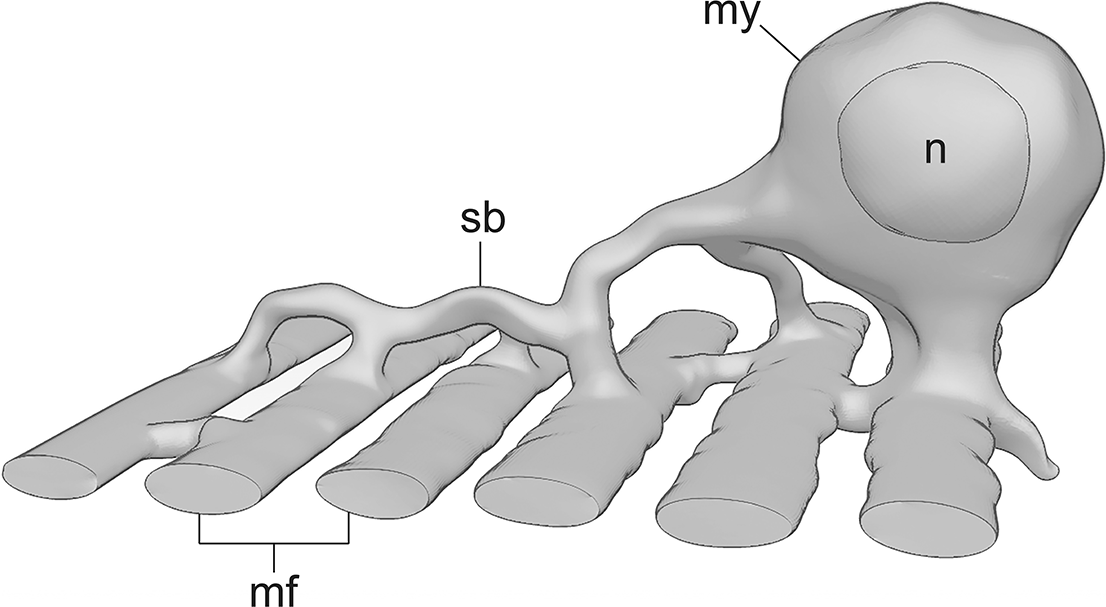
The three-dimensional shape of a body-wall muscle cell was reconstructed from a serial set of thin sections (Figure 6). As in all Digenea, the myocytes of B. progenetica mother sporocysts include a perikaryal region, often referred to as the myocyton, and a contractile part. The contractile part in our specimens consists of a single row of parallel myofibrils located just beneath the tegument (Figure 6). The myofibrils have a typical ultrastructure and contain myosin (thick) filaments surrounded by actin (thin) filaments (Figure 7a). Other components include microtubules, mitochondria, occasional elements of the sarcoplasmic reticulum, and sparsely distributed cisternae of rough endoplasmic reticulum (ER) concentrated near the cytoplasmic bridges connecting myofibrils with the perikaryon. The myofibrils are anchored to the basal lamina of the tegument via electron-dense hemidesmosomes (Figure 7a).
Schematic drawing of a muscle cell with multiple myofibrils (mf). Abbreviations: my, myocyton; n, nucleus; sb, sarcoplasmic bridge.

Ultrastructure of subtegumental myocytes in mother sporocysts of Bunocotyle progenetica. (a) Longitudinal section through the sporocyst body showing subtegumental myofibrils. (b, c, d) Serial sections through a muscle cell showing the myocyton with processes (boundaries marked with arrowheads) and associated myofibrils at different levels. Multiple connections between the myocyton and myofibrils are indicated by arrows, and sarcoplasmic connections between adjacent myofibrils are marked by asterisks. The same myofibrils in different sections are labelled with the same Arabic numerals. (e, f) Details of the myocyte sarcoplasm in the perinuclear region (e) and in a thick sarcoplasmic process (f). Scale bars: a, e, f – 0.5 μm; b–d – 1 μm. Abbreviations: bl, basal lamina; cmf, circular myofibril; go, Golgi apparatus; hd, hemidesmosome; lmf, longitudinal myofibril; mt, mitochondria; my, myocyton; n, nucleus; pa, parenchymal cells; rb, residual bodies; rer, rough ER; tc, tegumental cell; te, outer tegumental layer; ve, vesicles.

The myocyton lies some distance inward from the subtegumental layer of myofibrils (Figure 7b–d). It has a nucleus with a prominent nucleolus and a small amount of heterochromatin concentrated in a thin layer beneath the nuclear membrane (Figure 7b, e). The sarcoplasm of the myocyton contains an extensive network of rough ER with dilated cisternae, free ribosomes, Golgi apparatus, mitochondria, and microtubules (Figure 7b–f). Residual bodies, some multilamellar in structure, are always present, along with occasional endosome-like vesicles. Small granules with an electron-dense core and a surrounding electron-lucent rim are also common and are often located near the Golgi apparatus (Figure 7e, f). Thick sarcoplasmic processes of varying lengths, which are ultrastructurally similar to the perinuclear region, extend from the perikaryon toward the tegument (Figures 6, 7b–d). In our sections, their number ranges from one to four per cell. Apically, these cellular processes connect to the myofibrils, which are also linked together at certain points by thin horizontal sarcoplasmic strands (Figures 6, 7b–d). Each process joins the myocyton to 1–3 myofibrils. Our serial sections of a single muscle cell show that it has 8 myofibrils, with the myocyton connected to the contractile region by 4 thick processes.
The muscles surrounding the proximal portion of the birth canal are composed of similar cells: each myocyton is connected to several myofibrils, and the perinuclear sarcoplasm contains a well-developed endoplasmic reticulum with expanded cisternae. These muscle cells, however, differ from ‘subtegumental’ myocytes in that their myocytons are less distinctly separated from the myofibrils, lying almost at the myofibril layer (Figure 2d).
Parenchyma
Parenchymal cells, with their numerous cytoplasmic extensions, occupy a substantial portion of the sporocyst body wall (Figures 7c–d, 8a). Ther large (up to 12 μm) perikarya are located away from the tegument, often near the brood cavity lining (Figure 8a). The cell nuclei are euchromatic, with faint nucleoli. A thin layer of perinuclear cytoplasm appears denser because of abundant free ribosomes, which are interspersed with Golgi dictyosomes, large mitochondria, and cisternae of rough ER, some of which are expanded (Figure 8a). Small islands of similarly ‘dense’ cytoplasm are also present in the cytoplasmic extensions of parenchymal cells (Figure 2b). Most of the peripheral cytoplasm, including that of the processes, has low electron density and contains few organelles, most of which are lipid inclusions, mitochondria, and a variety of vesicular structures (Figure 8a). A commonly occurring type of vesicles are rounded bodies with contents of medium to high electron density (Figure 8a). Long, branching processes of parenchymal cells extend in various directions, filling the spaces between other cellular elements. They form numerous gap junctions with each other, as well as with tegumental cells, myocytes, and the syncytial lining of the brood cavity (Figures 2b, 8b, c).
Ultrastructure of parenchymal cells in mother sporocysts of Bunocotyle progenetica. (a) General view of a parenchymal cell and its cytoplasmic extensions. (b) Gap junction (arrow) between a parenchymal cell and a myofibril. (c) Gap junction (arrow) between extensions of a parenchymal cell. Scale bars: a – 1 μm; b, c – 0.5 μm. Abbreviations: go, Golgi apparatus; li, lipid bodies; mf, myofibril; mt, mitochondria; n, nucleus; pa, parenchymal cells and their extensions; rer, rough endoplasmic reticulum; syl, syncytial brood cavity lining; tc, tegumental cell; te, outer tegumental layer; ve, vesicular structures.

Nervous system
The nervous system is relatively well developed, with numerous neuronal cell bodies and an extensive network of neurites, but they show considerable individual variability in number and distribution, and only a few common patterns can be discerned in their arrangement. There is no evidence of cerebral or other ganglia; both 5HT- and FaRP-immunoreactive (IR) nerve cells lie separately in the anterior region and throughout the body. Serotonin antibody staining reveals an anterior nerve ring (Figure 9a, b) in the body wall located at the level of the proximal region of the birth canal. From this ring, several fine longitudinal neurites (Figure 9b) arranged in a radially symmetrical pattern project anteriorly toward the birth pore, and 4–6 neurites run posteriorly. One pair of these posterior nerves (Figure 9a–d) is generally thicker than the others and stains more intensely; in some individuals, it includes two or three 5HT-IR neurites (Figure 9c). This pair continues posteriorly for about two-thirds to three-fourths of the body length, where the neurites divide successively into several branches (Figure 9d). Neurons associated with the longitudinal nerves are bipolar cells located at various points along the length of these nerves. The remaining neurites belong to neuronal cells distributed throughout the body; these neurites do not show any distinct pattern in their arrangement and form a loose network (Figure 9a), which varies in configuration among individuals.
Nervous system of mother sporocysts of Bunocotyle progenetica. Confocal images of 5HT (a–e) and FMRFamide (f–h) immunoreactivity staining. The anteroposterior axis is labelled as a–p. (a) General view of the 5HT-IR nervous system. Arrows indicate neuronal somata arranged in a row in the posterior region of the body. (b) Anterior end of a sporocyst showing the anterior nerve ring (ar) and nerves (an) projecting toward the birth pore. (c) Midportion of the body showing the main longitudinal cords, each composed of two or more neurites as indicated by arrows. (d) Posterior region of the body showing successively branching (arrows) neurites of a main longitudinal cord. (e) Posterior end of the body showing bipolar neurons (pb) and their neurites. (f) General view of the FaRP-IR nervous system showing the main longitudinal cords (ml) and a third, weakly stained longitudinal nerve (arrow). (g, h) Anterior end of the body showing bipolar neurons (bb) and their neurites (nb) associated with the birth canal. Scale bars: a, d, f – 50 μm; b, c, e, g, h – 20 μm. Abbreviations: an, anterior nerves; ar, anterior nerve ring; bb, bipolar FaRP-IR neurons of birth canal; bc, birth canal; bp, birth pore; ml, main longitudinal nerves; nb, nerve net surrounding middle birth canal region; pb, bipolar 5HT-IR cells in posterior body region; tc, transverse commissure.

The overall number of detectable 5HT-IR cell bodies ranges from 44 to 52 in different individuals. About 10–20 cells lie at the anterior end of the body around the birth canal and 2–4 cells in the midportion of the body, but most (15–25) 5HT-IR neurons are located in the posterior fourth of the body (Figure 9e). Most of the anterior cells appear to be unipolar or bipolar, with one neurite projecting anteriorly toward the birth pore. The posteriorly located neurons are predominantly bipolar (Figure 9e), but a few appear multipolar, with four or five neurites. The posterior cells are irregularly distributed, but some are arranged in transverse rows (Figure 9a).
Anti-FMRFamide staining reveals a much less extensive system of neurites and nerve cells compared to the 5HT-IR nervous system. The only neuronal cell bodies distinctly stained by FMRFamide antibodies are 5–7 bipolar cells (Figure 9f–h) located at the anterior end of the body around the proximal region of the birth canal. These cells have one anterior and one posterior neurite; the anterior neurites of these cells form a nerve net (Figure 9f–h) around the wall of the middle region of the birth canal.
Only two longitudinal FaRP-IR nerves (Figure 9f) are generally discernible, although a faint third longitudinal neurite (Figure 9f) is visible in some of the preparations. These two nerves run through much of the body but branch out in the posterior fourth of the body. Anteriorly, they fade out at the level of the proximal region of the birth canal, and their association with other structures is unclear. The nerves are connected by a transverse commissure (Figure 9f) located just posterior to the birth canal.
The ultrastructural examination of the FaRP-IR neuronal somata located within the cellular mass at the anterior end of the body (Figure 10a) shows that these small perikarya (up to 5 μm) contain oval nuclei with a prominent nucleolus and clusters of condensed chromatin that form small, scattered clumps and a thin layer beneath the nuclear envelope. The perinuclear cytoplasm is rich in free ribosomes and contains sparse flattened cisternae of rough ER, Golgi dictyosomes, small electron-dense mitochondria, and microtubules. The neurons have two types of membrane-bound, spherical secretory vesicles, both measuring 55–70 nm in diameter but differing in content (Figure 10a). The vesicles of the first type, known in the literature as dense core vesicles (DCVs), contain electron-dense material separated from the membrane by a thin electron-lucent rim. The second type of vesicles is filled with a medium-density, slightly non-homogeneous material that is usually in direct contact with the membrane (Figure 10a, inset). The neurites containing these vesicles constitute the bulk of the dense nerve net surrounding the middle region of the birth canal (Figure 10c). These neurites are found in abundance, either singly or as part of thin nerves, near the muscles surrounding the birth canal, where they often form neuromuscular contacts with these muscles (Figure 10b). Single neurites containing DCVs are also observed near the muscle cells of the body wall (Figure 10e). In the longitudinal nerves located in the sporocyst body wall, which typically include 4–6 neurites (Figure 10d), DCVs are present in small quantities, but there are often clusters of small electron-lucent synaptic vesicles (20–30 nm) and less frequent, medium-sized electron-lucent vesicles (65–70 nm).
TEM images showing the nervous system in mother sporocysts of Bunocotyle progenetica. (a) Perikaryon of a FaRP-IR neuron with two types of vesicles shown enlarged in the inset. (b) Innervation of muscles associated with the proximal region of the birth canal. The arrowhead marks the neuromuscular junction and the arrow indicates the synapse between neurons. (c) Nerve net surrounding the middle region of the birth canal. (d) Oblique section through a longitudinal nerve in the body wall. (e) Micrograph showing a neurite adjacent to a myocyte in the body wall. (f) Section through the apical portion of the non-ciliated T1 sensory bulb showing an electron-dense ring and electron-dense ‘cap’ (arrowhead). (g) Longitudinal section through the uniciliated T2 sensory ending. Note two dense rings, a rootlet, and a cilium inside the cavity filled with vesicles. (h) Apical portion of the uniciliated T3 sensory ending. Scale bars: a–g – 1 μm; h – 0.5 μm. Abbreviations: ci, cilium; dcv, dense core vesicles; dr, dense ring; lv, lucent vesicles; mdv, vesicles of moderate electron density; mep, epithelium of middle part of birth canal; mf, myofibril; mt, mitochondria; my, myocyte; n, nucleus; ne, neuron; nr, neurite; nu, nucleolus; pep, epithelium of proximal part of birth canal; rl, rootlet; sj, septate junction; vin, vacuole-like invaginations of plasma membrane.

Three types of tegumental sensory endings are identified in the B. progenetica mother sporocysts; these differ in the presence or absence of ciliary structures and their mode of communication with the external environment (Figure 10f–h).
The highest concentration of sensory receptors is found in the anterior region of the sporocyst body. One type of these tegumental receptors is non-ciliated sensory endings (T1), which have a relatively large bulb lacking a cilium, kinetosome, and rootlets, and are completely embedded in the outer layer of the tegument (Figure 10f). The cytoplasm in the apical-central region of the bulb has low electron density and may contain sparse lucent vesicles, a few microtubules, and mitochondria. In the periphery and the basal portion of the bulb, the cytoplasm becomes denser and contains deep vacuole-like invaginations of the outer plasma membrane, sometimes enclosing cytoplasmic extensions of the tegument. The apical portion of the sensillum is reinforced by a thin electron-dense ring and a small ‘cap’, both composed of similar electron-dense material (Figure 10f). A weakly discernible septate junction anchors the sensillum to the tegument at the level of the ‘cap’.
The other type of tegumental receptors located at the anterior end of the body are uniciliated sensory endings (T2). These receptors are unusual in that they do not open directly onto the body surface: their short cilium projects into a cavity located within the upper layer of the external syncytium and enclosed by its plasma membrane (Figure 10g). This cavity is not completely isolated but communicates with the exterior through internal tegumental channels – deep, branched invaginations of the outer plasma membrane (see Podvyaznaya and Filimonova Reference Podvyaznaya and Filimonova2024). The cavity is densely packed with spherical vesicles 45–65 nm in diameter containing electron-lucent material (Figure 10g). A small, distinct rootlet extends from the basal body of the cilium into the sensory bulb. The cytoplasm of the bulb contains sparse microtubules and a small number of vesicles, similar to those found in the tegumental cavity, and some of these vesicles make contact with the apical plasma membrane of the sensory ending (Figure 10g). Each sensillum of this type has two supporting electron-dense rings; the larger apical ring is aligned with the septate junction that connects the sensory bulb to the tegument. The basal region of the sensory bulb contains vacuole-like invaginations of its outer plasma membrane, which resemble those seen in non-ciliated sensory endings, but are fewer in number and less deep.
Uniciliated sensory endings that open directly to the exterior (type T3) occur in the midportion of the sporocyst body. Each of these has a short cilium emerging from the base of a small pit in the tegument, which often contains a few electron-lucent vesicles (Figure 10h). In other respects, these sensilla are similar to the T2 sensory endings.
Discussion
Although comparative data on the morphology of mother sporocysts are very scarce, the prevailing view in the literature describes them as highly simplified organisms, even compared to their miracidium larvae (see, for example, Ataev Reference Ataev2017; Diaz Soria et al. Reference Diaz Soria, Attenborough, Lu, Fontenla, Graham, Hall, Thompson, Andrews, Rawlinson, Berriman and Rinaldi2024; Esch et al. Reference Esch, Barger and Fellis2002). This life cycle stage is often characterised as simple, primitive, or amorphous germinal sacs, an interpretation apparently derived from studies of the structure and development of mother sporocysts in schistosomes (Bahia et al. Reference Bahia, Avelar, Vigorosi, Cioli, Oliveira and Mortara2006; Coustau et al. Reference Coustau, Ataev, Jourdane and Yoshino1997; Diaz Soria et al. Reference Diaz Soria, Attenborough, Lu, Fontenla, Graham, Hall, Thompson, Andrews, Rawlinson, Berriman and Rinaldi2024; Meuleman et al. Reference Meuleman, Holzmann and Peet1980; Meuleman et al. Reference Meuleman, Lyaruu, Khan, Holzmann and Sminia1978; Pan Reference Pan1996; Poteaux et al. Reference Poteaux, Gourbal and Duval2023). Schistosome miracidia hatch from eggs in the external aquatic environment, actively seek the first intermediate host and penetrate the mollusc’s tissue upon contact. These are fairly large larvae with complex behaviour and structure, having a well-developed nervous system, subepidermal musculature, and a glandular penetration apparatus (Pan Reference Pan1980). Once the miracidium enters the mollusc and forms the tegument, the provisional glands and terebratorium degenerate, as does a significant portion of the nervous system, musculature, and parenchymal tissue (Bahia et al. Reference Bahia, Avelar, Vigorosi, Cioli, Oliveira and Mortara2006; Diaz Soria et al. Reference Diaz Soria, Attenborough, Lu, Fontenla, Graham, Hall, Thompson, Andrews, Rawlinson, Berriman and Rinaldi2024; Meuleman et al. Reference Meuleman, Holzmann and Peet1980; Pan Reference Pan1996; Poteaux et al. Reference Poteaux, Gourbal and Duval2023). These tissues are replaced by a brood cavity containing embryos of the next parthenogenetic generation. Apart from the tegument, no other specialised structures have been observed to form in developing mother sporocysts of schistosomes.
An entirely different type of development occurs in the mother sporocysts examined in this study. The miracidia of B. progenetica, like those of other hemiuroid digeneans, employ a passive mode of host infection: enclosed in an eggshell, they enter the mollusc’s intestine with ingested food, hatch there, and penetrate the host through the intestinal epithelium (Galaktionov and Dobrovolskij Reference Galaktionov and Dobrovolskij2003; Levakin Reference Levakin2008). These passively infecting larvae are typically small, with a much lower number of cellular elements, which results in significant simplification of most organ systems (Galaktionov and Dobrovolskij Reference Galaktionov and Dobrovolskij2003). For instance, in the recently studied miracidium of one of the hemiuroid species, Derogenes varicus (Müller, 1784), which has a body length of about 15 μm, the excretory system was shown to be absent and only one cell was identified as a neuron (Smirnov and Krupenko Reference Smirnov and Krupenko2023). The miracidia of B. progenetica show similar structural simplicity, as demonstrated at the light microscopical level by Galaktionov and Dobrovolskij (Reference Galaktionov and Dobrovolskij2003) and Levakin (Reference Levakin2008). These tiny larvae, only about 30 μm in length, contain a small cluster of undifferentiated cells located in the posterior region of the body (Galaktionov and Dobrovolskij Reference Galaktionov and Dobrovolskij2003, Figure 3F). Once the miracidium penetrates the mollusc and its tegument undergoes transformation, these cells begin to actively divide and differentiate, generating entirely new structures of mother sporocysts that were absent in the original miracidium. These newly formed structures include a brood cavity lining containing incorporated germinal cells, a highly specialised muscular birth canal replacing the miracidial apical gland, a well-developed nerve net with sensory endings, and most of the subtegumental musculature. During development, the body length of the sporocyst increases by a factor of 40–50 compared to the miracidium. Mature parthenitae retain a small reserve of undifferentiated cells, which are used, in particular, to replenish the population of tegumental cells (Podvyaznaya and Filimonova Reference Podvyaznaya and Filimonova2024), but it is also possible that these undifferentiated cells have broader potency, beyond tegument renewal. The mature, highly mobile mother sporocysts of B. progenetica significantly surpass their larvae in terms of overall organisational complexity and cannot be viewed as amorphous, sac-like organisms.
Brood cavity and birth canal
Our study has shown that in the mother sporocysts of B. progenetica, the birth canal and the brood cavity, which has its own lining, are structurally and functionally linked, effectively forming a reproductive apparatus differentiated into distinct regions (Figure 11a). In some respects, it resembles the reproductive system of mature rediae of the same species, which we have studied earlier (Podvyaznaya et al. Reference Podvyaznaya, Petrov and Galaktionov2020), but there are also significant differences.
Schematic drawing of the birth canal in the mother sporocyst of Bunocotyle progenetica, showing its epithelial lining (a) and associated musculature (b). Abbreviations: abd, anterior birth canal dilator muscles; bc, brood cavity; bcp, birth canal protractor muscles; bcr, birth canal retractor muscles; bp, birth pore; dbi, distal region of birth canal; mbi, middle region of birth canal; mc, circular muscles of middle birth canal region; mep, epithelium of middle birth canal region; pbi, proximal birth canal region; pd, dilator muscles of proximal birth canal region; pep, epithelium of proximal birth canal region; te, tegument.

The fine structure of the syncytial lining of the brood cavity in both sporocysts and rediae shows certain similarities, despite differences in its functional roles. In both cases, nutrients are transported across the syncytium into the cavity, which contains developing embryos. This explains frequent contacts between the syncytium and parenchymal cells and the presence of folds and invaginations on the side facing the body wall, which serve to increase the syncytium’s surface area. In rediae, the cavity lining actively absorbs and digests degeneration products of early embryos, which are especially numerous during the winter months (Galaktionov and Podvyaznaya Reference Galaktionov and Podvyaznaya2019). In sporocysts, however, this function is much less pronounced: only a few embryos undergo degeneration, and the intensity of this process does not depend on the season. Instead, the syncytium in mature sporocysts supports a small number of incorporated germinal cells at various maturation stages, along with early embryos. These embryos exit into the cavity at the germinal ball stage and complete their embryonic development there. A similar pattern of germinal development has been observed in late embryonic rediae of the same species (Podvyaznaya and Galaktionov Reference Podvyaznaya and Galaktionov2021). These rediae, however, also contain an inactive primordial germinal mass associated with the cavity lining, which becomes fully formed and functional during the post-embryonic period when the cavity wall is cleared of germinal elements (Podvyaznaya and Galaktionov Reference Podvyaznaya and Galaktionov2021; Podvyaznaya et al. Reference Podvyaznaya, Petrov and Galaktionov2020). Mother sporocysts of B. progenetica lack this stage of germinal development, which significantly limits their reproductive potential, as evidenced by the small size of infrapopulations of parthenitae in molluscs. According to Levakin (Reference Levakin2008), this number averages 12 ± 1 individuals in P. ulvae and 8 ± 1 individuals in E. ventrosa. We suggest that the low fecundity of the mother sporocysts of B. progenetica is largely due to the very small size of their host molluscs: in our material, the shell height of E. ventrosa was about 2.5–3.5 mm and that of P. ulvae was about 3–4.5 mm. A similar relationship between the fecundity of parthenitae and host size has been reported previously by Rondelaud and Barthe (Reference Rondelaud and Barthe1987) and Ataev (Reference Ataev2017). Another possible factor limiting the number of rediae in the host mollusc is the relatively high pathogenicity of these motile histiophagous parasites (Levakin Reference Levakin2008). By contrast, light microscopy observations by Ameel et al. (Reference Ameel, Cort and Van der Woude1949) suggest that the first-generation parthenitae may exhibit high fecundity in some hemiuroid digeneans. According to these authors, the mother sporocysts of Halipegus eccentricus possess both a well-developed germinal mass and separate germinal elements ‘attached to the body wall’ (Ameel et al. Reference Ameel, Cort and Van der Woude1949; Cort et al. Reference Cort, Ameel and Van der Woude1954).
Judging by the simultaneous presence of embryos at different maturity stages in the brood cavity, the offspring of B. progenetica mother sporocysts appear to develop asynchronously, with rediae leaving the parental organism as they reach maturity, exiting through the birth canal and birth pore. The prolonged and gradual release of highly mobile, histiophagous rediae probably mitigates their pathogenic impact on the host. This may be especially important during the cold winter period when the development of mother sporocysts in mud snails slows down but does not cease entirely (Levakin Reference Levakin2008).
At the ultrastructural level, the birth canal and birth pore have so far been studied only in the daughter parthenitae of several digenean species, including the rediae of B. progenetica (Denisova et al. Reference Denisova, Shunatova, Lebedenkov and Shchenkov2023; Galaktionov and Dobrovolskij Reference Galaktionov and Dobrovolskij2003; Irwin et al. Reference Irwin, Threadgold and Howard1978; Matthews Reference Matthews1980; Podvyaznaya et al. Reference Podvyaznaya, Petrov and Galaktionov2020). Our study shows that the epithelial wall of the birth canal in mother sporocysts is virtually indistinguishable ultrastructurally from that described in the rediae of the same species (Podvyaznaya et al. Reference Podvyaznaya, Petrov and Galaktionov2020). There are differences, however, in the shape of the birth canal and the relative lengths of the regions lined with the same type of epithelium. In sporocysts, the distal region of the canal, formed by the continuation of the tegument, is significantly longer than in rediae. The proximal region of the birth canal, which is lined with squamous epithelial cells bearing long irregular projections, forms an elongated duct in rediae, but expands into a spacious, cup-shaped chamber in sporocysts.
The morphology of the birth canal and the arrangement of the associated musculature (Figure 11) allow some inferences to be made about the way rediae are released to the exterior. Late embryonic rediae entering the cup-shaped chamber of the proximal birth canal are probably held there by gentle contractions of the circular musculature and by folds of the epithelial wall. Once a redia reaches the anterior end of the canal’s proximal region, the middle and distal regions may open widely to allow its passage to the exterior. This is probably accomplished by extrinsic radial muscles attached to the posterior portion of the canal’s distal region and the posterior end of the middle region (Figure 11b); the contraction of these muscles pulls the walls of the canal outward and is accompanied by the relaxation of the circular sphincter muscles. Longitudinally oriented protractor muscles inserted at the junction between the proximal and middle regions may contract to reduce the distance between the cup-shaped chamber and the birth pore, thereby facilitating passage of the emerging redia. Another possibility is that once a redia enters the middle region of the canal, the muscular sphincter closes behind it acting as a valve, after which the protractor muscles contract to shorten this part of the canal and expel the redia to the exterior.
Our results indicate that FaRP-IR neurons and their neurites may be involved in the control of the musculature of the birth canal. This is supported by the high concentration of these elements near the intrinsic muscles of the canal and by the presence of numerous neuromuscular contacts. The innervation of the birth canal musculature by FaRP-IR neurons has recently been reported in the daughter sporocysts of Podocotyle sp. (Denisova et al., Reference Denisova, Shunatova, Lebedenkov and Shchenkov2023), but similar data for the mother generation of parthenitae have so far been lacking.
Overall, the high level of specialisation observed in the epithelial lining and musculature of the birth canal in the sporocysts of B. progenetica suggests that it performs multiple functions: opening and closing the entrance to the brood cavity, facilitating the movement of rediae towards the birth pore, and probably also sorting them by maturity before their release from the parent’s body.
Muscle system
The subtegumental musculature in sporocysts of B. progenetica, with an orthogonal network of outer circular and inner longitudinal layers, has the same overall organisation as that described for schistosome mother parthenitae (Bahia et al. Reference Bahia, Avelar, Vigorosi, Cioli, Oliveira and Mortara2006; Bulantová et al. Reference Bulantová, Chanová, Houžvičková and Horák2011) and daughter parthenitae in all studied digenean species (see, for example, Denisova et al. Reference Denisova, Shunatova, Lebedenkov and Shchenkov2023; Klag et al. Reference Klag, Niewiadomska and Czubaj1997; Køie Reference Køie1971; Krupenko et al. Reference Krupenko, Krapivin and Gonchar2016; Podvyaznaya and Galaktionov Reference Podvyaznaya and Galaktionov2012; Reader Reference Reader1974; Šebelová et al. Reference Šebelová, Stewart, Mousley, Fried, Marks and Halton2004). A correlation has previously been noted between the degree of muscle development in the body wall of parthenitae and their motility (Krupenko et al. Reference Krupenko, Krapivin and Gonchar2016; Pojmanska and Machaj Reference Pojmanska and Machaj1991). The mother sporocysts of B. progenetica support this correlation: they have a dense network of subtegumental muscles and are capable of active locomotion. This trait of mother sporocysts, combined with their vermiform body shape, has been observed in several species of Hemiuroidea (Køie Reference Køie1991; Krull Reference Krull1935; Madhavi Reference Madhavi1978; Thomas Reference Thomas1939) and may be characteristic of this group.
In recent years, muscles surrounding the brood cavity have been found in daughter parthenitae of several digenean species from different taxonomic groups, including Hemiuroidea (Denisova et al. Reference Denisova, Shunatova, Lebedenkov and Shchenkov2023; Krupenko et al. Reference Krupenko, Krapivin and Gonchar2016). These muscles, however, are almost entirely absent in mother sporocysts of B. progenetica. The few muscle fibres observed around the anterior region of the brood cavity, near its junction with the birth canal, represent the posteriormost portion of the canal’s musculature that overlaps a short distance with the cavity.
Our results on the fine structure of myofibrils and perikarya of muscle cells in sporocysts of B. progenetica are generally consistent with ultrastructural data on parthenitae of other studied digenean species (Denisova et al. Reference Denisova, Shunatova, Lebedenkov and Shchenkov2023; Klag et al. Reference Klag, Niewiadomska and Czubaj1997; Køie Reference Køie1971; Podvyaznaya and Galaktionov Reference Podvyaznaya and Galaktionov2012; Reader Reference Reader1974). A new finding is the complex shape of the myocytes, which was observed through the examination of serial sections. We have demonstrated the connection of a single myocyton with a group of myofibrils and the interconnection of some adjacent myofibrils via thin sarcoplasmic bridges (Figure 6). These bridges are present only in limited areas of the cell and therefore are often overlooked; they are more pronounced in young myocytes. Some elements of this organisation of muscle cells have been noted in the body wall musculature of daughter sporocysts in Diplostomum pseudospathaceum (Klag et al. Reference Klag, Niewiadomska and Czubaj1997, Figure 15), the musculature of the cirrus tube in adult flukes of Prosthodendrium ascidia (Podvyaznaya Reference Podvyaznaya1993), and the differentiating subtegumental myoblasts of developing metacercariae of D. chromatophorum (=D. pseudospathaceum) (Galaktionov and Dobrovolskij Reference Galaktionov and Dobrovolskij2003; Podvyaznaya and Dobrovolsky Reference Podvyaznaya and Dobrovolsky2001). Muscle cells of this morphology are apparently present at different stages of the digenean life cycle; they are probably characteristic of small and medium-sized muscles arranged in layers.
Parenchymal cells
The parenchymal cells of the mother sporocysts of B. progenetica have a distinct set of ultrastructural traits, including electron-lucent peripheral cytoplasm, abundant lipid inclusions, long and variably oriented cytoplasmic processes, and multiple gap junctions joining adjacent parenchymal cells and linking them with other cellular elements. In our opinion, these cells are equivalent to the ‘vacuolar’ cells of daughter sporocysts in D. pseudospathaceum (Klag et al. Reference Klag, Niewiadomska and Czubaj1997) and to the process-bearing parenchymal cells containing lipids and glycogen, or only lipids, in the sporocyst body walls of C. buccini, S. mansoni, Postharmostomum gallinum, and P. borealis (Fournier and Théron Reference Fournier and Théron1985; Køie Reference Køie1991; Podvyaznaya and Galaktionov Reference Podvyaznaya and Galaktionov2012; Žďárská and Soboleva Reference Žďárská and Soboleva1985). These connective tissue elements are attributed various functions, including the transport of substances within the body wall of parthenitae, storage of reserve substances, participation in carbohydrate and lipid metabolism, and the synthesis of components for the extracellular matrix.
Parenchymal cells of certain parthenitae, including the sporocysts of B. progenetica and P. borealis that we studied, show a close resemblance to the cells described in hermaphroditic adults as parenchymal cells (Threadgold and Arme Reference Threadgold and Arme1974; Threadgold and Gallagher Reference Threadgold and Gallagher1966) and to the so-called ‘storage’ myocytons described by Conn (Reference Conn1988, Reference Conn1993) in cestodes. We specifically examined the cytoplasmic processes of parenchymal cells in B. progenetica and P. borealis for possible cytoplasmic continuity with myofibrils on a series of ultrathin sections but found no evidence of such connections. However, in adults of Prostodendrium ascidia, we previously observed small, rare bundles of myofilaments in the processes of cells identified as parenchymal (Podvyaznaya Reference Podvyaznaya1993), consistent with the descriptions of Threadgold and Gallagher (Reference Threadgold and Gallagher1966). Clearly, the question of the existence of different types of muscle cells in digeneans remains unresolved and requires further investigation.
Nervous system
The overall organisation of the nervous system in mother sporocysts of B. progenetica is generally consistent with that described in daughter sporocysts of other species (Czubaj and Niewiadomska Reference Czubaj and Niewiadomska1990; Denisova et al. Reference Denisova, Shunatova, Lebedenkov and Shchenkov2023; Grabda-Kazubska and Moczoń Reference Grabda-Kazubska and Moczoń1991; Niewiadomska et al. Reference Niewiadomska, Czubaj, Maślińska and Pomorski1999; Niewiadomska and Moczoń Reference Niewiadomska and Moczoń1990; Solis-Soto and De Jong Brink Reference Solis-Soto and De Jong Brink1994; Theron and Fournier Reference Theron and Fournier1982). A common pattern includes a concentration of nerve elements surrounding the birth canal, from which several small nerves project anteriorly and two main longitudinal cords extend posteriorly through most of the sporocyst body. Additional longitudinal nerves are present, but these are less prominent and cannot be traced throughout the length of the body. In addition to these features common to all studied sporocysts, the mother sporocysts of B. progenetica show a number of distinct traits. Although there is a concentration of cell bodies near the birth pore, all cells in the mother sporocysts of B. progenetica lie separately from one another rather than forming a single cerebral ganglion or a pair of ganglia connected by a transverse commissure as described in daughter sporocysts. In the previously studied daughter sporocysts, the cells of the cerebral ganglion usually lie on one side of the birth canal provisionally identified as dorsal, since the cerebral ganglion is dorsal in digeneans, which allows the main longitudinal cords to be identified as lateral. In B. progenetica, the nerve cells and neurites associated with the birth canal are arranged either irregularly or in a radial pattern around the birth canal, and therefore, the dorso-ventral polarity is difficult or impossible to determine.
A substantial aggregation of nerve cells is observed at the posterior end of the B. progenetica mother sporocysts, an arrangement not previously reported in other parthenitae. The overall cellular arrangement appears less regular compared to other sporocysts, resulting in a more diffuse organisation of the nervous system. For example, the daughter sporocysts of Podocotyle sp. have a distinct bilaterally symmetrical distribution of serotonergic cells (Denisova et al. Reference Denisova, Shunatova, Lebedenkov and Shchenkov2023), while in B. progenetica, neither 5HT-IR nor FaRP-IR cells show bilateral arrangement or any other clear patterns in their distribution, and only in some specimens do neurons appear to form transverse rows in the posterior region of the body.
One of the most unusual features of the B. progenetica sporocysts is their exceptionally high number of detectable serotonergic nerve cells (44–52). This is in sharp contrast to only eight cells reported in Podocotyle sp. (Denisova et al. Reference Denisova, Shunatova, Lebedenkov and Shchenkov2023) and fluorescent images showing the serotonergic nervous system in daughter sporocysts of Diplostomum pseudospathaceum (Niewiadomska et al. Reference Niewiadomska, Czubaj, Maślińska and Pomorski1999) suggest a similarly low count. The number of serotonergic cells in B. progenetica is not only higher than in the other studied parthenitae but may also surpass those in the hermaphroditic generation: for example, cercariae of 16 digenean species have only 12 to 20 5HT-IR cells (Terenina and Movsesyan Reference Terenina and Movsesyan2018). The arrangement of 5HT-IR neurons in the mother sporocysts of B. progenetica also shows significant individual variability, but this is probably explained, at least in part, by age-related differences. By contrast, the counts of FaRP-IR cells are not exceptional (5–7 cells) and comparable to those in the daughter sporocysts of Podocotyle sp. (10 cells; Denisova et al. Reference Denisova, Shunatova, Lebedenkov and Shchenkov2023), the only other sporocysts studied for FaRP immunoreactivity.
Serotonin has previously been shown to modulate body-wall muscle contractions and stimulate motor activity in digeneans and other parasitic flatworms (Boyle et al. Reference Boyle, Zaide and Yoshino2000; McKay et al. Reference McKay, Halton, Allen and Fairweather1989; Patocka et al. Reference Patocka, Sharma, Rashid and Ribeiro2014; Pax et al. Reference Pax, Siefker and Bennett1984; Thompson and Mettrick Reference Thompson and Mettrick1989; Tolstenkov et al. Reference Tolstenkov, Prokofiev, Pleskacheva, Gustafsson and Zhukovskaya2017), and it can therefore be presumed that the abundance of serotonergic cells in mother sporocysts of B. progenetica is related to their mobile lifestyle and prolonged existence within the hosts. However, serotonin’s myoexcitatory function is probably not the only reason for the complexity of the serotonergic nervous system in this species. In free-living flatworms, serotonin has been implicated in stimulating stem cell maturation during asexual reproduction and regeneration (Franquinet Reference Franquinet1979; Franquinet and Martelly Reference Franquinet and Martelly1981; Sarkar et al. Reference Sarkar, Mukundan, Sowndarya, Dubey, Babu, Lakshmanan, Rangiah, Panicker, Palakodeti, Subramanian and Subramanian2019; Villar and Schaeffer Reference Villar and Schaeffer1993), in cestodes it has been demonstrated to regulate developmental processes (Camicia et al. Reference Camicia, Herz, Prada, Kamenetzky, Simonetta, Cucher, Bianchi, Fernández, Brehm and Rosenzvit2013; Herz and Brehm Reference Herz and Brehm2021) and in digeneans to exhibit control over reproduction, at least in the hermaphroditic generation (Fairweather et al. Reference Fairweather, Maule, Mitchell, Johnston and Halton1987). This may explain the unusually high concentration of 5HT-IR neuronal somata in the posterior body region of the B. progenetica mother sporocysts, as these cells may regulate renewal of the tegumental cells and proliferation of the germinal material in response to external cues.
The ultrastructure of receptors in mother sporocysts remains poorly understood, even in well-studied schistosomes. A recent study by Poteaux et al. (Reference Poteaux, Gourbal and Duval2023) used scanning electron microscopy to demonstrate that some uniciliated and multiciliated sensory endings in the tegument of juvenile mother sporocysts of S. mansoni represent ‘vestigial remnants’ of the miracidium’s sensory apparatus. By contrast, in B. progenetica, all or nearly all sensory endings are likely to form de novo during the development of the mother sporocyst and must meet the specific functional demands of this life cycle stage. Receptors located in the anterior portion of the body may help the sporocysts in navigating narrow lacunae within the haemocoel. The non-ciliated sensory endings embedded in the tegument (type T1) are likely to function as tangoreceptors. These structures are characterised by pronounced, wide invaginations of the outer plasma membrane that fill a significant portion of the bulb and probably confer structural rigidity in the absence of rootlets. Similar structures have previously been noted in the uniciliated sensory endings of the rediae of Notocotylus attenuatus (Czubaj and Niewiadomska Reference Czubaj and Niewiadomska1988) and in the non-ciliated sensilla of mature flukes of Prosthodendrium ascidia (Podvyaznaya Reference Podvyaznaya1995) and Crepidostomum metoecus (Žďárská and Nebesárová Reference Žďárská and Nebesárová2003).
The T2 and T3 sensory endings bear short cilia, which is a characteristic feature of ciliary sensory endings in parthenitae (Czubaj and Niewiadomska Reference Czubaj and Niewiadomska1988, Reference Czubaj and Niewiadomska1991; Denisova et al. Reference Denisova, Shunatova, Lebedenkov and Shchenkov2023; Podvyaznaya and Galaktionov Reference Podvyaznaya and Galaktionov2012). Such breviciliate receptors are commonly believed to respond to mechanical (tactile) stimuli and some may also receive chemical signals (Biserova and Korneva Reference Biserova and Korneva1999; Czubaj and Niewiadomska Reference Czubaj and Niewiadomska1996). The presence of vesicles in the extracellular space surrounding the cilia of the T2 and T3 sensory endings suggests an additional secretory function. The T2 sensory endings appear to release secretion through the internal channels in the tegument, and the discharge of this secretion is probably triggered by the mechanical pressure on the receptor.
Similar secretory activity in sensory cells has been observed in daughter sporocysts and cercariae of D. pseudospathaceum (Czubaj and Niewiadomska Reference Czubaj and Niewiadomska1991, Reference Czubaj and Niewiadomska1996), the rediae of N. attenuatus (Czubaj and Niewiadomska Reference Czubaj and Niewiadomska1988), and certain species of cestodes (reviewed in Biserova Reference Biserova2015). The chemical composition of the secreted material remains unclear, but one hypothesis (Biserova Reference Biserova2015; Biserova and Korneva Reference Biserova and Korneva1999) suggests that it contains neuroactive substances influencing the host.
Conclusions
The mother sporocysts of B. progenetica have been shown to have limited reproductive potential, as they lack a germinal mass and have a fairly small number of germinal elements. The low fecundity of these parthenitae is most likely an adaptation to the small size of their mud snail hosts.
At the same time, the sporocysts of B. progenetica appear well adapted for an active and prolonged existence within hosts living in the harsh and constantly fluctuating conditions of the littoral zone. The presence of a well-innervated birth canal enables them to prolong their reproductive processes and regulate the release of offspring in response to external cues. A complex, well-differentiated nervous system supports both the active locomotion of mother sporocysts within the host and the controlled, intermittent release of offspring. The highly specialised tegument, adapted for absorbing a wide spectrum of nutrients, continues to function throughout the year (Podvyaznaya and Filimonova Reference Podvyaznaya and Filimonova2024), supporting the growth, development, and vital activities of these parthenitae. The prolonged functional activity of the tegument is sustained by the renewal of the population of tegumental cells from a reserve of undifferentiated cells that persists in mature mother sporocysts. Since serotonergic neurons are implicated in the morphogenesis and proliferation of stem cells in flatworms, the role of the unusually abundant 5HT-IR neurons in the posterior body region of these sporocysts is likely to control the replacement of tegumental cells and the proliferation of germinal material.
In the littoral zone of the White Sea, mass infection of mud snails by B. progenetica occurs during spring warming and, to a lesser extent, in autumn before the onset of the cold season (Levakin Reference Levakin2008). The high adaptability of the mother sporocysts of B. progenetica allows them to develop and reproduce, albeit at varying rates, during both the warm season and the unfavourable winter period (if the infection occurs in autumn), which seems to be crucial for maintaining their monoxenous life cycle.
Acknowledgements
We would like to thank Dr I.A. Levakin, Dr K.E. Nikolaev, and Prof K.V. Galaktionov for their help in collecting material during the winter period.
Financial support
This work was supported by budgetary funding from the Zoological Institute RAS (projects 125012800903-5 and 125012800894-6).
Competing interests
The authors declare none.