Introduction
Prominauris (prominent ears) occurs in approximately 2 per cent of the UK population,Reference Alexander, Stott, Sivakumar and Kang1 and can lead to low self-esteem, bullying, social isolation and significant psychological morbidity, particularly in childhood and adolescence.Reference Bradbury, Hewison and Timmons2 It is inherited in an autosomal dominant pattern, and commonly occurs as a result of two development defects: underdevelopment of the anti-helical fold and an enlarged conchal bowl.Reference Adamson and Strecker3
Otoplasty is the surgical technique used for correcting protruding ears. It is characterised by various operative techniques that can be classified into two broad categories: cartilage sculpting (cutting)Reference Mustardé4,Reference Furnas5 and cartilage-sparing (suturing).Reference Chongchet6,Reference Stenstroem7 The Mustardé suture technique is one of the most commonly used methods for creating an anti-helical fold and can often be used in conjunction with other techniques.Reference Mustardé4
Currently, otoplasty is taught either with cadavers or under the direct supervision of a consultant in the operating theatre. The introduction of the European Working Time Directive has limited surgical training opportunities,Reference Giles8 restricting operative exposure. Simulation training has emerged as a method to overcome this, offering trainees first-hand experience of complex procedures in a safe, controlled environment outside working hours, whilst minimising risk to patients. There are currently limited platforms available for otoplasty simulation, including three-dimensional printed and animal models which are yet to be validated.Reference Schneider, Voigt and Rettinger9,Reference Loh, Gunn, Pennell and Athanassopoulos10 This study aimed to perform a validation assessment of a novel porcine ex vivo model for otoplasty training.
Materials and methods
Materials
Nine porcine ears were obtained from local abattoirs and defrosted on the day of study. The pinna was secured to the operating platform using pins and screws.
Study design
This was a prospective cohort study. Nine Scottish otolaryngology trainees were recruited as participants as part of a facial plastics simulation day.
Participants were asked to complete a pre-simulation questionnaire in order to assess the level of operative experience – all participants had previously assisted in or performed an otoplasty procedure under supervision. Participants were asked to perform a standard approach otoplasty and complete a series of tasks, including posterior skin incision, anterior scoring, Mustardé suture placement and concha–mastoid suture placement. Consultant otolaryngologists provided technical guidance and assistance throughout the procedure.
After each task was completed, participants were asked to complete a validation questionnaire. The questionnaire focused on three key domains: (1) face validity (assessing the likeness of the model to otoplasty performed on a patient); (2) global content validity (assessing the overall usefulness of the model for teaching and training); and (3) task-specific content validity (assessing the model's ability to replicate each step of the procedure).
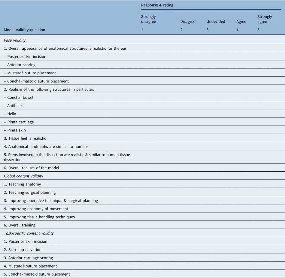
Model validity was assessed using a 17-item validation questionnaire, with 5-point Likert scales assessing the extent to which participants agreed or disagreed with each statement (Table 1). This questionnaire design was adapted from previous studies that assessed the validity of other surgical simulation models.Reference Milner, Okhovat, Clement, Wynne and Kunanandam11 Data were prospectively collected, and analysed. A median threshold score of 4 out of 5 was set as the requirement for validation, to ensure a high standard of agreement and reliable validity. Descriptive statistics were used to assess model outcomes.
Validation questionnaire

Ethical considerations
All the animal models were obtained from a licensed medical meat supplier compliant with all UK and EU regulations. Each model was utilised within the criteria of the Strengthening the Reporting of Observational Studies in Epidemiology (‘STROBE’) guidelines and the Animal Research: Reporting In Vivo Experiments (‘ARRIVE’) guidelines.12,13 Departmental approval was granted for the use of animal tissue. No animal was specifically slaughtered for this study.
Results
Participants
There were nine participants on the otoplasty simulation course. The participants were all otolaryngology trainees with surgical training experience ranging from one to seven years.
Feasibility assessment
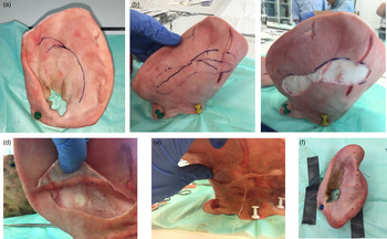
The porcine model was considered an excellent simulation tool for surgical simulation training, although some anatomical variations were identified in comparison to a cadaveric model (Figure 1). These include the shape and position of the ear with respect to the operating table (representing the cranium), and the size and thickness of the cartilage. The skin and soft tissue of the pinna was of adequate texture, with preservation of the perichondrial planes, facilitating wide dissection, elevation and re-positioning manoeuvres.
Images showing the steps involved in performing otoplasty on the porcine model: (a) marking out the anti-helical fold; (b) mapping out the posterior markings for skin excision; (c) elliptical skin excision; (d) elevation of the perichondrial flaps; (e) placement of the Mustardé sutures and closure of skin; and (f) final configuration of the pinna.

The absence of an anti-helical fold in the porcine model provided an ideal contour deformity for corrective surgery and reconstruction. Mustardé suture placement was accommodated with sufficient strength to prevent extrusion, with suitable pliability. Overall, this model is extremely practical and adaptable for simulation training using a variety of cartilage sculpting and suture techniques, for both adult and paediatric otoplasty.
Model validity
Face validity
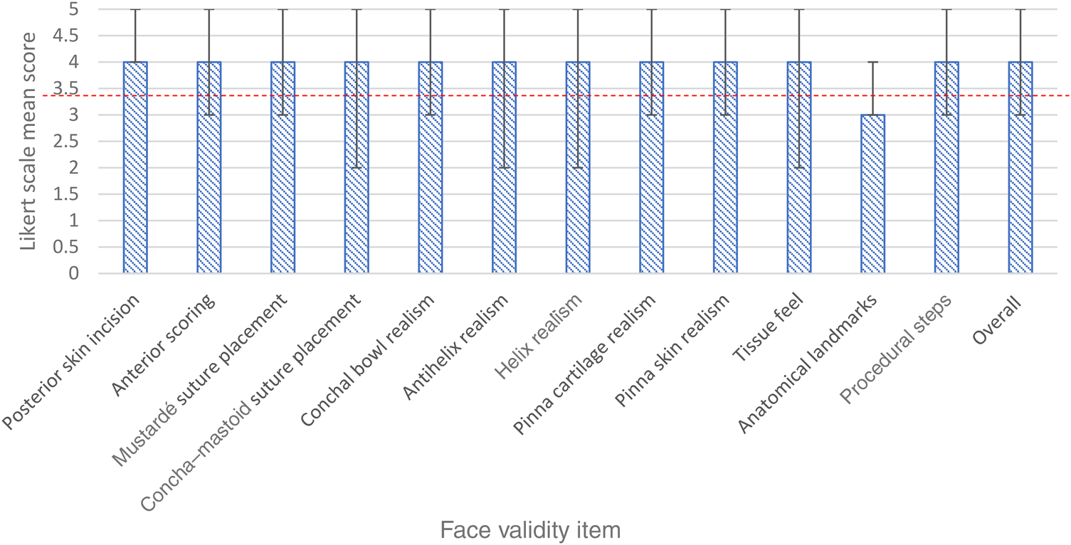
The porcine model achieved an overall face validity median score of 4. The model scored favourably across individual face validity items assessing tissue feel, anatomical organisation and dissection realism, achieving median scores of 4 (interquartile range, 3–4) (Figure 2). The only area where the model failed to achieve validation was in representing anatomical landmarks. Overall, the model achieved requisite scores to satisfy model validation.
Bar chart demonstrating the median Likert scores for face validity outcomes. Error bars indicate the range of scores for each category. The validation threshold is highlighted by the dotted line.

Global content validity
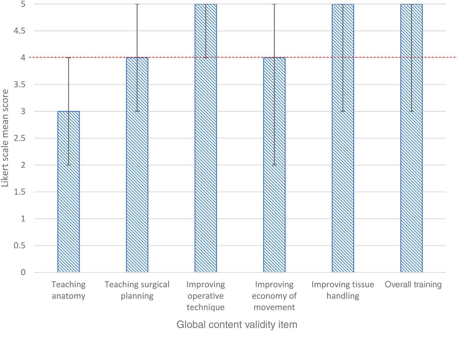
Overall, the median score for global content validity for the porcine model was 5 (interquartile range, 4–5). Median global content validity scores were 4 or higher for all individual items apart from teaching anatomy (Figure 3).
Bar chart demonstrating the median Likert scores for global content validity outcomes. Error bars indicate the range of scores for each category. The validation threshold is highlighted by the dotted line.

Task-specific content validity
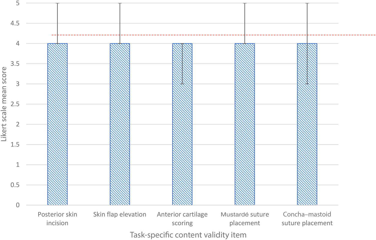
The porcine model received median task-specific content validity scores of 4 (interquartile range, 4–4) for all the steps involved in otoplasty, including: posterior skin incision, skin flap elevation, anterior cartilage scoring, Mustardé suture placement and concha–mastoid suture placement (Figure 4).
Bar chart demonstrating the median Likert scores for task-specific content validity outcomes. Error bars indicate the range of scores for each category. The validation threshold is highlighted by the dotted line.

Discussion
Simulation training has been widely accepted as an effective platform for training in otolaryngology surgery. It allows the delivery of expert training in a standardised fashion, and facilitates the acquisition of skills, knowledge and experience in a safe environment. The use of animal models offers a compromise between the scarcity of human cadavers and the unreal experience of synthetic models. This study is the first to formally validate the ex vivo porcine auricular model as a useful tool for teaching and training in otoplasty techniques for the correction of prominent ears.
Surgical correction of prominauris is challenging, and this is reflected in the variety of techniques and complications described in the literature.Reference Limandjaja, Breugem, Mink van der Molen and Kon14 Weerda identified 94 articles describing different otoplasty techniques, and indicated that the technique should be tailored to the individual patient.Reference Weerda and Clark15 The most commonly used approaches include a combination of cartilage sculpting and suture techniques. Achieving reliable results is difficult, with an associated learning curve, and years of experience and training are required to develop competency in this field.
Simulation training can therefore be an ideal platform for trainees to gain valuable knowledge and hands-on skills in otoplasty techniques, in a controlled manner, and can thus help to flatten the learning curve for this procedure. The diverse range of otoplasty techniques described in the literature also highlights the need for a reliable, validated simulation model, to allow trainees to develop different operative skills.
There are currently limited platforms available for otoplasty training.Reference Schneider, Voigt and Rettinger9,Reference Loh, Gunn, Pennell and Athanassopoulos10 Whilst cadaveric dissection plays an important role for many operations, its role in otoplasty is limited, with preserved ear cartilage becoming considerably softer with fixation, making the surgical steps more difficult to carry out.Reference Alberty, Filler, Schmal and Peuker16 Furthermore, most UK centres have limited access to cadaveric models because of the high costs and need for specific facilities, making this model less practical for implementation into training programmes.
Three-dimensional printed models have recently been described; these offer the advantage of wider availability, without the use of an anatomical dissection room. However, the materials used lack a realistic tissue feel, impacting their usefulness to simulation training in otoplasty, and as yet they have not been validated for this purpose.Reference Schneider, Voigt and Rettinger9
Animal models have been used for simulation training in otolaryngology surgery as an alternative to cadaveric tissue. Animal models offer a realistic, inexpensive and practical platform that is readily available and easily implemented into local training schemes.Reference Milner, Okhovat, Clement, Wynne and Kunanandam11
The ex vivo porcine auricular model has been previously described for use as a simulation model for otoplasty.Reference Loh, Gunn, Pennell and Athanassopoulos10 However, in order to establish the validity of the simulation model, it needs to be subjected to a comprehensive assessment of its realism (face validity) and usefulness for training (content validity). We have been able to validate the porcine model for use in otoplasty across all domains assessed. The porcine ears possess the realistic tissue feel, skin thickness and cartilaginous strength to accommodate all surgical steps involved in otoplasty.
The model had several limitations that restricted its applicability to clinical practice. The thick elastic nature of the cartilage meant that greater force was required to achieve anti-helical fold reconstruction. Furthermore, trainees did not feel that the model had anatomical realism, suggesting that it would not help develop the aesthetic nuances particular to operating on a human ear. Overall, trainees found this model extremely useful for improving surgical planning, tissue handling skills and the overall economy of movement.
• Prominauris (prominent ears) can result in significant psychological morbidity, particularly in childhood and adolescence
• Otolaryngology trainees are expected to demonstrate adequate competence in otoplasty during training
• The European Working Time Directive has limited surgical training opportunities, with simulation utilised to overcome this
• The porcine ex vivo model was validated as a high-fidelity practical tool for otoplasty simulation training
Whilst cadaveric models will always be the ‘gold standard’ for surgical dissection, commercially available ex vivo porcine models provide a low-cost, anatomically compatible and high-fidelity model for otoplasty training.
Study limitations
This investigation involved a prospective cohort study and therefore provides level 3 evidence. This study had a small number of participants because of the narrow recruitment process. There was no comparison to the gold standard human cadaveric model; however, this study aimed to assess the use of a porcine model as an alternative to the more costly human cadaveric model.
Junior participants’ limited experience may also have contributed to observation bias in the validation questionnaire. This suggests that the model is more useful in the early years of training. There are no definitive validation tools for otoplasty simulation training available; therefore, this validation questionnaire was derived from previous simulation studies.
Conclusion
Trainees in otolaryngology are expected to demonstrate adequate competence in otoplasty during their training. Simulation provides a high-fidelity means of delivering expert training to gain technical competence in a standardised, risk-free environment. This study has validated the porcine model as a high-fidelity practical tool for simulation training. We advocate incorporating the model into simulation training for otoplasty, in order to improve learning, enable the acquisition of specific surgical skills and improve operative outcomes.
Competing interests
None declared