Introduction
Eukaryotes are an important part of the Precambrian fossil record. Molecular clock calculations estimate the age of the last eukaryotic common ancestor (LECA) to between 1.9 and 1.0 Ga (Parfrey et al., Reference Parfrey, Lahr, Knoll and Katz2011; Eme et al., Reference Eme, Sharpe, Brown and Roger2014), and nonambiguous eukaryotic body fossils are reported from 1.65 Ga strata (Javaux et al., Reference Javaux, Knoll and Walter2004; Lamb et al., Reference Lamb, Awramik, Chapman and Zhu2009; Javaux, Reference Javaux2019; Miao et al., Reference Miao, Moczydłowska, Zhu and Zhu2019).
Although the Proterozoic record of fossil eukaryotes is dominated by organic-walled microfossils, it also preserves macroscopic carbonaceous compressions often interpreted as macroalgae (review in Bykova et al., Reference Bykova, LoDuca, Ye, Marusin, Grazhdankin and Xiao2020), but the eukaryotic nature or identity of some is unclear. Organic-walled microfossils from shale and siltstone of the 3.2 Ga Moodies Group in South Africa may represent much older stem eukaryotes, but they lack important diagnostic criteria (e.g., conspicuous surface ornamentation or processes) to distinguish them from prokaryotes (Javaux et al., Reference Javaux, Marshall and Bekker2010). A diversification model (Javaux, Reference Javaux2011; Javaux and Lepot, Reference Javaux and Lepot2018) and paleodiversity data (Knoll et al., Reference Knoll, Javaux, Hewitt and Cohen2006; Cohen and Macdonald, Reference Cohen and Macdonald2015; Riedman and Sadler, Reference Riedman and Sadler2017) indicate that the eukaryotic domain developed various biological innovations during the late Paleoproterozoic–early Mesoproterozoic (review in Javaux, Reference Javaux and Jékely2007, Reference Javaux2011; Knoll, Reference Knoll2014; Butterfield, Reference Butterfield2015a; Cohen and Macdonald, Reference Cohen and Macdonald2015; Javaux and Knoll, Reference Javaux and Knoll2017; Porter, Reference Porter2020), such as multicellularity (Butterfield, Reference Butterfield2009; Knoll and Lahr, Reference Knoll, Lahr, Niklas and Newman2016; Javaux and Knoll, Reference Javaux and Knoll2017), predation (Porter, Reference Porter2016; Cohen and Riedman, Reference Cohen and Riedman2018; Loron et al., Reference Loron, Rainbird, Turner, Greenman and Javaux2018), and photosynthesis (Butterfield, Reference Butterfield2015b), leading to the emergence of major crown groups before the end of the Mesoproterozoic. These views are supported by recent studies of late Mesoproterozoic–Neoproterozoic fossil assemblages (Baludikay et al., Reference Baludikay, Storme, François, Baudet and Javaux2016; Beghin et al., Reference Beghin, Storme, Blanpied, Gueneli, Brocks, Poulton and Javaux2017; Tang et al., Reference Tang, Hughes, McKenzie, Myrow and Xiao2017; Loron et al., Reference Loron, Rainbird, Turner, Greenman and Javaux2019a) and various reports of crown-group eukaryotes, such as probable (Loron et al., Reference Loron, François, Rainbird, Turner, Borensztajn and Javaux2019b; Berbee et al., Reference Berbee, Strullu-Derrien, Delaux, Strother, Kenrick, Selosse and Taylor2020) and putative (Berbee et al., Reference Berbee, Strullu-Derrien, Delaux, Strother, Kenrick, Selosse and Taylor2020; Bonneville et al., Reference Bonneville, Delpomdor, Préat, Chevalier, Araki, Kazemian, Steele, Schreiber, Wirth and Benning2020) fungi; testate amoebae (Porter and Knoll, Reference Porter and Knoll2000; Porter et al., Reference Porter, Meisterfeld and Knoll2003; Morais et al., Reference Morais, Fairchild, Lahr, Rudnitzki, Schopf, Garcia, Kudryavtsev and Romero2017; Riedman et al., Reference Riedman, Porter and Calver2018; Martí Mus et al., Reference Martí Mus, Moczydłowska and Knoll2020); red algae (Butterfield, Reference Butterfield2000; Gibson et al., Reference Gibson2017); possible and probable green algae (Butterfield et al., Reference Butterfield, Knoll and Swett1994, Reference Butterfield2015b; Arouri et al., Reference Arouri, Greenwood and Walter1999; Marshall et al., Reference Marshall, Javaux, Knoll and Walter2005; Dong and Xiao, Reference Dong, Xiao, Xiao and Kaufman2006; Moczydłowska and Willman, Reference Moczydłowska and Willman2009; Moczydłowska, Reference Moczydłowska2016; Brocks et al., Reference Brocks, Jarrett, Sirantoine, Hallmann, Hoshino and Liyanage2017; Tang et al., Reference Tang, Pang, Yuan and Xiao2020); possible xanthophyte algae (Butterfield, Reference Butterfield2004); foraminifera (Bosak et al., Reference Bosak, Macdonald, Lahr and Matys2011a, Reference Bosak, Lahr, Pruss, Macdonald, Dalton and Matysb; Brocks et al., Reference Brocks, Jarrett, Sirantoine, Hallmann, Hoshino and Liyanage2017); possible ciliates (Bosak et al., Reference Bosak, Lahr, Pruss, Macdonald, Gooday, Dalton and Matys2012); and possible stem metazoan eggs (Cornet et al., Reference Cornet, François, Compère, Callec, Roberty, Plumier and Javaux2019). However, the ciliate candidates proposed by Bosak et al. (Reference Bosak, Lahr, Pruss, Macdonald, Gooday, Dalton and Matys2012) are reinterpreted as red algal spores by Cohen et al. (Reference Cohen, Vizcaíno and Anderson2020), and Jacutianema (Butterfield, Reference Butterfield2004) lacks diagnostic characteristics and falls outside the molecular clock estimates for xanthophyte algae (Butterfield, Reference Butterfield2015b). Similarly, the fungal affinity of the filaments reported by Bonneville et al. (Reference Bonneville, Delpomdor, Préat, Chevalier, Araki, Kazemian, Steele, Schreiber, Wirth and Benning2020) are ambiguous (Berbee et al., Reference Berbee, Strullu-Derrien, Delaux, Strother, Kenrick, Selosse and Taylor2020), and their syngenicity is doubtful.
Although some of those examples have yet failed to find a scientific consensus and need further investigation, the overall record in Mesoproterozoic–Neoproterozoic successions supports the importance of this hinge period for eukaryotic crown-group evolution. All these data suggest that crown-group eukaryotes may have already been present by the early Mesoproterozoic and were minor, or overlooked, components of paleontological assemblages (Javaux, Reference Javaux2011; Knoll, Reference Knoll2014; Butterfield, Reference Butterfield2015a; Javaux and Knoll, Reference Javaux and Knoll2017; Javaux and Lepot, Reference Javaux and Lepot2018); alternatively, they may have emerged later, at the end of the Mesoproterozoic, shortly before their diversification (Porter, Reference Porter2020).
Recent studies of late Paleoproterozoic and early Mesoproterozoic shale-hosted assemblages of the ca. 1.74–1.41 Ga Ruyang Group (Yin et al., Reference Yin, Xunlai, Fanwei and Jie2005; Agić et al., Reference Agić, Moczydłowska and Yin2015, Reference Agić, Moczydłowska and Yin2017) and the ca. 1.67–1.63 Ga Changcheng Group (Miao et al., Reference Miao, Moczydłowska, Zhu and Zhu2019) from China, the ca. 1630 Vindhyan Supergroup from India (Prasad et al., Reference Prasad, Uniyal and Asher2005), the ca. 1.49 Ga Roper Group from Australia (Javaux et al., Reference Javaux, Knoll and Walter2001; Javaux and Knoll, Reference Javaux and Knoll2017), and the ca. 1.58–1.45 Ga lower Belt Supergroup, Montana (Adam et al., Reference Adam, Skidmore, Mogk and Butterfield2017) show that microfossils with eukaryotic characteristics were already moderately diverse at that time. However, with 14 eukaryotic taxa recognized in the Beidajian and Baicaoping formations, the Ruyang Group (Agić et al., Reference Agić, Moczydłowska and Yin2015, Reference Agić, Moczydłowska and Yin2017) is an exception for a time otherwise marked by a low eukaryotic diversity (≤8 taxa; Prasad et al., Reference Prasad, Uniyal and Asher2005; Nagovitsin, Reference Nagovitsin2009; Vorob'eva et al., Reference Vorob'eva, Sergeev and Petrov2015; Adam et al., Reference Adam, Skidmore, Mogk and Butterfield2017; Javaux and Knoll, Reference Javaux and Knoll2017; Miao et al., Reference Miao, Moczydłowska, Zhu and Zhu2019). This exceptional assemblage may constitute an example of primary eukaryotic radiation in the late Paleoproterozoic but is, for now, constrained to a particular geographic area (North China craton). Therefore, it is important to document late Paleoproterozoic–early Mesoproterozoic assemblages in other parts of the world.
This paper presents an assemblage of organic-walled microfossils from shales of the Dease Lake and Fort Confidence formations of the Dismal Lakes Group of Arctic Canada. Although microbialites and microfossils were previously recognized and studied in the Dismal Lakes Group (Horodyski and Donaldson, Reference Horodyski and Donaldson1980, Reference Horodyski and Donaldson1983; Horodyski et al., Reference Horodyski, Donaldson and Kerans1980; Bartley et al., Reference Bartley, Kah, Frank and Lyons2015), shales from the Dease Lake and Fort Confidence formations have not yet been investigated. The new assemblage described here presents a moderate diversity of 25 forms, including 11 eukaryotic taxa, one new species, and one as-yet-unnamed taxon. This new example of early Mesoproterozoic eukaryotic diversity, compared with other contemporaneous assemblages, supports that eukaryotic diversification had already begun by the early Mesoproterozoic.
Geological setting
The organic-walled microfossils studied here are extracted from the shale units of the Dismal Lakes Group in northwestern Canada.
Dismal Lakes Group
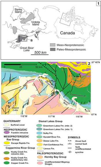
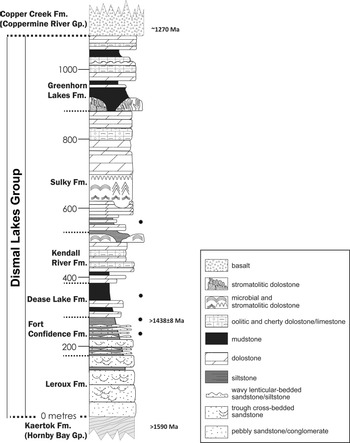
The Dismal Lakes Group is exposed in the Coppermine River basin, which straddles the Nunavut–Northwest Territories border of northern mainland Canada (Fig. 1). The Dismal Lakes Group unconformably overlies the Hornby Bay Group, which forms the base of the Hornby Bay sedimentary basin (Ross et al., Reference Ross, Kerans and Narraway1989). It consists of terrestrial to shallow-marine siliciclastic rocks (LeRoux and Fort Confidence formations), which pass up-section into shallow- and deeper-water carbonate rocks (Dease Lake, Kendall River, Sulky, and Greenhorn Lakes formations; Kerans et al., Reference Kerans, Ross, Donaldson, Geldsetzer, Campbell and Campbell1981; Fig. 2). The Fort Confidence Formation gradationally overlies the LeRoux Formation and has a maximum thickness exceeding 200 m on the basis of outcrop studies (Kerans, Reference Kerans1983). It is composed of interbedded wavy- and lenticular-bedded sandstone and carbonaceous mudstone, interpreted to represent extensive tidal-flat deposits. The shale samples HB07-41A 183m and HB07-41A 232m, from which most of the fossil specimens were recovered, contain folded sandstone dikelets representing infilled desiccation cracks. This is indicating an intermittent subaerial exposure (see Rainbird et al., Reference Rainbird, Rooney, Creaser and Skulski2020).
(1) Location of study area in northwestern Canada. (2) Geological map of the study area. Locations of the samples indicated by asterisks (see table in supplementary data for GPS coordinates); samples 13, 14, 15–18 were extracted from drill core. Modified from Baragar and Donaldson (Reference Baragar and Donaldson1973) and Ross et al. (Reference Ross, Kerans and Narraway1989).

Simplified stratigraphic column for the Dismal Lakes Group, modified from Kerans et al. (Reference Kerans, Ross, Donaldson, Geldsetzer, Campbell and Campbell1981) and Franck et al. (Reference Frank, Kah and Lyons2003). Black asterisks indicate approximate positions of the sampled strata.

The Dease Lake Formation is best exposed west of the Dismal Lakes (Fig. 1) where it comprises three members (Kerans, Reference Kerans1982; Ross et al., Reference Ross, Kerans and Narraway1989). Specimens collected for this microfossil study come from the middle member, which consists of cross-laminated sandy dolostone, microbially laminated to stromatolitic dolostone, and dark siltstone (Skulski at al., Reference Skulski, Rainbird, Turner, Meek, Ielpi, Halverson, Davis, Mercadier, Girard and Loron2018).
Age of the Dismal Lakes Group
The depositional age of the Dismal Lakes Group is broadly constrained to between 1590 Ma—the age of mafic sills that intrude the underlying Hornby Bay Group but not the Dismal Lakes Group (U–Pb; Hamilton and Buchan, Reference Hamilton and Buchan2010)—and 1270 Ma, the U–Pb baddeleyite age of Mackenzie diabase dikes that cross-cut the Dismal Lakes Group (LeCheminant and Heaman, Reference LeCheminant and Heaman1989; French et al., Reference French, Heaman and Chacko2002; Mackie et al., Reference Mackie, Scoates and Weis2009) (Fig. 2). A more specific age of 1438 ± 8 Ma (2σ) was obtained using Re–Os isotope geochronology of finely crystalline pyrite from a shale sample from the Fort Confidence Formation (HB-07-41) (Rainbird et al., Reference Rainbird, Rooney, Creaser and Skulski2020). The pyrite probably formed during early diagenesis of the shale, so its age is probably closer to the minimum age than to the maximum age. Because most of the microfossils from this study are reported from the same strata (HB-07-41A), the age of the present assemblage can be constrained between 1446 and 1430 Ma.
Materials and methods
The shale samples analyzed in this study were collected from drill core (uranium exploration core drilled by Unor Inc. in 2007; see Rainbird et al., Reference Rainbird, Rooney, Creaser and Skulski2020) and outcrop during a field expedition in summer 2017. Samples were crushed into fine fragments and demineralized by static maceration in hydrochloric and hydrofluoric acids in the Early Life Traces and Evolution–Astrobiology Laboratory of the University of Liège (Belgium) (Early life lab) following a low-manipulation and low-agitation protocol established by Grey (Reference Grey1999). The kerogenous residue obtained from acid treatments was filtered with 25 μm and 10 μm mesh-size filters and mounted on microscope slides. Microfossils were identified, measured, and imaged using an Axio Imager A1m microscope equipped with an AxioCam MRc5 digital camera (Carl Zeiss, Germany) in the Early life lab. Additional smear microscopic slides were prepared for scanning electron microscopy (SEM) and gold-coated using a Quorum q150T ES. Images were acquired using an Auriga microscope (Carl Zeiss, Germany) at the Institut de Physique du Globe de Paris (IPGP), Paris, France.
Eighteen samples were collected and prepared from the Sulky, Dease Lake, and Fort Confidence formations. With the exception of CL17-12, all samples yielded organic material, though with variable abundances (see Supplementary Table). Nine samples were fossiliferous (eight from the Fort Confidence Formation and one from the Dease Lake Formation; member d1; Fig. 3).
Microfossil distribution in shale of the Dismal Lakes Group, including early work from Horodyski et al. (Reference Horodyski, Donaldson and Kerans1980). Note that the highest diversity is in sample HB07-41A 183 m.

Repository and institutional abbreviation
All illustrated specimens are identified by slide number—England Finder coordinates and stored in the collection of the Early life lab at the University of Liège, Liège, Belgium (E.J. Javaux).
Systematic paleontology
The microfossils are described following the International Code of Nomenclature for Algae, Fungi, and Plants (Shenzhen Code; ICBN) (Turland et al., Reference Turland2018). Taxa conventionally organized in arbitrary size-class species and previously abundantly described in the literature are left under open nomenclature and are discussed in the text (Siphonophycus spp.; Leiosphaeridia spp., Oscillatoriopsis spp., Tortunema spp., Synsphaeridium spp., Symplassiosphaeridium spp.). In the following, we describe 14 taxa interpreted as eukaryotes or possible eukaryotes. Specimens that are morphologically identical but may differ in size are grouped within a single “species.” Such species are morpho-species, with no necessarily real biological identity, as usual in paleontology. Some specimens placed in distinct species might represent different developmental stages of a same microorganism, but in absence of complementary analyses, they remain interpreted as separate entities. New genera and species are erected for specimens that show clearly different morphologies from those previously known. The taxa are listed in alphabetical order under the designation “Organic-walled microfossils.”
Organic-walled Microfossils
Genus Dictyosphaera Xing and Liu, Reference Xing and Liu1973
Type species
Dictyosphaera macroreticulata Xing and Liu, Reference Xing and Liu1973.
Remarks
The genus was revised by Agić et al. (Reference Agić, Moczydłowska and Yin2015), and the five species D. macroreticulata, D. sinica Xing and Liu, Reference Xing and Liu1973, D. delicata Yin et al., Reference Yin, Xunlai, Fanwei and Jie2005, D. gyrorugosa Hu and Fu, Reference Hu and Fu1982, and D. incrassata Yan and Zhu, Reference Yan and Zhu1992 were synonymized under the type and senior species, D. macroreticulata. Subsequently, Tang et al. (Reference Tang, Pang, Yuan, Wan and Xiao2015) described D. tacita for vesicles with a smooth external wall and smaller hexagonal plates located on the inner vesicle surface, on the basis of two specimens. The present material contains specimens of D. macroreticulata and specimens of D. smaugi n. sp. The latter exhibits a wall partially made of polygonal plates, bears no excystment structure, and therefore is distinct from D. macroreticulata.
Dictyosphaera macroreticulata Xing and Liu, Reference Xing and Liu1973
Figure 4.7–4.9
- Reference Xing and Liu1973
Dictyosphaera macroreticulata Xing and Liu, p. 22, pl. I16, I17.
- Reference Javaux, Knoll and Walter2001
Dictyosphaera sp.; Javaux et al., p. 67, fig. 1e.
- Reference Yin, Xunlai, Fanwei and Jie2005
Dictyosphaera delicata; Yin et al., p. 52, fig. 2.1, 2.2, 2.5, 2.7, 2.9, 2.10.
- Reference Agić, Moczydłowska and Yin2015
Dictyosphaera macroreticulata; Agić et al., p. 32, fig. 2.1–2.9.
- Reference Javaux and Knoll2017
Dictyosphaera macroreticulata; Javaux and Knoll, p. 6, fig. 2.15.
- Reference Agić, Moczydłowska and Yin2017
Dictyosphaera macroreticulata; Agić et al., p. 108, figs. 3A–F, 4A–C, 14G.
- Reference Adam, Skidmore, Mogk and Butterfield2017
Dictyosphaera macroreticulata; Adam et al., p. 388, fig. 3A–C.
- Reference Loron, Rainbird, Turner, Greenman and Javaux2019a
Dictyosphaera macroreticulata; Loron et al., p. 368, fig. 4M.
- Reference Miao, Moczydłowska, Zhu and Zhu2019
Dictyosphaera macroreticulata; Miao et al., p. 185, fig. 4a–f.
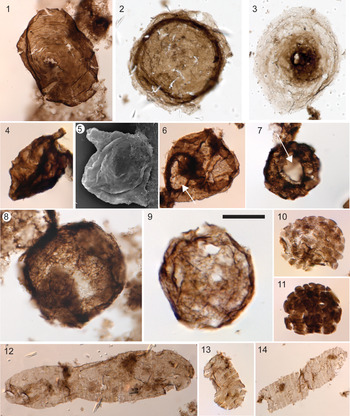
(1) Valeria lophostriata, 76521-t32, 4. (2) Simia annulare, 76091-r27. (3) Pterospermopsimorpha insolita, 76091-v52. (4, 5) Germinosphaera bispinosa: (4) bearing one large process (76801-h28,4); (5) bearing two large processes (DLFC-25; SEM). (6) Osculosphaera hyalina, with a large pylome opening (excystment structure; arrow), 76801-s37,2. (7–9) Dictyosphaera macroreticulata: (7) 76092-n30,1 bearing a pylome opening (arrow); (8) 76567-f45; (9) 75514-o58. (10, 11), Satka favosa: (10) 76803-u34; (11) 76514-m45. (12–14) Spiromorpha segmentata: (12) 76091-j35; (13) 76522-f54, 4; (14) 76511-y41. All photomicrographs taken under transmitted, plane-polarized light. (1–7, 9–14) are from sample HB07-41A 183 m; (8) is from sample HB07-41A 232 m. Scale bar in (9) = 20 μm for (4, 5, 9–11, 13), 30 μm for (1–3, 6, 8, 12), 40 μm for (7), and 50 μm for (14).

See Agić et al. (Reference Agić, Moczydłowska and Yin2015) and Miao et al. (Reference Miao, Moczydłowska, Zhu and Zhu2019) for extended synonymy.
Lectotype
D. macroreticulata Xing and Liu, Reference Xing and Liu1973 (pl. 1, fig. 18) from the Chuanlingguo Formation, northern China, Mesoproterozoic. The species was originally described as Dictyosphaera sinica Xing and Liu, Reference Xing and Liu1973, junior synonym of D. macroreticulata (Agić et al., Reference Agić, Moczydłowska and Yin2015)
Occurrence
Paleoproterozoic of the Chuanlinggou Formation, Changcheng Group, China (Xing and Liu, Reference Xing and Liu1973; Miao et al., Reference Miao, Moczydłowska, Zhu and Zhu2019); Paleoproterozoic–Mesoproterozoic of the Baicaoping and Beidajian formations, Ruyang Group, China (Yin et al., Reference Yin, Xunlai, Fanwei and Jie2005; Agić et al., Reference Agić, Moczydłowska and Yin2015, Reference Agić, Moczydłowska and Yin2017); Mesoproterozoic of the Velkerri Formation, Roper Group, Australia (Javaux et al., Reference Javaux, Knoll and Walter2001; Javaux and Knoll, Reference Javaux and Knoll2017); Mesoproterozoic Greyson Formation, Belt Supergroup, Montana (Adam et al., Reference Adam, Skidmore, Mogk and Butterfield2017); Mesoproterozoic of the Fort Confidence Formation, Dismal Lakes Group, Canada (this study); and late Mesoproterozoic–early Neoproterozoic Escape Rapids and Grassy Bay formations, Shaler Supergroup, Canada (Loron et al., Reference Loron, Rainbird, Turner, Greenman and Javaux2019a). See Agić et al. (Reference Agić, Moczydłowska and Yin2015) for extended occurrences.
Description
Spheroidal vesicles, 37.5 to 132.5 μm in diameter (n = 13). The wall is composed of >1 μm tessellate polygonal platelets. Excystment structure (pylome) is present on one specimen (11.9 μm in diameter).
Materials
Fifty-five specimens in sample CL17-15, HB07-41A 183 m, HB07-41A 232 m, and 08RAT-K106.
Remarks
Although D. macroreticulata is interpreted as a taxon characteristic of Mesoproterozoic rocks (Javaux et al., Reference Javaux, Knoll and Walter2001; Agić et al., Reference Agić, Moczydłowska and Yin2015, Reference Agić, Moczydłowska and Yin2017; Adam et al., Reference Adam, Skidmore, Mogk and Butterfield2017; Javaux and Knoll, Reference Javaux and Knoll2017), it has recently been reported from the <1013–892 ± 13 Ma Grassy Bay Formation, Shaler Supergroup, Canada (Loron et al., Reference Loron, Rainbird, Turner, Greenman and Javaux2019a), extending its biostratigraphic range to at least the entire Mesoproterozoic and probably into the early Neoproterozoic.
Moczydłowska et al. (Reference Moczydłowska, Landing, Zang and Palacios2011) and Agić et al. (Reference Agić, Moczydłowska and Yin2015) suggested that D. macoreticulata could have been a cyst, opening through an operculate pylome excystment structure, as previously observed by Yin et al. (Reference Yin, Xunlai, Fanwei and Jie2005). The specimen bearing a circular pylome reported here (Fig. 4.7) supports this suggestion, although no operculum is evidenced. The presence of this elaborate structure and the tessellated nature of the wall of D. macroreticulata indicate that it was unambiguously a member of the total group eukaryote.
Dictyosphaera smaugi new species
Figure 5
Holotype
Specimen 76091-R37, sample HB07-41A 183m; illustrated in Figure 5.1.
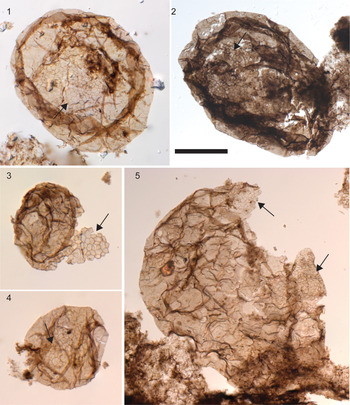
Dictyosphaera smaugi n. sp. with a mostly smooth wall but a restricted area made of polygonal platelets (arrows). (1) Holotype, 76091-r37. (2) 76085-o27. (3) 76514-m44. (4) 76514-j34,2. (5) 76520-p53. All photomicrographs taken under transmitted, plane-polarized light. (1, 3–5) are from sample HB07-41A 183 m; (2) is from sample CL17-14. Scale bar in (2) = 20 μm for (1, 4), 30 μm for (3), and 45 μm for (2, 5).

Diagnosis
Spheroidal to subspheroidal vesicles with wall partly consisting of hexagonal platelets on less than one-third of the surface (most commonly one-sixth of the surface). The area made of platelets is irregular in shape and may vary in form and size, as well as number of plates (two dozen and more), from one specimen to another.
Occurrence
Samples CL17-14, CL17-15, and HB07-41A 183 m, shale of the Fort Confidence Formation, Dismal Lakes Group, in the Dismal Lakes area, Nunavut, Canada.
Description
Vesicles are 55.0 to 270.0 μm in diameter (average = 112.4 μm, n = 25). Platelet sizes are 1.5–2.5 μm.
Etymology
From the J.R.R. Tolkien legendarium, “Smaug, the Golden” dragon, covered by an impenetrable scaled armor, save for his underbelly, in reference to the incompleteness of the platelet pattern on the microfossil vesicle surface.
Materials
Fifty specimens recovered from samples CL17-14, CL17-15, and HB07-41A 183 m.
Remarks
The surface ornamentation is consistent over the 50 specimens reported from the three different strata. This partial cover of platelets is not observed in any other microfossils from these samples. Therefore, it is unlikely that this ornamentation results from superimposition of different materials. The cover is consistently less than a third of the surface in each specimen reported for each stratum. Such consistency is unlikely to result from diagenetic processes as it would have implied that the degradations have stopped at the same stage and time for all specimens of the three samples.
D. smaugi differs from D. macroreticulata by its wall surface. In D. macroreticulata, the whole vesicle wall is made of small tessellated platelets, whereas in D. smaugi the wall is smooth except on one-third to one-sixth of its surface, where it consists of small polygonal tessellated platelets.
The complex microscale wall ornamentation of D. smaugi indicates an affinity of these microfossils to the total group eukaryote (Javaux et al., Reference Javaux, Knoll and Walter2003).
Genus Gangasphaera (Prasad and Asher, Reference Prasad and Asher2001), emend.
Type species
Gangasphaera bulbousus Prasad and Asher, Reference Prasad and Asher2001 (p. 70).
Emended diagnosis
Vesicles spheroidal to subspheroidal bearing one or several prominent spheroidal or subspheroidal bulbous protrusions or extensions of the vesicle wall. The protrusions are rounded and closed at the distal end and freely communicate with the main vesicle.
Remarks
The genus Gangasphaera was erected by Prasad and Asher (Reference Prasad and Asher2001) to accommodate spheroidal to subspheroidal vesicles bearing one or two bulbous protrusions. The wall surface texture characterized by Prasad and Asher (Reference Prasad and Asher2001, p. 70) as “chagrinate to microgranulate” and the described “irregular wrinkles or folds” probably resulted from taphonomy and should not be included in the genus diagnosis. The amended diagnosis used here accommodates specimens from other locations bearing more than two bulbous protrusions (see the following).
Gangasphaera bulbousus (Prasad and Asher, Reference Prasad and Asher2001), emend.
Figure 6.10–6.12
- Reference Jankauskas, Mikhailova and Hermann1989
Своеобразная форма с тремя пленчатымн придатками [peculiar form with three membranous appendages]; Jankauskas et al., p. 168, pl. 42, fig. 1.
- Reference Hermann1990
Densely packed cells widely ranging in size; Hermann, p. 18, pl. 4, fig. 10.
- Reference Hermann1990
Cell under gemmation; Hermann, p. 20, pl. 5, fig. 11.
- Reference Luo1991
Coneosphaera inaequalalis Luo, p. 189, pl. 1, figs 1–3, 7.
- nonReference Luo1991
Coneosphaera inaequalalis Luo, p. 189, pl. 1, figs 4–6.
- Reference Hofmann and Jackson1994
Coneosphaera sp.; Hofmann and Jackson, p. 30, fig. 18.14, 18.15.
- Reference Yin and Guan1999
Trachysphaeridium cf. T. laufeldi; Yin and Guan, p. 135, fig. 5.1, 5.8, 5.10.
- Reference Prasad and Asher2001
Gangasphaera bulbousus Prasad and Asher, p. 69, pl. 11, figs. 1–5.
- Reference Baludikay, Storme, François, Baudet and Javaux2016
Coneosphaera sp.; Baludikay et al., p. 170, fig. 8M, N.
- Reference Loron, Rainbird, Turner, Greenman and Javaux2019a
Coneosphaera sp.; Loron et al., p. 353, fig. 2S.
- Reference Beghin, Storme, Blanpied, Gueneli, Brocks, Poulton and Javaux2017
Coneosphaera cf. C. sp.; Beghin et al., p. 68, pl. 1I.
- Reference Li, Pang, Chen, Zhou, Han, Yang and Wang2019
Sphaeromorphs likely in various stages of cell division; Li et al., p. 270, fig. 7I-7R.
- nonReference Li, Pang, Chen, Zhou, Han, Yang and Wang2019
Sphaeromorphs likely in various stages of cell division; Li et al., p. 270, fig. 7S, 7T.
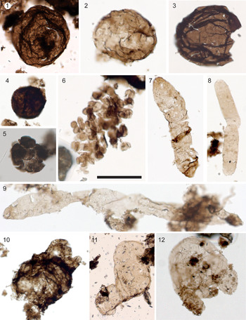
(1–3) Leiosphaeridia spp., nonornamented spheroidal vesicles: (1) L. tenuissima, 76090-w40; (2) L. minutissima, 76548-k53-2; (3) L. tenuissima, 76088-p32. (4) Leiosphere, unidentified spheroidal vesicle, 76092-w45. (5) Synsphaeridium sp., 75377-j48. (6) Symplassiosphaeridium sp., 76092-q40,2. (7–9) Navifusa majensis: (7) 76520-e41,2; (8) 76514-e56,3; (9) 76803-s28,2. (10–12) Gangasphaera bulbousus: (10) 76511-s36; (11) 76506-v28; (12) 76801-j34. All images taken under transmitted, plane-polarized light. (1, 4, 6–12) are from sample HB07-41A 183 m; (2) is from sample HB07-41A 232 m; (3) is from sample CL17-15; (5) is from sample 08RAT-K106. Scale bar in (6) = 30 μm for (2, 5, 6), 50 μm for (1, 3, 4, 7, 8, 12), 100 μm for (9, 10), and 150 μm for (11).

Holotype
Specimen UJN-D-A, DC-13 in Prasad and Asher (Reference Prasad and Asher2001, pl. 11, fig. 1).
Emended diagnosis
Spheroidal to subspheroidal vesicles bearing one to several spheroidal to subspheroidal bulbous protrusions. Protrusions are rounded at distal end and communicate freely with the vesicle interior.
Occurrences
This form-taxa is ubiquitous and long ranging, spanning from the early Mesoproterozoic (Prasad and Asher, Reference Prasad and Asher2001; this study) to the Tonian (Jankauskas et al., Reference Jankauskas, Mikhailova and Hermann1989; Hermann, Reference Hermann1990; Li et al., Reference Li, Pang, Chen, Zhou, Han, Yang and Wang2019).
Description
The vesicles are 43.1–103.9 μm in minimum diameter (n = 7). Bulbous protrusions are 8.4–82.5 μm in diameter (n = 10).
Material
Eight specimens in sample HB07-41A 183 m.
Remarks
The genus Gangasphaera was created by Prasad and Asher (Reference Prasad and Asher2001) to accommodate spheroidal to subspheroidal specimens bearing one or two bulbous protrusions. They differ from the specimens reported as Coneosphaera sp. by Hofmann and Jackson (Reference Hofmann and Jackson1994), Baludikay et al. (Reference Baludikay, Storme, François, Baudet and Javaux2016), Beghin et al. (Reference Beghin, Storme, Blanpied, Gueneli, Brocks, Poulton and Javaux2017), and Loron et al. (Reference Loron, Rainbird, Turner, Greenman and Javaux2019a) only in the number of protrusions. The original diagnosis of the genus Coneosphaera by Luo (Reference Luo1991) referred to “aggregated colonies of small spheroids surrounding a single, larger spheroid” (Luo, Reference Luo1991, p. 189; Hofmann and Jackson, Reference Hofmann and Jackson1994, p. 30), which is not compatible with specimens from the aforementioned authors. These specimens fit the diagnosis of the genus Gangasphaera (Prasad and Asher, Reference Prasad and Asher2001), and so they are synonymized under its type species, G. bulbousus (Prasad and Asher, Reference Prasad and Asher2001) emend.
The original diagnosis of G. bulbousus (Prasad and Asher, Reference Prasad and Asher2001) included both size and taphonomic information. The wall surface texture characterized by Prasad and Asher (Reference Prasad and Asher2001, p. 70) as “chagrinate to microgranulate” and the report of “irregular wrinkles or folds” are irrelevant for taxonomic diagnosis because they almost certainly result from taphonomy.
The large range of morphology and size documented in these microfossils does not exclude possible polyphyletism. In addition, the morphology of G. bulbousus, although certainly complex in its variability, does not show any strong, undeniably eukaryotic trait (Javaux et al., Reference Javaux, Knoll and Walter2003) and cannot confidently be designated as either prokaryote or eukaryote (incertae sedis).
Genus Germinosphaera Mikhailova (Reference Mikhailova and Sokolov1986) Butterfield in Butterfield et al., Reference Butterfield, Knoll and Swett1994
Type species
Germinosphaera bispinosa Mikhailova, Reference Mikhailova and Sokolov1986, p. 33.
Remarks
The genus Germinosphaera was created to accommodate process-bearing spheroidal vesicles with one or two tubular processes that communicate freely with the vesicle interior and are distributed on a single “equatorial” plane. On the basis of the number of processes, two distinct species were originally erected by Mikhailova (Reference Mikhailova and Sokolov1986): G. unispinosa and G. bispinosa. Butterfield et al. (Reference Butterfield, Knoll and Swett1994) reported specimens with a highly variable number of processes (one to six processes) and showed that the variable number of processes was intraspecific. The genus was emended, and the two species were subsumed into one type species, G. bispinosa.
In 2019, Miao and colleagues erected G. alveolata, a distinct species of Germinosphaera, on the basis of differences in the wall surface structure. The species is emended in the following to include the results of new SEM evidence.
The presence of processes, and surface ornamentation for G. alveolata, indicates that Germinosphaera is unambiguously eukaryotic (Javaux et al., Reference Javaux, Knoll and Walter2003; Butterfield, Reference Butterfield2015a). Although long, filamentous branching protrusions have recently been discovered on one new archaeon species (Imachi et al., Reference Imachi2020), and have been known in Planctomycetes–Verrucomicrobia–Chlamydiae (PVC) bacteria, showing that prokaryotes may show complex morphologies supported by their cytoskeleton, the size of Germinosphaera, and of many other Proterozoic organic-walled acanthomorphic microfossils, strongly differs by several orders of magnitude from this 0.5 μm archaeon and few-microns-sized bacteria. Moreover, these prokaryotes are unknown so far in the fossil record.
Germinosphaera alveolata (Miao et al., Reference Miao, Moczydłowska, Zhu and Zhu2019), emend.
Figure 7.4–7.10
- Reference Miao, Moczydłowska, Zhu and Zhu2019
Germinosphaera alveolata (Miao et al., Reference Miao, Moczydłowska, Zhu and Zhu2019, p. 187, fig. 5g–k).
Holotype
Specimen PB22506, ChL-CQ0501, Q/36 in Miao et al. (Reference Miao, Moczydłowska, Zhu and Zhu2019, fig. 5g).
Emended diagnosis
“Spheroidal to slightly elongate vesicle with a single robust process extending gradually from the vesicle wall. Process is hollow, having a broad base, slightly tapering towards the end, and communicating freely with the vesicle cavity” (Miao et al., Reference Miao, Moczydłowska, Zhu and Zhu2019, p. 187). Vesicle and process surface are ornamented with irregularly overlapping scale-like structures.
Occurrence
Late Paleoproterozoic Chuanlinggou Formation, Changchang Group, China (Miao et al., Reference Miao, Moczydłowska, Zhu and Zhu2019) and Mesoproterozoic Fort Confidence Formation, Dismal Lakes Group, Canada (this study).
Description
Vesicles are 25.9–57.0 μm in diameter (n = 20). Processes are 5.0–14.3 μm wide (n = 20). The length of the processes is difficult to evaluate due to preservation (breakage). Scales are <1 μm in size.
Materials
Materials include 144 specimens from sample HB07-41A 183 m.
Remarks
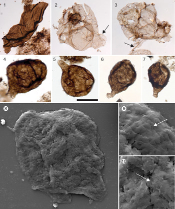
This species constitutes an example of the limitations of optical microscopy in taxonomy. The “alveolar” surface pattern described by Miao et al. (Reference Miao, Moczydłowska, Zhu and Zhu2019) in the type specimens is similar in optical microscopy to the surface pattern of the present specimens from the Dismal Lakes Group, and the microfossils undoubtedly belong to the same species, but only electron microscopy revealed that this pattern actually corresponds to overlapping organic scales forming the surface structures (Fig. 7.9, 7.10). For such complex taxa, electron microscopy constitutes a useful taxonomic tool.
(1) Lineaforma elongata, 76517-f28. (2, 3) Tappania? sp.: (2) 76553-o30; (3) 76515-o58. (4–10) Germinosphaera alveolata emend.: (4) 76091-n29,3; (5) 76522-r59; (6) 76804-n37; (7) 76092-h45, 3; (8–10) DLFC-25; SEMs show the wall structure of the microfossils, made of overlapping polygonal scale-like plates (arrows in (9) and (10)). (1–7) Taken under a plane-polarized, transmitted light; (1, 3–10) are from sample HB07-41A 183 m; (2) is from sample HB07-41A 232 m. Scale bar in (5) = 2 μm for (9, 10), 5 μm for (8), 20 μm for (4–7), and 30 μm for (1–3).

Germinosphaera bispinosa (Mikhailova, Reference Mikhailova and Sokolov1986) Butterfield in Butterfield et al., Reference Butterfield, Knoll and Swett1994
Figure 4.4, 4.5
- Reference Mikhailova and Sokolov1986
Germinosphaera bispinosa Mikhailova, p. 33, fig. 6.
- Reference Mikhailova and Sokolov1986
Germinosphaera unispinosa Mikhailova, p. 33, fig. 5.
- Reference Butterfield, Knoll and Swett1994
Germinosphaera bispinosa; Butterfield in Butterfield et al., p. 38, fig. 16D, E.
- Reference Loron, Rainbird, Turner, Greenman and Javaux2018
Germinosphaera bispinosa; Loron and Moczydłowska, p. 24, pl. 1, fig. 3.
- Reference Loron, Rainbird, Turner, Greenman and Javaux2019a
Germinosphaera bispinosa; Loron et al., p. 364, fig. 8E, F.
See Loron and Moczydłowska (Reference Loron and Moczydłowska2018) for extended synonymy.
Holotype
Specimen no. 882/2 in Mikhailova (Reference Mikhailova and Sokolov1986, fig. 6).
Occurrence
Neoproterozoic (upper Riphean) Dashka Formation, East Siberian Platform, Siberia (Mikhailova, Reference Mikhailova and Sokolov1986); Neoproterozoic Svanbergfjellet Formation, Akademikerbreen Group Spitsbergen (Butterfield et al., Reference Butterfield, Knoll and Swett1994); Neoproterozoic upper formation, Visingsö Group, Sweden (Loron and Moczydłowska, Reference Loron and Moczydłowska2018); late Mesoproterozoic Escape Rapids Formation and early Neoproterozoic Grassy Bay Formation, Shaler Supergroup, Canada (Loron et al., Reference Loron, Rainbird, Turner, Greenman and Javaux2019a); Mesoproterozoic of the Fort Confidence Formation, Dismal Lakes Group, Canada (this study); and various other occurrences from the early Mesoproterozoic to early Cambrian (see Loron and Moczydłowska, Reference Loron and Moczydłowska2018).
Description
Smooth spheroidal vesicle (16.0–25.8 μm in diameter; n = 3) bearing one or two unbranched processes (10.5–12 μm in width; n = 4) that communicate freely with the vesicle interior.
Materials
Three specimens from sample HB07-41A 183 m.
Remarks
Butterfield et al. (Reference Butterfield, Knoll and Swett1994) emended the species to include specimens with one to four processes, distributed on the equatorial plan of the vesicle, and synonymized G. bispinosa and G. unispinosa into G. bispinosa (according to name priority).
Genus Lineaforma Vorob'eva et al., Reference Vorob'eva, Sergeev and Petrov2015
Type species
Lineaforma elongata Vorob'eva et al., Reference Vorob'eva, Sergeev and Petrov2015.
Lineaforma elongata Vorob'eva et al., Reference Vorob'eva, Sergeev and Petrov2015
Figure 7.1
- Reference Javaux, Knoll and Walter2004
Large striated tubes, Javaux et al., p. 126, fig 3g–k.
- Reference Vorob'eva, Sergeev and Petrov2015
Lineaforma elongata Vorob'eva et al., p. 216, fig. 7.1–7.5.
- Reference Javaux and Knoll2017
Lineamorpha elongata; Javaux and Knoll, p. 13, fig. 5.1–5.4.
- Reference Adam, Skidmore, Mogk and Butterfield2017
Lineaforma elongata; Adam et al., p. 388, fig. 2G, I.
Holotype
Specimen GINPC 14711-804 in Vorob'eva et al. (Reference Vorob'eva, Sergeev and Petrov2015, fig. 7.4).
Occurrence
Early Mesoproterozoic Jalboi, Crawford, and Mainoru formations, Roper Group, Australia (Javaux et al., Reference Javaux, Knoll and Walter2004; Javaux and Knoll, Reference Javaux and Knoll2017); Fort Confidence Formation, Dismal Lakes Group, Canada (this study); Belt Supergroup, Montana (Adam et al., Reference Adam, Skidmore, Mogk and Butterfield2017); and Mesoproterozoic Kotuikan Formation, Siberia (Vorob'eva et al., Reference Vorob'eva, Sergeev and Petrov2015).
Description
Longitudinally striated filament, 18.0–73.9 μm wide (n = 6). No complete filaments are found.
Materials
Six fragmental specimens in samples CL17-14, HB07-41A 183m, and HB07-41A 232m.
Remarks
Morphological and ultrastructural analyses (optical microscopy, SEM, transmission electron microscopy) of L. elongata specimens from the Roper Group, Australia, have shown that the striated surface sculpture of these large hollow filamentous tubes reflects original compositional heterogeneities in the tube wall, indicating complex physiological controls on wall formation different from bundles of filaments or fibrous wall ultrastructure known in prokaryotes (Javaux et al., Reference Javaux, Knoll and Walter2004). The combination of an ornamented wall and a complex ultrastructure supports a eukaryotic interpretation for L. elongata tubes (Javaux et al., Reference Javaux, Knoll and Walter2003, Reference Javaux, Knoll and Walter2004; Javaux and Knoll, Reference Javaux and Knoll2017).
Genus Osculosphaera (Butterfield in Butterfield et al., Reference Butterfield, Knoll and Swett1994) emend.
Type species
Osculosphaera hyalina Butterfield in Butterfield et al., Reference Butterfield, Knoll and Swett1994.
Original diagnosis
Psilate, hyaline spheroidal vesicles with a single, rimmed, circular opening, 25–50% the diameter of the vesicle (Butterfield et al., Reference Butterfield, Knoll and Swett1994, p. 43)
Emended diagnosis
Smooth-walled vesicles with a single, circular opening. This opening might be operculate or not, and the operculum might be smooth-walled or ornamented.
Remarks
Butterfield et al. (Reference Butterfield, Knoll and Swett1994) erected this genus to accommodate spheroidal microfossils preserved in chert and bearing a rimmed circular opening. The circular opening of Osculosphaera is interpreted as a pylome, a regular circular excystment aperture that opens to liberate the cyst content. In microfossils, such openings might be operculate (e.g., L. kulgunica (Jankauskas et al., Reference Jankauskas, Mikhailova and Hermann1989) in Loron et al., Reference Loron, Rainbird, Turner, Greenman and Javaux2019a, fig. 6E, F), operculate with an ornamented operculum (e.g., Kaibabia gemmulella Porter and Riedman, Reference Porter and Riedman2016 (fig. 7.1–7.9)), or not operculate (e.g., L. kulgunica in Jankauskas et al., Reference Jankauskas, Mikhailova and Hermann1989, pl. 11, figs. 8–10; O. hyalina in Butterfield et al., Reference Butterfield, Knoll and Swett1994, fig. 15F–J). The absence of an operculum might be due to preservation (detached and lost opercula) or original absence (formation of a pylome by enzymatic digestion of the wall). In the latter cases, it is possible that such a morphospecies actually includes several different biological entities.
Cell wall rupture to liberate the cell content can be observed in some pleurocapsalean cyanobacteria (Waterbury and Stanier, Reference Waterbury and Stanier1978), but the morphological complexity of a pylome (operculate or not) and required genetic and cellular machinery are unknown in prokaryotes (Javaux et al., Reference Javaux, Knoll and Walter2003) and indicate a eukaryotic affinity.
Smooth-walled pylome-bearing forms are not rare in the Precambrian, and the presence of a circular excystment opening constitutes the only substantial morphological distinction from specimens of the genus Leiosphaeridia. Some leiospheres were certainly precursors of these pylome-bearing microfossils (Butterfield et al., Reference Butterfield, Knoll and Swett1994). However, the presence of a pylome (a eukaryotic trait; Javaux et al., Reference Javaux, Knoll and Walter2003) constitutes a criterion to erect a distinct genus from Leiosphaeridia, a basket genus gathering arbitrarily sorted smooth-walled spheroidal microfossils of uncertain biological affinity.
Following Butterfield et al. (Reference Butterfield, Knoll and Swett1994), we propose here to recombine all smooth-walled pylome-bearing microfossils under the genus Osculosphaera and recognize three distinct morphospecies: (1) O. gemmulella (Porter and Riedman, Reference Porter and Riedman2016) n. comb., bearing an operculate pylome ornamented with numerous ~1 μm granulae; (2) O. kulgunica (Jankauskas et al., Reference Jankauskas1980) Butterfield et al., Reference Butterfield, Knoll and Swett1994, bearing a smooth-walled operculate pylome (operculum can be absent); and (3) O. hyalina Butterfield et al., Reference Butterfield, Knoll and Swett1994, bearing a non-operculate, rimmed, circular opening. In O. kulgunica, opercula were suggested lost by Jankauskas et al. (Reference Jankauskas, Mikhailova and Hermann1989) and were detected only in microfossils reported by Loron et al. (Reference Loron, Rainbird, Turner, Greenman and Javaux2019a).
Loron and Moczydłowska (Reference Loron and Moczydłowska2018) erected the new species L. gorda on the basis of a smooth-walled specimen bearing a large, polygonal-shaped pylome opening. Only two specimens are illustrated, and one of them (pl. 2, fig. 4) might be modern contamination. They might constitute a distinct species of Osculosphaera, but this will require the observation of more convincing specimens.
A circular, sometimes operculated, excystment structure (pylome) requires the presence of a complex cytoskeleton, sophisticated genetic programming, and enzymatic machinery to digest and open the recalcitrant wall along a predetermined circular line and, thereby, constitutes an unambiguous character at the level of eukaryotic cellular complexity (Javaux et al., Reference Javaux, Knoll and Walter2003, Reference Javaux, Knoll and Walter2004; Javaux, Reference Javaux and Jékely2007, Reference Javaux2011; Porter, Reference Porter2020). Polyphyly remains possible within these form species, but it is now constrained within the domain eukaryote.
Osculosphaera hyalina Butterfield in Butterfield et al., Reference Butterfield, Knoll and Swett1994
Figure 4.6
- Reference Butterfield, Knoll and Swett1994
Osculosphaera hyalina Butterfield in Butterfield et al., p. 43, fig. 15F–J.
Holotype
Specimen HUPC 627 1 6, illustrated by Butterfield et al. (Reference Butterfield, Knoll and Swett1994, fig. I SF).
Occurrence
Mesoproterozoic of the Fort Confidence Formation, Dismal Lakes Group, Canada (this study); Neoproterozoic of the Svanbergfjellet Formation, Spitsbergen (Butterfield et al., Reference Butterfield, Knoll and Swett1994).
Description
Smooth-wall spheroidal vesicle ranging from 16.0 to 55.0 μm in minimum diameter (n = 5) and bearing a circular opening 6.7–19.5 μm in diameter (n = 5).
Materials
Five specimens in samples CL17-14 and HB07-41A 183m.
Remarks
Specimens of Osculosphaera hyalina reported by Nagovitsin (Reference Nagovitsin2009, fig. 5d, e) do not display a rimmed opening as diagnostic of the species and are, here, recognized as O. kulgunica.
Genus Pterospermopsimorpha (Timofeev, Reference Timofeev1966) Mikhailova in Jankauskas et al., Reference Jankauskas, Mikhailova and Hermann1989
Type species
Pterospermopsimorpha pileiformis Timofeev, Reference Timofeev1966, emend. Mikhailova in Jankauskas et al., Reference Jankauskas, Mikhailova and Hermann1989.
Remarks
The genus was described as morphotaxa corresponding to a vesicle enclosing another vesicle (disphaeromorph), with a smooth wall (P. insolita [Timofeev, Reference Timofeev1969]) or granular wall (P. pileiformis). Sixteen different taxa were described for this genus (Fensome et al., Reference Fensome, Williams, Barss, Freeman and Hill1990; Jachowicz-Zdanowska, Reference Jachowicz-Zdanowska2013), but some of them were subsequently synonymized and subsumed in P. insolita (see Loron and Moczydłowska, Reference Loron and Moczydłowska2018). Opening of the outer vesicle through medial split is observed in some specimens (e.g., Loron et al., Reference Loron, Rainbird, Turner, Greenman and Javaux2019a, fig. 8C).
The morphology of Pterospermopsimorpha is similar to the phycoma (resting stage) of prasinophyte algae, and affinity to this clade of eukaryote has been proposed by several authors (e.g., Tappan, Reference Tappan1980; Inouye et al., Reference Inouye, Hori and Chihara1990; Guy-Ohlsson, Reference Guy-Ohlson, Jansonius and McGregor1996; Samuelsson et al., Reference Samuelsson, Dawes and Vidal1999; Moczydłowska et al., Reference Moczydłowska, Landing, Zang and Palacios2011; Moczydłowska, Reference Moczydłowska2016). However, the possibility of morphological convergence cannot be discarded, and without further information about their ultrastructure and wall chemistry, specimens of Pterospermopsimorpha cannot be interpreted as unambiguous crown-group eukaryotes. The presence of a recalcitrant vesicle within another recalcitrant vesicle has been suggested to indicate the presence of cytoskeleton and organelles (Parke et al., Reference Parke, Boalch, Jowett and Harbour1978; Graham and Wilcox, Reference Graham and Wilcox2000), unknown in prokaryotes. Regardless of taxonomy, we interpret Pterospermopsimorpha as a member of the total group eukaryote because of the combination of a disphaeromorph morphology and the presence of granular ornamentation on the external vesicle and of medial split openings in some specimens. The combination of these characters differs from bacterial colonial envelopes enclosing several small cells.
Pterospermopsimorpha insolita (Timofeev, Reference Timofeev1969) Mikhailova in Jankauskas et al., Reference Jankauskas, Mikhailova and Hermann1989
Figure 4.3
- Reference Timofeev1969
Pterospermopsimorpha insolita Timofeev, p. 16, pl. 3, fig. 8.
- Reference Jankauskas, Mikhailova and Hermann1989
Pterospermopsimorpha insolita; Mikhailova in Jankauskas et al., Reference Jankauskas, Mikhailova and Hermann1989.
- Reference Riedman and Porter2016
Pterospermopsimorpha insolita; Riedman and Porter, p. 873, fig. 12.6–12.9.
- Reference Baludikay, Storme, François, Baudet and Javaux2016
Pterospermopsimorpha insolita; Baludikay et al., p. 170, fig. 7I–L.
- Reference Beghin, Storme, Blanpied, Gueneli, Brocks, Poulton and Javaux2017
Pterospermopsimorpha insolita; Beghin et al., p. 73, pl. 3., figs. b–d.
- Reference Agić, Moczydłowska and Yin2017
Pterospermopsimorpha insolita; Agić et al., p. 113, fig. 10A–C.
- Reference Loron and Moczydłowska2018
Pterospermopsimorpha insolita; Loron and Moczydłowska, p. 18, pl. 4, figs. 1–6.
- Reference Loron, Rainbird, Turner, Greenman and Javaux2019a
Pterospermopsimorpha insolita; Loron et al., p. 358, fig. 8B, C.
- Reference Miao, Moczydłowska, Zhu and Zhu2019
Pterospermopsimorpha insolita; Miao et al., p. 189, fig. 7a–d.
For extended synonymy see Loron and Moczydłowska (Reference Loron and Moczydłowska2018) and Miao et al. (Reference Miao, Moczydłowska, Zhu and Zhu2019).
Holotype
Specimen with preparation number 16/5 illustrated by Timofeev (Reference Timofeev1969, p. 16, pl. 3, fig. 8). Jankauskas et al. (Reference Jankauskas, Mikhailova and Hermann1989) reported the holotype as lost. Lectotype was selected from the same location and illustrated: preparation number 16/42 in Jankauskas et al. (1989, p. 49, pl. 3, fig. 6).
Occurrence
This long-ranging taxon is cosmopolitan in the Precambrian. It has been reported worldwide from the late Paleoproterozoic (Agić et al., Reference Agić, Moczydłowska and Yin2017; Miao et al., Reference Miao, Moczydłowska, Zhu and Zhu2019) to mid-Neoproterozoic (Riedman and Porter, Reference Riedman and Porter2016).
Description
Smooth-walled spheroidal vesicle enclosing another, more opaque, vesicle. The outer vesicle is 55.5–292.5 μm in minimum diameter, and the inner vesicle is 35.0–116.5 μm in minimum diameter (n = 5).
Materials
Five specimens reported from samples HB07-41A 183m and HB07-41A 232m.
Remarks
It is possible that P. insolita represents a developmental variant of Leiospheria spp. that are simple smooth-walled vesicles and could correspond to the empty outer vesicle for P. insolita or to its inner vesicle without the external envelope.
Genus Satka (Jankauskas, Reference Jankauskas1979) Loron et al., Reference Loron, Rainbird, Turner, Greenman and Javaux2019a
Type species
Sakta favosa (Jankauskas, Reference Jankauskas1979).
Remarks
The genus Satka was originally divided into six species (Jankauskas et al., Reference Jankauskas, Mikhailova and Hermann1989). Satka elongata Jankauskas et al., Reference Jankauskas, Mikhailova and Hermann1989 and S. granulosa Jankauskas et al., Reference Jankauskas, Mikhailova and Hermann1989 were synonymized with S. favosa by Javaux and Knoll (Reference Javaux and Knoll2017), and Satka colonialica Jankauskas, Reference Jankauskas1979 was combined with the genus Squamosphaera (Tang et al., Reference Tang, Pang, Yuan, Wan and Xiao2015). Loron et al. (Reference Loron, Rainbird, Turner, Greenman and Javaux2019a) followed the reevaluation of Tang et al. (Reference Tang, Pang, Yuan, Wan and Xiao2015) and emendation by Porter and Riedman (Reference Porter and Riedman2016) and recognized Squamosphaera as a distinct genus from Satka. Loron et al. (Reference Loron, Rainbird, Turner, Greenman and Javaux2019a) proposed to remove Satka undosa Jankauskas, Reference Jankauskas1979 from the genus and to recombine it with Synsphaeridium. Similarly, they recombined Satka squamosphaera Pyatiletov, Reference Pyatiletov1980 with the genus Squamosphaera. The genus Satka originally included lobate forms (now Squamosphaera) and forms made of polygonal plates (Satka favosa). For this reason, it was emended to conform with the description of its type species: Satka favosa (Loron et al., Reference Loron, Rainbird, Turner, Greenman and Javaux2019a).
Sakta favosa Jankauskas, Reference Jankauskas1979
Figure 4.10, 4.11
- Reference Jankauskas1979
Satka favosa Jankauskas, pl. 4, fig. 2.
- Reference Jankauskas, Mikhailova and Hermann1989
Satka favosa; Jankauskas et al., p. 51, pl. 4, figs. 1, 2?
- Reference Jankauskas, Mikhailova and Hermann1989
Satka elongata; Jankauskas et al., p. 51, pl. 4, figs. 3, 5.
- Reference Jankauskas, Mikhailova and Hermann1989
Satka granulosa; Jankauskas et al., p. 51, pl. 4, fig. 8.
- Reference Jankauskas, Mikhailova and Hermann1989
Satka squamifera; Jankauskas et al., p. 51, pl. 5, figs. 3, 8.
- Reference Hofmann and Jackson1994
Satka spp.; Hofmann and Jackson, pl. 18, figs. 26–31.
- Reference Javaux and Knoll2017
Satka favosa; Javaux and Knoll, p. 15, figs. 5.6–5.9.
- Reference Loron, Rainbird, Turner, Greenman and Javaux2019a
Sakta favosa; Loron et al., p. 371, fig. 7A–D.
See Loron et al. (Reference Loron, Rainbird, Turner, Greenman and Javaux2019a) for extended synonymy and discussion.
Holotype
Specimen with preparation number 16–1815-635 in Jankauskas et al. (Reference Jankauskas, Mikhailova and Hermann1989, pl. 4, fig. 2).
Occurrence
Mesoproterozoic Fort Confidence Formation, Dismal Lakes Group, Canada (this study); Greyson Formation, Belt Supergoup, Montana (Adam et al., Reference Adam, Skidmore, Mogk and Butterfield2017); Eqalulik Formation, Bylot Supergroup, Canada (Hofmann and Jackson, Reference Hofmann and Jackson1994); Mainoru Formation, Roper Group, Australia (Javaux and Knoll, Reference Javaux and Knoll2017); Kamov, Chuktukon, Terina and Brus formations of the southern Urals (Jankauskas et al., Reference Jankauskas, Mikhailova and Hermann1989); and late Mesoproterozoic–early Neoproterozoic Grassy Bay and Nelson formations, Shaler Supergroup, Canada (Loron et al., Reference Loron, Rainbird, Turner, Greenman and Javaux2019a).
Description
Hollow spheroidal vesicles, ranging from 27.6 to 41.2 μm in minimal diameter (n = 5), with a wall made of tessellated polygonal plates. The plates are 3.7–7.0 μm in size (n = 13).
Materials
Seventeen specimens from samples CL17-07, HB07-41A 183m, and 08RAT-K106.
Remarks
The wall of S. favosa, made of tessellated organic plates, implies the presence of a complex cellular machinery that is indicative of its eukaryotic nature (Javaux et al., Reference Javaux, Knoll and Walter2003). By comparison, the simpler bulging envelopes of Squamosphaera could be prokaryotic in origin. The original genus Satka was, before reassignment by Loron et al. (Reference Loron, Rainbird, Turner, Greenman and Javaux2019a), most probably gathering polyphyletic biological species belonging to either domain (Eukaryota and Bacteria). Within the form species Satka favosa, although polyphyletism remains possible, it is constrained within eukaryotes.
Genus Simia Mikhailova and Jankauskas in Jankauskas et al., Reference Jankauskas, Mikhailova and Hermann1989
Type species
Simia simica (Jankauskas, Reference Jankauskas1980) Jankauskas, Reference Jankauskas, Mikhailova and Hermann1989.
Simia annulare (Timofeev, Reference Timofeev1969) Mikhailova in Jankauskas et al., Reference Jankauskas, Mikhailova and Hermann1989
Figure 4.2
- Reference Timofeev1969
Pterospermopsimorpha annulare Timofeev, p. 17, pl. 3, fig. 9.
- Reference Vorob'eva, Sergeev and Knoll2009
Ostiumsphaeridium complitum; Vorob'eva et al., p. 186, fig. 14.1–14.5.
- Reference Tang, Pang, Xiao, Yuan, Ou and Wan2013
Simia annulare; Tang et al., p. 162, fig. 4G.
- Reference Riedman and Porter2016
Simia annulare; Riedman and Porter, p. 874, fig. 12.1–12.5.
- Reference Agić, Moczydłowska and Yin2017
Simia annulare; Agić et al., p. 118, figs. 10FI, 14H, I.
- Reference Beghin, Storme, Blanpied, Gueneli, Brocks, Poulton and Javaux2017
Simia annulare; Beghin et al., p. 73, pl. 3, fig. e.
- Reference Loron and Moczydłowska2018
Simia annulare; Loron and Moczydłowska, p. 21, pl. 5, figs. 1–3.
- Reference Miao, Moczydłowska, Zhu and Zhu2019
Simia annulare; Miao et al., p. 192, fig. 7e–g.
- Reference Loron, Rainbird, Turner, Greenman and Javaux2019a
Simia annulare; Loron et al., p. 358, fig. 8A.
Holotype
Specimen with preparation number 147/4 illustrated by Timofeev (Reference Timofeev1969, pl. 3, fig. 9).
Occurrence
This long-ranging fossil is present from the late Paleoproterozoic–early Mesoproterozoic (Miao et al., Reference Miao, Moczydłowska, Zhu and Zhu2019) to the Ediacaran (Vorob'eva et al., Reference Vorob'eva, Sergeev and Knoll2009).
Description
Spheroidal vesicles bearing an equatorial flange. The vesicle diameter is ranging from 62.5 to 105.5 μm and the flange is 5–7.5 μm wide (n = 7).
Materials
Nineteen specimens recovered from the samples HB07-41A 183m, HB07-41A 232m, and 08RAT-K106.
Remarks
As mentioned by Riedman and Porter (Reference Riedman and Porter2016), there was much confusion between the pteromorph (vesicle bearing an equatorial flange) genus Simia and the disphaeromorph (vesicle enclosing another vesicle) genus Pterospermopsimorpha. Because of the equatorial development of its flange, Simia resembles the genus Pterospermella known in the Phanerozoic successions since the Cambrian (Eisenack, Reference Eisenack1972; Moczydłowska, Reference Moczydłowska2016) and reported from the Neoproterozoic (Loron and Moczydłowska, Reference Loron and Moczydłowska2018), but Pterospermella is a disphaeromorph and not a pteromorph (see discussion in Loron and Moczydłowska, Reference Loron and Moczydłowska2018).
Many authors have interpreted vesicles of Simia (along with Pterospermopsimorpha and Pterospermella) as members of the class Prasynophyceae (e.g., Tappan, Reference Tappan1980; Inouye et al., Reference Inouye, Hori and Chihara1990; Playford, Reference Playford2003; Moczydłowska et al., Reference Moczydłowska, Landing, Zang and Palacios2011; Moczydłowska, Reference Moczydłowska2016). The large equatorial flange ornamenting the vesicles of Simia annulare is a complex morphology that ascribes them with confidence among eukaryotes (Javaux et al., Reference Javaux, Knoll and Walter2003). However, despite morphological resemblance with modern prasinophytes (e.g., Pterosperma), further ultrastructural and chemical analyses are required to identify Simia as a stem or crown eukaryote.
Genus Spiromorpha Yin et al., Reference Yin, Xunlai, Fanwei and Jie2005
Type species
Spiromorpha segmentata (Prasad and Asher, Reference Prasad and Asher2001) Yin et al., Reference Yin, Xunlai, Fanwei and Jie2005.
Spiromorpha segmentata (Prasad and Asher, Reference Prasad and Asher2001) Yin et al., Reference Yin, Xunlai, Fanwei and Jie2005
Figure 4.12–4.14
- Reference Prasad and Asher2001
Navifusa segmentatus Prasad and Asher, p. 77, pl. 5, figs. 4, 5, 14, 15.
- Reference Yin, Xunlai, Fanwei and Jie2005
Spiromorpha segmentata; Yin et al., p. 57, fig. 5.1, 5.4–5.8.
- Reference Nagovitsin2009
“short trichomes containing terminal lenticular and medial arcuate cells”; Nagovitsin, p. 143, fig. 5h, i.
- Reference Pang, Tang, Yuan, Wan and Xiao2015
Spiromorpha sp.; Pang et al., p. 254, fig. 2C.
- Reference Beghin, Storme, Blanpied, Gueneli, Brocks, Poulton and Javaux2017
Spiromorpha segmentata; Beghin et al., p. 73, pl. 3, figs. o, p.
Holotype
Specimen KDM-A, 5204-07m, originally Navifusa segmentatus (Prasad and Asher, Reference Prasad and Asher2001, pl. 5, fig. 5).
Occurrence
Late Paleoproterozoic–early Mesoproterozoic Beidajian Formation, Ruyang Group, China (Yin et al., Reference Yin, Xunlai, Fanwei and Jie2005; Pang et al., Reference Pang, Tang, Yuan, Wan and Xiao2015); Mesoproterozoic (early middle Riphean) Sarda and Avadh formations, Bahraich Group, India (Prasad and Asher, Reference Prasad and Asher2001); Mesoproterozoic of the Fort Confidence Formation, Dismal Lakes Group, Canada (this study); Mesoproterozoic Yurubchen and Dzhelindukon formations, Kamo Group, Siberia (Nagovitsin, Reference Nagovitsin2009); and late Mesoproterozoic Khatt Formation, Atar/El Mreïti Group, Mauritania (Beghin et al., Reference Beghin, Storme, Blanpied, Gueneli, Brocks, Poulton and Javaux2017).
Description
Smooth-walled oval-shaped vesicle, 40.0–114.5 μm long and 15.0–38.5 μm wide (n = 13). Grooves are spirally distributed from one extremity of the vesicle to the other, each groove is ≤1 μm wide.
Materials
Thirty-nine specimens reported from sample HB07-41A 183m.
Remarks
The grooves present on the vesicle surface of S. segmentata are not septae but a surface sculpture (Beghin et al., Reference Beghin, Storme, Blanpied, Gueneli, Brocks, Poulton and Javaux2017). Here we agree and recognize S. segmentata as a total group eukaryote on the basis of the presence of this microscale surface ornamentation (Javaux et al., Reference Javaux, Knoll and Walter2003).
The morphology, with spiral grooves, of S. segmentata closely resembles the basal algal taxon Spirotaenia (Yin et al., Reference Yin, Xunlai, Fanwei and Jie2005). Further investigations of their ultrastructure and chemistry might unravel a crown-group affinity to these microfossils.
Genus Tappania Yin, Reference Yin1997
Type species
Tappania plana Yin, Reference Yin1997.
Tappania? sp.
Figure 7.2, 7.3
- Reference Baludikay, Storme, François, Baudet and Javaux2016
?cf. Tappania sp. Baludikay et al., p. 173, figs. 6R, 7A, B.
Description
Spheroidal to subspheroidal smooth-walled vesicles bearing trapezoidal protrusions and neck-like extensions. One possible process is present on one specimen (Fig. 7.3).
Materials
Six specimens from samples CL17-15, HB07-41A 183m, and HB07-41A 232m.
Remarks
The present specimens resemble specimens of Tappania plana with neck-like extensions but lack the diagnostic tubular processes of the species (see Javaux and Knoll, Reference Javaux and Knoll2017, fig. 6.5, 6.6). In our material, one specimen (Fig. 7.3) bears what might be a process, but it is possible that this results from taphonomic superimposition. In the absence of all diagnostic characters of the species, we cannot unambiguously recognize these microfossils as Tappania plana.
In Tappania? sp. the neck-like extensions are trapezoidal, differing from the bulbous spheroidal protrusion of Gangasphaera bulbousus.
The complex morphology of Tappania indicates the presence of a dynamic cytoskeleton and endomembrane system, which allow their unambiguous placement within eukaryotes (see discussion in Javaux et al., Reference Javaux, Knoll and Walter2001; Javaux and Knoll, Reference Javaux and Knoll2017). In the absence of taxonomic certainty, we cautiously recognize our specimens of Tappania? sp. as probable eukaryotes. Examination of more specimens of this material is necessary to confirm their identification.
Genus Valeria Jankauskas, Reference Jankauskas1982
Type species
Valeria lophostriata (Jankauskas, Reference Jankauskas1979) Jankauskas, Reference Jankauskas1982.
Valeria lophostriata (Jankauskas, Reference Jankauskas1979) Jankauskas, Reference Jankauskas1982
Figure 4.1
- Reference Jankauskas, Mikhailova and Hermann1979
Kildinella lophostriata Jankauskas, p. 153, fig. 1.13–1.15.
- Reference Jankauskas1982
Valeria lophostriata; Jankauskas, p. 109, pl. 39, fig. 2.
- Reference Jankauskas, Mikhailova and Hermann1989
Valeria lophostriata; Jankauskas et al., p. 86, pl. 16, figs. 1–5.
- Reference Javaux, Knoll and Walter2004
Valeria lophostriata; Javaux et al., fig. 2F–I.
- Reference Nagy, Porter, Dehler and Shen2009
Valeria lophostriata; Nagy et al., fig. 1A, B.
- Reference Tang, Pang, Yuan, Wan and Xiao2015
Valeria lophostriata; Tang et al., p. 315, fig. 11.
- Reference Riedman and Porter2016
Valeria lophostriata; Riedman and Porter, p. 10, fig. 4.1.
- Reference Porter and Riedman2016
Valeria lophostriata; Porter and Riedman, fig. 19.1–19.3.
- Reference Baludikay, Storme, François, Baudet and Javaux2016
Valeria lophostriata; Baludikay et al., p. 170, fig. 7H.
- Reference Beghin, Storme, Blanpied, Gueneli, Brocks, Poulton and Javaux2017
Valeria lophostriata; Beghin et al., p. 73, pl. 4, fig. j, k.
- Reference Agić, Moczydłowska and Yin2017
Valeria lophostriata; Agić et al., p. 119, fig. 12I.
- Reference Loron, Rainbird, Turner, Greenman and Javaux2019a
Valeria lophostriata; Loron et al., p. 356, fig. 4E.
- Reference Miao, Moczydłowska, Zhu and Zhu2019
Valeria lophostriata; Miao et al., p. 194, fig. 11a–f.
Holotype
Specimen number 16-62-4762/16, sp. 1 illustrated by Jankauskas (Reference Jankauskas1979, fig. 1.14).
Occurrence
This long-ranging taxon is found worldwide throughout the Proterozoic, from the late Paleoproterozoic (Javaux et al., Reference Javaux, Knoll and Walter2004; Agić et al., Reference Agić, Moczydłowska and Yin2017; Miao et al., Reference Miao, Moczydłowska, Zhu and Zhu2019) to the Cryogenian (Nagy et al., Reference Nagy, Porter, Dehler and Shen2009).
Description
Large spheroidal vesicle (35.0–205.0 μm minimum diameter; n = 13) bearing conspicuous surface ornamentation made of concentric ridges (“archery target” pattern). Medial split opening of some vesicles is observed.
Materials
Materials include 111 specimens reported from samples CL17-15, HB07-41A 183m, HB07-41A 232m, and 08RAT-K106.
Remarks
The distinctive micron-scale regularly spaced concentric striations on the inner surface of the recalcitrant wall of Valeria, combined with the common occurrence of medial split excystment structure, indicate its eukaryotic affinity as such combination is unknown in prokaryotes (Javaux et al., Reference Javaux, Knoll and Walter2003, Reference Javaux, Knoll and Walter2004). Butterfield (Reference Butterfield2015a) pointed out the similarity of this surface pattern with the one observed on the wall of the cyanobacteria Glaucocystis, formed by cellulose fibrils. However, in Glaucocystis, the fibrils are at the nanoscale (~10 nm wide), are cross-linked, and constitute the ultrastructure of the cell wall rather than an ornamentation (Willison and Brown, Reference Willison and Brown1978).
Unnamed sp. A
Figure 8.8
Description
Filamentous microfossil, 21.4 μm wide and 209 μm long, with one flattened and thickened extremity. The opposite extremity is separated into three lobes with rounded shapes.
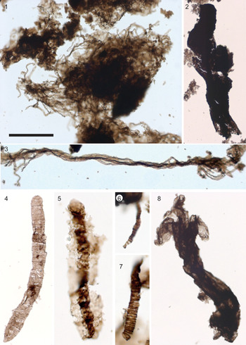
Filamentous forms. (1) Mat of Siphonophyccus spp., 76553-u49. (2) Syphonophyccus gigas, 76085-l46. (3) Polythrichoides lineatus. (4) Oscillatoriopsis sp., 76085-e53,3. (5) Palaeolyngbya sp., 76092-n44,4. (6) Tortunema sp., 76802-p31-1. (7) Cephalonyx sp., 76804-j36. (8) Unnamed species A with trilobate extremity, 76084-p41. All photomicrographs taken under transmitted, plane-polarized light. (1) is from sample HB07-41A 232 m; (2, 3, 8) are from sample CL17-14; (5–7) are from sample HB07-41A 183 m. Scale bar in (1) = 30 μm for (5–7), 60 μm for (1, 4), 120 μm for (3, 8), and 200 μm for (2).

Material
One single filament in sample CL17-14.
Remarks
Such a complex morphology would allow the placement of this specimen among eukaryotes but needs to be confirmed by examination of more specimens to discard possible taphonomic artefact; therefore, it is left in open nomenclature.
Shale-hosted microfossils of the Dismal Lakes Group
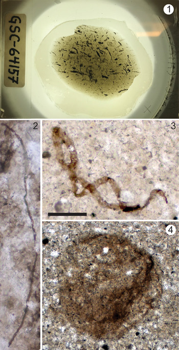
Previous investigations of the Dismal Lakes Group biota reported a very low diversity of organic-walled microfossils. A study of chert samples from the Kendall River, Dease Lake, and Greenhorn River formations yielded a low diversity of prokaryotic filamentous and coccoidal microfossils (Horodyski and Donaldson, Reference Horodyski and Donaldson1980, Reference Horodyski and Donaldson1983). Thin sections of shale from the lower Greenhorn Formation revealed spheroidal and filamentous microfossils of Leiospheridia and Siphonophycus genera (Horodyski et al., Reference Horodyski, Donaldson and Kerans1980). In previous reports, no fossils with eukaryotic attributes were described. Our reinvestigation of this historical material (thin sections GSC-64157, GSC-64158, and GSC-67159) yielded identical conclusions (Figs. 3, 9).
New images of historical material from Horodyski at al. (1980). (1) Photomicrograph of shale in thin-section GSC-64157; note the abundance of organic material (black). (2, 3) Siphonophycus spp. (4) Leiosphaeridia sp. All images are of thin-sectioned (thick section) shale taken under transmitted, plane-polarized light; (2–4) are from thin-section GSC-64159. Scale bar in (3) = 1,500 μm for (1), 150 μm for (3), and 50 μm for (2, 4).

The present work describes 24 different taxa, one new species, and one unnamed form recovered from shales of the middle Dease Lake and Fort Confidence formations, strata that were not investigated for paleontology by Horodyski and Donaldson (Reference Horodyski and Donaldson1980, Reference Horodyski and Donaldson1983).
Filamentous forms
Filamentous forms are present in all of the fossiliferous samples with the exception of the sample from the Dease Lake Formation. Nonseptate empty sheaths of Siphonophycus (Schopf, Reference Schopf1968) Knoll, Swett, and Mark, Reference Knoll, Swett and Mark1991 are the most abundant (Fig. 8.1, 8.2) and may be assigned to seven of the size-class species described by Butterfield et al. (Reference Butterfield, Knoll and Swett1994) and Tang et al. (Reference Tang, Pang, Yuan, Wan and Xiao2015): Siphonophycus septatum (Schopf, Reference Schopf1968) Knoll, Swett, and Mark, Reference Knoll, Swett and Mark1991 (1.0–2.0 μm wide); S. robustum (Schopf, Reference Schopf1968) Knoll, Swett, and Mark, Reference Knoll, Swett and Mark1991 (2.1–4.0 μm wide); S. typicum (Hermann, Reference Hermann and Timofeev1974) Butterfield in Butterfield et al., Reference Butterfield, Knoll and Swett1994 (4.1–8.0 μm wide); S. kestron Schopf, Reference Schopf1968 (8.1–16.0 μm wide); S. solidum (Golub, Reference Golub and Sokolov1979) Butterfield in Butterfield et al., Reference Butterfield, Knoll and Swett1994 (16.1–32.0 μm wide); S. punctatum Maithy, Reference Maithy1975 (32.1–64.0 μm wide); and S. gigas Tang et al., Reference Tang, Pang, Yuan, Wan and Xiao2015 (64.1–128.0 μm wide). Bundles of tightly packed parallel filamentous sheaths are present and identified as Polytrichoides lineatus (Hermann, Reference Hermann and Timofeev1974) (Fig. 8.3). Uniseriate trichomes of Oscillatoriopsis (Schopf, Reference Schopf1968) Butterfield in Butterfield et al., Reference Butterfield, Knoll and Swett1994 and pseudoseptate filaments of Tortunema (Hermann, Reference Hermann and Timofeev1974) Butterfield in Butterfield et al., Reference Butterfield, Knoll and Swett1994 are also present (Fig. 8.4, 8.6) as well as a single specimen of Palaeolyngbya (Schopf, Reference Schopf1968) Butterfield in Butterfield et al., Reference Butterfield, Knoll and Swett1994, with the sheath enclosing remains of the original cells (Fig. 8.5). A small filament with regularly distributed annular bulges is recognized as Cephalonyx Weiss, Reference Weiss1984 (5.5 μm wide, n = 1; Fig. 8.7), and a large unknown form with a trilobate extremity is reported (unnamed species A; Fig. 8.8).
Unornamented microfossils
Spheroidal unornamented vesicles of Leiosphaerida spp. constitute the main abundance of the assemblage (Fig. 6.1–6.3). In this assemblage, the arbitrary size-class species of Leiosphaeridia crassa (Naumova, Reference Naumova1949) Jankauskas et al., Reference Jankauskas, Mikhailova and Hermann1989: L. minutissima (Naumova, Reference Naumova1949) Jankauskas in Jankauskas et al., Reference Jankauskas, Mikhailova and Hermann1989; L. jacutica (Timofeev, Reference Timofeev1966) Jankauskas in Jankauskas et al., Reference Jankauskas, Mikhailova and Hermann1989; and L. tenuissima Eisenack, Reference Eisenack1958 can be recognized on the basis of the size of the vesicles (<70 μm for L. minutissima and L. crassa; >70μm for L. jacutica and L. tenuissima) and shape of the folds indicating wall flexibility (sinuous for L. minutissima and L. tenuissima; lanceolate for L. crassa and L. jacutica) following the principles of Javaux and Knoll (Reference Javaux and Knoll2017). However, a large number of specimens in different samples are opaque and do not display conspicuous taphonomic folds or cracks (leiospheres; Fig. 6.4). These size-class species are morphotaxa that are probably polyphyletic and do not necessarily coincide with biological species, but they are a useful tool to describe diversity of forms within an assemblage and to compare with previous micropaleontological studies.
Aggregates of smooth vesicles are also common in the assemblages, including clusters of closely attached (Synsphaeridium spp.; Fig. 6.5) and loosely attached (Symplassiosphaeridium spp.; Fig. 6.6) vesicles.
Elongated vesicles of Navifusa majensis Pyatiletov, Reference Pyatiletov1980 (25.0–246.8 μm long and 2.5–40.0 μm wide; n = 12) are common (Fig. 6.7–6.9). In sample HB07-41A 183m (Fort Confidence Formation), many of the small specimens of this species (Fig. 6.8) are very similar to ovoidal specimens of Archeoellipsoides preserved in chert (Horodyski and Donaldson, Reference Horodyski and Donaldson1980, Reference Horodyski and Donaldson1983) and may, possibly, constitute their shale-hosted equivalent.
In addition, spheroidal and subspheroidal vesicles with one or several bulbous protrusions are recognized as Gangasphaera bulbousus, a form species with a large morphological variability (Fig. 6.10–6.12).
Ornamented microfossils
The long-ranging taxon Valeria lophostriata, with characteristic circular ridges on the wall inner surface, is abundant throughout the assemblage (Fig. 4.1). Rare specimens of Simia annulare ornamented with an equatorial flange and the disphaeromorph (vesicle enclosing another vesicle) Pterospermopsimorpha insolita are also recognized (Fig. 4.2, 4.3). In addition, five specimens of Osculosphaera hyalina, with a pylome excystment structure (circular opening) are documented (Fig. 4.6). Vesicles of Dictyosphaera macroreticulata (Fig. 4.7–4.9) and Satka favosa (Fig. 4.10, 4.11) are also present. D. macroreticulata has a wall made of tessellate hexagonal plates, whereas wall plates of S. favosa are larger, fewer, and have a polygonal or quadrate shape (see Javaux and Knoll, Reference Javaux and Knoll2017 and Loron et al., Reference Loron, Rainbird, Turner, Greenman and Javaux2019a for discussion of this species). Oval vesicles ornamented with spiral grooves, S. segmentata (Fig. 4.12–4.14), are abundant, and rare fragments of Lineaforma elongata, a large tube with longitudinal striations on its wall surface, also occur. We also report the presence of a new taxa, Dictyosphaera smaugi n. sp. (Fig. 5). These microfossils possess a smooth wall except for a localized area of their wall made of hexagonal platelets (see Fig. 5.1–5.5). As opposed to D. macroreticulata, these structures do not form the whole vesicle wall but are present only on one irregularly shaped area representing less than one-third of the vesicle wall surface.
Acanthomorphic (processes-bearing) microfossils
Three specimens of Germinosphaera bispinosa are present, one of them bearing two equatorial processes (Fig. 4.4, 4.5). The Fort Confidence assemblage also includes the second report of Germinosphaera alveolata (Fig. 7.4–7.10). SEM reveals that the microfossils are covered with small overlapping scale-like structures (Fig. 7.8–7.10) and not alveoli as suggested in its original description (Miao et al., Reference Miao, Moczydłowska, Zhu and Zhu2019).
Six specimens of Tappania? sp. were recovered from samples CL17-15, HB07-41A 183 m, and HB07-41A 232 m (32.0–65.3 μm in size; n = 6); these microfossils are irregularly shaped and have neck-like expansions. One specimen might bear a process (Fig. 7.2, 7.3).
Discussion
The moderate diversity of organic-walled microfossils recovered from the shale units of the Dismal Lakes Group, especially from the Fort Confidence Formation, provide new insights on early Mesoproterozoic life. The 24 described taxa most certainly include members of both eukaryotic and prokaryotic domains.
Affinity of Dismal Lakes Group microfossils
Several authors have proposed criteria to recognize eukaryotic fossils within assemblages. The presence of surface ornamentation, complex wall ultrastructure, complex excystment structure (e.g., a pylome, or circular opening, with occasionally an operculum preserved, to release the cell content), as well as the presence of processes, complex multicellularity, and eukaryotic biopolymers making up the wall, constitute characteristics unknown in prokaryotes (Javaux et al., Reference Javaux, Knoll and Walter2001, Reference Javaux, Knoll and Walter2003, Reference Javaux, Knoll and Walter2004; Javaux and Marshall, Reference Javaux and Marshal2006; Knoll et al., Reference Knoll, Javaux, Hewitt and Cohen2006). As discussed, although long filamentous branching protrusions have recently been discovered on one new archaeon species (Imachi et al., Reference Imachi2020) and verrucae and processes are known in some PVC bacteria, showing that prokaryotes may show complex morphologies supported by their cytoskeleton, the size of the vesicle and ornamentation of Proterozoic organic-walled acanthomorphic microfossils strongly differ by several orders of magnitude from these smaller-than-a-micron- to a-few-microns-sized prokaryotes. Moreover, these prokaryotes are not known to form kerogenous walls fossilized in the geological record. These examples illustrate why most of the criteria listed in the preceding need to be used in combination to discriminate eukaryotic from prokaryotic microfossils.
Similarly, Butterfield (Reference Butterfield2015a) suggests that conspicuous surface ornamentations, processes, and “true” multicellularity (with specialized cells) may unambiguously classify a microfossil as eukaryotic but went further by suggesting that they would indicate a placement among crown eukaryotes. The size of the microfossils, although informative when combined with other criteria, is not valid on its own since the existence of giant bacteria and micro-eukaryote is now well established (Javaux et al., Reference Javaux, Knoll and Walter2003). Among the 24 taxa reported from the assemblage, we interpret 11 of them as being unambiguously eukaryotic because they have one particularly complex character or combine several of these characters (see Table 1).
Microfossil features characteristic of eukaryotic affinity.

The characters observed on the ornamented fossils V. lophostriata, S. annulare, P. insolita, O. hyalina, D. smaugi n. sp., D. macroreticulata, S. favosa, S. segmentata, L. elongata, and process-bearing G. bispinosa and G. alvaeolata indicate the evolution of various biological innovations, showing a eukaryotic grade of cellular complexity such as the presence of a complex cytoskeleton and possibly an endomembrane system (Javaux et al., Reference Javaux, Knoll and Walter2003, Reference Javaux and Marshal2006; Javaux and Marshall, Reference Javaux and Marshal2006; Butterfield, Reference Butterfield2015a). According to criteria from Butterfield (Reference Butterfield2015a), some of the most complex ornamented and process-bearing forms reported here (Dictyosphaera, Satka favosa, Germinosphaera alveolata) might even represent crown eukaryotes. However, without further complementary investigations of their wall ultrastructure and chemistry, and in the absence of taxonomically diagnostic characters, their stem or crown nature cannot be confirmed unambiguously. Nevertheless, the degree of morphological complexity achieved by the reported specimens indicates that the lineages represented by these microfossils have radiated after the first eukaryotic common ancestor (FECA), and although it is unknown whether they belong to stem (before LECA) or crown (after LECA) groups, they are members of the total group eukaryotes and, therefore, no longer considered prokaryotic.
By comparison, specimens of Leiosphaeridia spp., Navifusa majensis, Gangasphaera bulbousus, and the colonial forms Synsphaeridium spp. and Symplassiosphaeridium spp. cannot unequivocally be interpreted as eukaryotes as they do not preserve any diagnostic eukaryotic morphological characters. Careful multi-proxy studies of Leiospharidia wall ultrastructure and composition in the Proterozoic and in the Cambrian have shown that some of them were probably eukaryotic in origin (e.g., Arouri et al., Reference Arouri, Greenwood and Walter2000; Talyzina and Moczydłowska, Reference Talyzina and Moczydlowska2000; Javaux et al., Reference Javaux, Knoll and Walter2004; Moczydłowska and Willman, Reference Moczydłowska and Willman2009), but such analyses have not yet been conducted on the Dismal Lakes microfossils. These few examples illustrate a possible hidden diversity within leiospheres but cannot be extrapolated to all leiospheres through the geological record without further extensive investigations.
In addition, because of the simple but still variable morphology and size distribution of these microfossils, it is possible that they were polyphyletic or that the specimens recovered represent developmental variants of the same biological entity. Unnamed sp. A might present a complex morphology, but this needs to be confirmed to discard possible taphonomic artefact since only a single specimen was observed. The possible eukaryotic affinity of Tappania? sp. remains ambiguous as they display only the neck-like extension diagnostic of this species but no unequivocal processes. More specimens need to be discovered to correctly assign them to the species Tappania plana and the eukaryotic domain.
The remaining simple filamentous forms are globally too simple in their morphology to be recognized as eukaryotic and are usually interpreted as remains of mat-building cyanobacteria (Butterfield et al., Reference Butterfield, Knoll and Swett1994; Demoulin et al., Reference Demoulin, Lara, Cornet, François, Baurain, Wilmotte and Javaux2019).
Biostratigraphic significance of Dismal Lake Group acritarchs
The shale-hosted biota of the Dismal Lakes Group is typical of Proterozoic assemblages, containing abundant sphaeromorphs and filaments. In addition, this assemblage contains diverse ornamented and processes-bearing taxa, identified as eukaryotes (at least 11) and is comparable in diversity only to the biota of the Ruyang Group in China for the late Paleoproterozoic–early Mesoproterozoic period. Most of these eukaryotes are widespread in Proterozoic assemblages worldwide (Valeria, Pterospermopsimorpha) or characteristic of Mesoproterozoic assemblages (Lineaforma, Satka favosa, Dictyosphaera macroreticulata; but see Loron et al., Reference Loron, Rainbird, Turner, Greenman and Javaux2019a for younger occurrences of S. favosa and D. macroreticulata). One species is new (D. smaugi), and finally, this is the second occurrence of Germinosphaera alveolata, and the first in the early Mesoproterozoic, as it was previously reported only from shales of the late Paleoproterozoic Changzhougou Formation, China (Miao et al., Reference Miao, Moczydłowska, Zhu and Zhu2019). The Dismal assemblage also contains one of the oldest known specimens of O. hyalina, a pylome-bearing microfossil previously reported only from the Neoproterozoic (Butterfield et al., Reference Butterfield, Knoll and Swett1994). In addition, the pylome excystment structure documented here on Dictyosphaera macroreticulata (Fig. 4.7) confirms the suggestion that this species opened in a more complex way than simple medial splitting (Yin et al., Reference Yin, Xunlai, Fanwei and Jie2005; Moczydłowska et al., Reference Moczydłowska, Landing, Zang and Palacios2011; Agić et al., Reference Agić, Moczydłowska and Yin2015).
The high diversity of eukaryotes in the Dismal Lakes assemblage is unusual for early Mesoproterozoic strata. Other contemporaneous successions of shale-hosted microfossils record no more than six eukaryotic taxa—Bahraich Group (Prasad and Asher, Reference Prasad and Asher2001); Billyakh Group (Vorob'eva et al., Reference Vorob'eva, Sergeev and Petrov2015); Roper Group (Javaux and Knoll, Reference Javaux and Knoll2017); Belt Supergroup (Adam et al., Reference Adam, Skidmore, Mogk and Butterfield2017); Changcheng Group (Miao et al., Reference Miao, Moczydłowska, Zhu and Zhu2019); lower Vindhyan Supergroup (Prasad et al., Reference Prasad, Uniyal and Asher2005)—with the exception of the Ruyang Group (Yin et al., Reference Yin, Xunlai, Fanwei and Jie2005; Agić et al., Reference Agić, Moczydłowska and Yin2015, Reference Agić, Moczydłowska and Yin2017) and the Kamo Group (Nagovitsin, Reference Nagovitsin2009) (Table 2). The assemblages from China, Australia, Laurentia (USA and Canada), and India generally contain the same taxa, whereas Siberian assemblages are more distinct but similar to assemblages described from younger successions, with the exception of Spiromorpha segmentata and Tappania plana in the Kamo Group (Nagovitsin, Reference Nagovitsin2009) and Lineaforma in the Billyakh Group (Vorob'eva et al., Reference Vorob'eva, Sergeev and Petrov2015). The presence of common taxa indicates that independent oceanic basins were connected. Conversely, the paleogeographic distance of the Siberian localities from the other documented successions might explain their differences, along with facies variations (although all are from shallow-water marine successions). The Siberian successions are, however, poorly dated and may be younger than early Mesoproterozoic. In addition, favorable taphonomic conditions have inevitably played a role in the apparent diversity of fossils in the China, Australia, and Laurentia successions, and sampling bias must also be considered (Cohen and Macdonald, Reference Cohen and Macdonald2015).
Eukaryotic diversity of contemporaneous successions of late Paleoproterozoic–early Mesoproterozoic. In bold are taxa shared with the Dismal Lakes Group.

Implication for early eukaryotic evolution
LECA is thought to have possessed a complex system of endomembrane, a nucleus, a sophisticated cytoskeleton of actin and tubulin, and a complex cellular machinery typical of modern protists. It was capable of phagotrophy, meiosis, and aerobic metabolism and was probably heterotroph (see López-García and Moreira, Reference López-García and Moreira2015 for review; Koonin, Reference Koonin2010; Koumandou et al., Reference Koumandou, Wickstead, Ginger, Van Der Giezen, Dacks and Field2013). In the fossil record, the silicified remains of Bangiomorpha pubescens Butterfield, Reference Butterfield2000 from the 1047 +0.013/–0.017 Ga Hunting Formation, Canada (Butterfield, Reference Butterfield2000; Gibson et al., Reference Gibson2017) were interpreted as multicellular red algae. Recently, large benthic multicellular organic remains of Proterocladus antiquus Tang et al., Reference Tang, Pang, Yuan and Xiao2020 from the 1056 ± 22 to 947.8 ± 7.4 Ma Nanfen Formation, North China (Tang et al., Reference Tang, Pang, Yuan and Xiao2020) were recognized as a member of the crown group Chlorophyta (green algae). Together, they provide a minimum age for LECA and for eukaryotic photosynthesis (a crown trait), implying an earlier evolution of unicellular algae and an older origin for a unicellular and nonphototroph LECA (Javaux, Reference Javaux and Jékely2007). Older 1.6 Ga fossils, also interpreted as red algae (Bengtson et al., Reference Bengtson, Sallstedt, Belivanova and Whitehouse2017), and molecular clocks (Parfrey et al., Reference Parfrey, Lahr, Knoll and Katz2011; Eme et al., Reference Eme, Sharpe, Brown and Roger2014) suggest an older minimum age for LECA, but the age and identity of the fossils are debated (Gibson et al., Reference Gibson2017; Betts et al., Reference Betts, Puttick, Clark, Williams, Donoghue and Pisani2018). An alternative view proposes that Bangiomorpha provides a maximum age for LECA (Porter, Reference Porter2020).
Stem/crown groups’ dynamic and molecular-clock estimates indicate that crown groups diverge in the 300 Ma following the appearance of a last common ancestor (Eme et al., Reference Eme, Sharpe, Brown and Roger2014), or even faster (Budd and Mann, Reference Budd and Mann2019). On the basis of these rates, an early appearance of LECA at the end of the Paleoproterozoic, followed by a very discreet presence of crown groups before the end of the Mesoproterozoic (as observed in the fossil record) seems to be inconsistent with these estimations. Conversely, a late LECA, in the late Mesoproterozoic, implies that eukaryotic microfossils reported before that time were all, in fact, stem eukaryotes, possessing some, but not all, crown eukaryote attributes (Porter, Reference Porter2020). Porter (Reference Porter2020) suggested that the discrepancy between crown/stem mathematical models and the fossil/biomarker record supports the hypothesis of LECA emerging not before 1100 Ma (maximum age), with the crown group being fully installed by 800 Ma. An alternative hypothesis could be that crown groups are present already in the early fossil record but unrecognized before the late Mesoproterozoic. These early crown-group members may have remained undetected because of many biases and the incompleteness of the fossil and sampling record (Cohen and MacDonald, Reference Cohen and Macdonald2015), the taphonomic conditions, the lack of resolution and gap of knowledge in proxies, biology, and biomarkers despite potential, but debated, candidates for early multicellular crown-group eukaryotes in the early Mesoproterozoic, such as Rafatazmia and Ramathallus (Bengtson et al., Reference Bengtson, Sallstedt, Belivanova and Whitehouse2017) and Palaeoastrum diptocranum (Vorob'eva et al., Reference Vorob'eva, Sergeev and Petrov2015; Butterfield, Reference Butterfield2015b, who also points out that convergence is possible with other nonphotosynthetic protists). Moreover, fossils such as Bangiomorpha, Proterocladus, and the aforementioned potential earlier candidates are multicellular complex forms that certainly do not represent the basal taxa of any branches of the eukaryotic tree.
In the fossil record, complex excystment structures, along with vesicles bearing regularly distributed processes, are characteristic of Neoproterozoic and Paleozoic organic-walled microfossils (Butterfield, Reference Butterfield1997). The presence of microfossils with evenly distributed processes in late Paleoproterozoic–early Mesoproterozoic rock (Shuiyousphaeridium and Gigantosphaeridium in the Ruyang Group; Javaux et al., Reference Javaux, Knoll and Walter2003; Yin et al., Reference Yin, Xunlai, Fanwei and Jie2005; Agić et al., Reference Agić, Moczydłowska and Yin2015, Reference Agić, Moczydłowska and Yin2017), with complex excystment structures, such as O. hyalina and D. macroreticulata in the present work, or possibly Tappania plana in Javaux and Knoll (Reference Javaux and Knoll2017) and Prasad et al. (Reference Prasad, Uniyal and Asher2005), indicates that by the early Mesoproterozoic, eukaryotes had already acquired the cellular complexity to develop such features. Moreover, sedimentology for the Fort Confidence Formation indicates deposition in a shallow tidal-influenced environment. The presence of infilled desiccation cracks in the samples suggests occasional aerial exposure (Rainbird et al., Reference Rainbird, Rooney, Creaser and Skulski2020). Although atmospheric and oceanic redox conditions were fluctuating temporally and spatially in the Mesoproterozoic, the report of eukaryotic microfossils from such a shallow photic environment suggests that some of these early protists may have lived in slightly oxygenated waters, may have resisted oxidative stress, and therefore may have already possessed mitochondria. However, this remains to be tested by paleoredox proxies and paleoecological analyses.
Together with their diversity, the degree of morphological complexity and the speculative possibility of aerobic metabolism suggest that these eukaryotes evolved shortly before or after LECA. However, only further analyses of their ultrastructure, chemistry, and ecology may provide arguments supporting this hypothesis. Therefore, the Dismal Lake Group assemblage may include both stem eukaryotes and stem or crown clades within the crown eukaryotic supergroups, as proposed for the contemporaneous Roper Group assemblage (Javaux and Knoll, Reference Javaux and Knoll2017). It is plausible that this radiation was linked to particular environmental conditions and/or ecological interactions and biological innovations in the late Paleoproterozoic–early Mesoproterozoic, although paleoenvironmental data remain too sparse to establish a robust correlation. The hypothesis of an early crown radiation, coupled with biological innovations, was suggested by |Javaux (Reference Javaux and Jékely2007, Reference Javaux2011) as the second stage of a three-stage model of early eukaryotic evolution. Butterfield (Reference Butterfield2015a) and Parfrey et al. (Reference Parfrey, Lahr, Knoll and Katz2011) also suggested an older LECA. Agić et al. (Reference Agić, Moczydłowska and Yin2017) suggested an initial eukaryotic diversification in the Mesoproterozoic, linked to innovations in eukaryotic body plans, that set the stage for a second diversification event in the Tonian.
Conclusions
Previous studies of the ca. 1600–1430 Ma Dismal Lakes Group in the Canadian Arctic yielded simple microfossils but no eukaryotic forms. This new study builds on this earlier work with the first systematic investigation of shale-hosted fossils of the Dease Lake and Fort Confidence formations. The moderate diversity of 24 taxa includes 11 unambiguous eukaryotes that were not previously documented in these strata, including one new species (Dictyosphaera smaugi). This level of eukaryotic diversity is similar to that reported from slightly older successions in China and contributes to the growing understanding of eukaryotic diversity in the early Mesoproterozoic. The morphological complexity of the eukaryotic fossils (e.g., pylomes, processes, organic plates, ridges and scales) and their diversity in the Dismal Lakes Group (Canada), China, India, Australia, and USA collectively support the emerging view that eukaryotes first diversified in the late Paleoproterozoic to early Mesoproterozoic, shortly before, or possibly shortly after, LECA. Although this early diversification may have been associated with unique paleogeographic or paleoenvironmental conditions, the possible controls for the mechanisms driving early eukaryotic diversification remain to be rigorously investigated.
Acknowledgments
This research was supported by the Agouron Institute, the FRS-FNRS, the ERC Stg ELiTE FP7/308074, and the FRS-FNRS-FWO EOS ET-HOME. We gratefully thank M. Giraldo, A. Lambion (U. Liege), and S. Borensztajn (IPGP, France) for their technical support, the Geological Survey of Canada's Geomapping for Energy and Minerals Program for the fieldwork logistics, and B. Davis (Geological Survey of Canada, Ottawa), J. Mercadier (CNRS, France), and V. Cumming for assistance during sample collection.
Data availability statement
Data available from the Dryad Digital Repository: https://doi.org/10.5061/dryad.zpc866t8b.