INTRODUCTION
Fulvia fragilis (Forsskål in Niehbur 1775) is a Cardiidae (Mollusca: Bivalvia) belonging to a genus poorly known. The studies indicating F. fragilis by its correct name are rare and very recent. Moreover, Vidal (Reference Vidal1994) underlined many confusions and misidentifications. The Fulvia genus includes 4 species according to Fisher-Piette (Reference Fisher-Piette1977), 2 species according to Oliver (Reference Oliver1992), whereas Vidal (Reference Vidal1994) mentioned 25 species. Fulvia fragilis originates from the Indo-Pacific Ocean (Barash & Danin, Reference Barash and Danin1972). This cockle is common in the Red Sea and its presence in the Mediterranean Sea is probably due to its migration via the Suez Canal, i.e. lessepsian migration sensu Golani (Reference Golani2002). The first occurrence in the Mediterranean of this non-native species, on the Israeli coasts and the peninsula of Sinai (Barash & Danin, Reference Barash and Danin1972), was in 1955, almost a century after the opening of the Suez Canal in 1869. Since this date, F. fragilis has extended its geographical distribution. Currently, it is present in the Mediterranean Sea sighted in Turkey in 1988 (Lindner, Reference Lindner1988; Ozturk & Poutiers, Reference Ozturk and Poutiers2005), in Tunisia in 1994 (Passamonti, Reference Passamonti1996), in Greece in 1998 (Vardala-Theodorou, Reference Vardala-Theodorou1999), in Italy in 2003 (Crocetta, Reference Crocetta2005), in Spain in 2003 (Zenetos et al., Reference Zenetos, Gofas, Russo and Templado2003) and in Malta in 2008 (Goudand & Mifsud, Reference Goudand and Mifsud2009). Several authors (Streftaris et al., Reference Streftaris, Zenetos and Papathanassiou2005; Zenetos et al., Reference Zenetos, Koutsoubas and Vardala-Theodorou2005; Goudand & Mifsud, Reference Goudand and Mifsud2009) note that the most probable vector of its introduction would be the maritime traffic because most first occurrences were observed close to the ports.
‘Bioinvasion’ is a relatively young science; many studies were carried out on the non-indigenous aquatic species during the three last decades. Several terms are used in the literature indicating the status of each species and these did not always constitute synonyms. Occhipinti-Amborgi & Galil (Reference Occhipinti-Amborgi and Galil2004) reviewed a few thousand studies about non-indigenous species and proposed definitions for the key terms. We adopted this reference to define the ecological status of F. fragilis in Tunisia. Thus, F. fragilis is ‘a non-indigenous, exotic, allochthonous or alien’ bivalve because it is a non- native species originating from the Indo-Pacific Ocean and ‘established’ because it reproduces and gives rise to populations which are established durably outside of their original geographical area. Fulvia fragilis can also be considered as ‘invasive’. The term ‘invasive’ indicates the species whose populations are propagated quickly out of their geographical surface and who present an impact on the receiver environment. Indeed, in Tunisia, F. fragilis was collected for the first time in 1994 on the south coasts of Tunisia and precisely in the Gulf of Gabès (Passamonti, Reference Passamonti1996). Starting from this date, this species has extended its geographical range to the north coasts: Bay of Tunis in 2001 (Ben Souissi et al., Reference Ben Souissi, Rezig and Zaouali2003) and the lagoon of Bizerte in 2006 (Mansour et al., Reference Mansour, Rifi, Ben Souissi and Zaouali2007). Fulvia fragilis entered in competition with the sciaphilous species Acanthocardia pausicostata, in the Tunis southern lagoon (Ben Souissi et al., Reference Ben Souissi, Rezig and Zaouali2003) and showed a strong competitive ability against the autochthonous species Cerastoderma glaucum, in the Gulf of Gabès (Zaouali, Reference Zaouali2008).
The only available data on this bivalve are related to its biogeography and its use as a biomarker (Mahmoud et al., Reference Mahmoud, Dellali, El Bour, Aissa and Mahmoud2010). Insofar as a study of the bivalve gametogenic cycle correlated with the environmental variables it is essential to evaluate its adaptation to the local hydro-climatic conditions and to estimate the success of its installation on the Tunisian coasts. This paper is the first study on the reproductive strategy of F. fragilis. It has 3 main objectives: (i) the development of a specific scale of sexual maturity of F. fragilis; (ii) the description of the course of its reproductive cycle; and (iii) the evaluation of the impact of environmental variables on its reproduction. All these data contribute to explain the success of the installation of this invasive species, mainly in the Tunisian marine and brackish waters and more generally on the Mediterranean coasts.
MATERIALS AND METHODS
Sample collection
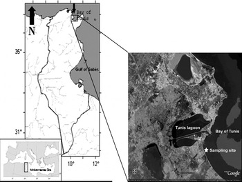
Specimens of F. fragilis were sampled on the littoral of the Bay of Tunis also known as the small Gulf of Tunis, between the latitude 36°47′N and the longitude 10°17′E (Figure 1). Sampling was in the intertidal zone, in very shallow depth between 0.5 m and 1.5 m in sandy–mud sediment.
Location of the Bay of Tunis (northern Tunisia) and the sampling site (map in satellite view copied from Google maps).

The Bay of Tunis is located in the southern part of the Gulf of Tunis and limited by the 30 m isobath. It occupies, approximately, a maritime surface of 350 km2 (Ben Charrada & Moussa, Reference Ben Charrada and Moussa1997; Ben Charrada et al., Reference Ben Charrada, Moussa and Zaouali1997). The Gulf of Tunis is located 200 km south-west of Sicily and represents the southern limit of the Tunisian–Siculo channel. The Tunisian–Siculo Strait, which is a ship transit line, constitutes hydrological, climatic and geographical borders between the east and the west of the Mediterranean basin (Pérès, Reference Pérès1967). The Bay of Tunis is near the two most important commercial harbours in north Tunisia (called respectively Radès and La Goulette) assuring the major sea traffic in Tunisia.
Specimens of F. fragilis were monthly collected from April 2006 until October 2007 except for June, July and August 2007 when sampling was twice a week. Each time, we collected 30 individuals for the histological study, except in October 2007 when we collected only 7 individuals due to its rarefaction. Samplings were carried out using drags and SCUBA diving.
Environmental variables
The temperature of the sea surface was taken by a thermometer with accuracy of 1/10°C and a multi-type Lab (WTW) for the measurements of other parameters such us pH, salinity and dissolved oxygen. Seawater samples were kept in 1.5 l opaque plastic bottles preserved at +4°C and transferred to the laboratory for the proportioning of chlorophyll-a concentration according to the method described by Aminot & Chaussepied (Reference Aminot and Chaussepied1983).
Metric measurements and histology
Fulvia fragilis samples were kept in aquaria containing seawater from the site of the study, at room temperature. All treatments and measurements were carried out on living specimens, within 24 hours following their having been fished. We measured the various morphological parameters such us length, height and thickness of the valves using a digital calliper accurate to 0.01 mm, and fresh total weight, fresh weight of the flesh and the shell with a 0.01g precision balance.
The visceral mass (gonad, digestive gland and foot) was dissected and fixed in alcohol-based Bouin's solution during 48 hours. Tissues were then dehydrated in a series of increasing concentration alcohol baths, in toluene and finally impregnated and included in paraffin wax; 5 µm thickness cuts were coloured with the haematoxylin–eosin (Gabe, Reference Gabe1968). Five sections were carried out for each individual. Sections were examined with an optical microscope. The scale of Lubet (Reference Lubet1959) was used to differentiate the gametogenic stages.
Data analysis
The differences of the frequencies of the gametogenic stages, between male and female follicles, were tested by analysis of variance (ANOVA) (P < 0.05). We used ‘analyses of the principal components’ in studying correlations between the sexual stages and the different environmental parameters. This method is frequently involved in environmental studies in order to reduce the dimensional space of the data, thus facilitating the establishment of the relationships between the studied variables. The Statgraphics Centurion software was used for data analysis treatment.
RESULTS
Study period
Our period of sampling was between April 2006 and September 2007, with interruptions following episodes of massive summer mortalities. The first episode of summer mortalities took place in July 2006 explaining the interruption of sampling during three months: July, August and September 2006. The second episode occurred in October 2007; only 7 individuals have been found alive and used for histological study.
Environmental parameters
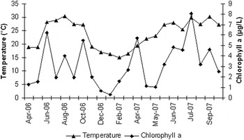
Sea surface temperature fluctuated between two extreme values (15.1°C and 30.9°C) throughout the sampling period with an average of 23.9°C. It ranged between 16.5°C and 18.9°C in spring, reaching maximum values in summer (30.9°C in July 2006), remained relatively high in the early autumn (average of 27°C) and decreased in late autumn to reach minimum values in winter (15.1°C in February 2007) (Figure 2).
Monthly evolution of the sea surface temperature and chlorophyll-a (April 2006–October 2007).

Recorded values of chlorophyll-a were highly variable. The average value was 3.16 µg/l. The lowest values were recorded during winter and early spring, respectively in December 2006 (0.65 µg/l) and March 2007 (0.79 µg/l) with a minimum value reached in January 2007 (0.28 µg/l). During the rest of the year, several peaks were noted; summer peaks were the most important, i.e. in June 2006 (6.21 µg/l) and July 2007 (8.09 µg/l) (Figure 2).
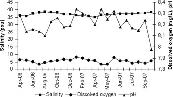
The mean salinity value was 37.08 psu. The minimum value was recorded in April 2006 (35.1 psu) and the maximum in August 2006 (38.5 psu) (Figure 3).
Monthly evolution of the salinity, concentration of the dissolved oxygen and pH (April 2006–October 2007).

The average concentration of dissolved oxygen was 5.59 mg/l. This parameter decreased in hot months and presented a minimum in May 2006 (3.23 mg/l) and increased during the colder months with a maximum in January 2007 (8.3 mg/l) (Figure 3).
The average pH was 8.21. A few fluctuations were monitored from 7.98 to 8.34 (Figure 3).
Biometric analyses
A total of 557 individuals were examined. The average length of the analysed individuals was 40.06 ± 4.82 mm (23.21–53 mm), the average height was 39.15 ± 4.59 mm (23.93–57 mm), the average thickness was 26.90 ± 3.31 mm (18.98–39.55 mm), the average total weight was 17.44 ± 4.29 mg (4.29–38.34 mg) and the average weight of the fresh flesh was 5.03 ±1.85 mg (1.37–12.15 mg).
Gametogenic scale
The gonad of F. fragilis is diffuse and extended between the digestive gland and the pedal constriction. During the reproductive activity, and especially during the maturation until the gamete emission, the foot volume which has a brownish colour normally increased in size and then became whitish.
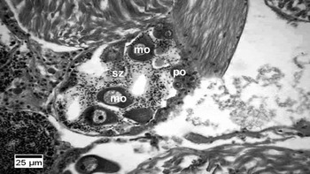
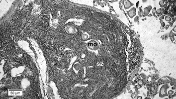
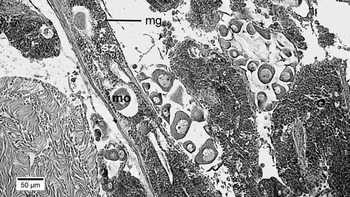
Examination of the histological cuts of the gonadic tissue of F. fragilis revealed that the species is strictly hermaphrodite. We observed oogenic, spermatogenic and a few hermaphroditic intermingled follicles. There were two types of hermaphroditic follicles; the first and the more frequently present were essentially female gametes surrounded by spermatozoa (Figure 7) and the second show some mature oocytes in follicle filled with spermatozoa (Figure 8). In some male gonoducts, we detected mature oocytes surrounded by spermatozoa (Figure 10). No case of gonochorism was noted. Only the germinal cells were taken into account to determine the gametogenesis scale. Indeed, F. fragilis presented a lack of reserve tissue in the gonad. Inter-follicular spaces were occupied by the haemocytes, fibroblasts and collagen fibres. We established 8 gametogenic stages.
Stage 0: sexual rest
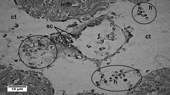
During the sexual rest, the foot of F. fragilis had, macroscopically, a brownish colour. Microscopically, the gonadic male and female follicles were very few, isolated and compressed consecutively to the last spawn. These follicles contained stem cells and respectively spermatogonia or oogonia. The determination of the follicular sex appeared difficult at this stage, except when some residual gametes in certain follicles were present. Many scattered haemocytes were present (Figure 4).
Image showing the Fulvia fragilis sexual rest stage. F, follicle; mfb, muscular fibres; ct, connective tissue; h, haemocytes (circled in the figure); sc, stem cells.

Stage 1: initiation of gametogenesis
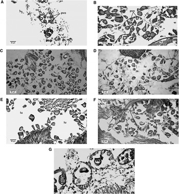
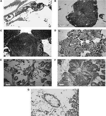
Macroscopically, the aspect of the foot was similar to the description given in the preceding stage. Gonadic follicles increased in size. Within the female follicle, oogonia were observed on the follicle wall and the first previtellogenic oocytes appear (Figure 5A). Within the male follicle, there were spermatogonia on the follicle wall and first spermatocytes were developed (Figure 6A).
Images of the different stages of development of the female follicle of Fulvia fragilis: (A) initiation of gametogenesis; (B) advanced gametogenesis; (C) ripe; (D) partial spawning; (E) advanced spawning; (F) restoration; (G) spent. ao, atretic oocyte; cf, collagen fibres; ct, connective tissue; ff, female follicle; h, haemocytes; ift, inter-follicular tissue, lu, lumen; mo, mature oocytes; mf, male follicles; oo, oogonia; pvo, previtellogenic oocyte; sc, stem cells; vso, vitellogenic stalked oocytes.

Images showing stages in the development of the male follicle of Fulvia fragilis: (A) initiation of gametogenesis; (B) advanced gametogenesis; (C) ripe; (D) partial spawning; (E) advanced spawning; (F) restoration; (G) spent. cf, collagen fibres; ct, connective tissue; h, haemocytes; ff, female follicle; sg, spermatogonia; sc, spermatocytes; sz, spermatozoa.

Stage 2: advanced gametogenesis
The follicles increased in size and were more numerous. All germinal lineage cells were represented at this stage. Within the female follicles, previtellogenic oocytes and vitellogenic stalked oocytes were adherent to the follicular wall. Mature free oocytes were present in the lumen of the follicle (Figure 5B). Within the male follicles, the spermatogonia occupied the peripheral base and the other categories of the male germinal cells (spermatocytes, spermatids and spermatozoa) have a centripetal laminated structure. Spermatozoa occupied the centre of the follicle and presented a centripetal radiate arrangement with tails directed towards the follicular light (Figure 6B). Some scattered haemocytes were still present.
Stage 3A: ripe
The volume of the foot increased and became whitish. The volume of the gonadic tissue also increased. It extended from digestive gland to pedal constriction. Follicles increased in number and grew in size. Inter-follicular spaces were reduced at this stage. Within female follicles, the number of previtellogenic and vitellogenic oocytes decreased gradually whereas the number of free mature oocytes increased. The latter occupied the lumen of the follicle and have a polygonal-shaped profile due to packing (Figure 5C). Within male follicles, spermatogonia occupied the periphery and spermatocytes number was reduced. Spermatozoa occupied the near total of the follicular surface and lost their radiate arrangement when maturation reached its maximum (Figure 6C). Scattered haemocytes were present in reduced number.
Stage 3B1: partial spawning
Macroscopically, the foot aspect remained similar to the preceding stage. It is important to note that when the maturation of male or female gametes was reached, spawning started quickly. Indeed, stages 3A (ripe) and 3B1 (partial spawning) were simultaneously present at the same individual. Within the female follicles, the density of mature oocytes decreased in the lumen and empty spaces consecutive to the gamete emission were present (Figure 5D). In some female follicles, we observed, at this stage, an intra-follicular tissue and spermatozoa surrounding the oocytes (Figure 7). The male follicles seemed to contract and have irregular shapes. Spermatozoa occupied the major portion of follicular volumes which presented empty zones following partial spawning (Figure 6D). Scattered haemocytes were observed in the connective tissue.
Image showing hermaphroditic follicle with majority of female cells. mo, mature oocyte; po, previtellogenic oocytes; sz, spermatozoa.

Image showing hermaphroditic follicle with majority of male cells. mo, mature oocyte; sz, spermatozoa.

Stage 3B2: advanced spawning
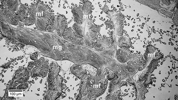
The foot volume decreased but kept its whitish colour. Microscopically, the gonadic tissue is reduced and the connective tissue became abundant. Female follicles were dilated and some of them presented broken walls. There were still a few stalked vitellogenic oocytes adhering to the walls and rare free oocytes. The follicular lumen was empty. Lysis of oocytes may occur (Figure 5E). Male follicles decreased in size and were compressed. Emission spaces became more important and the number of spermatozoa decreased compared to the preceding stage (Figure 6E). It has to be noted that at the spawning stages (3B1 and 3B2), volumes of ciliate male gonoducts increased and several male follicles surrounded them and dump their spermatozoa (Figure 9). The haemocytes became abundant. The latter are present around and in some follicles.
Image showing male gonoduct. mf, male follicle; mg, male gonoduct.

Image showing oocytes in male gonoduct. mg, male gonoduct; mo, mature oocyte; sz, spermatozoa.

Stage 3C: restoration
The size of female follicular lumen decreased in comparison with spawning stages. Within the female follicles, the number of previtellogenic oocytes increased along the follicular walls and contrasted with the reduction in the number of the free mature oocytes in the lumen (Figure 5F). Within the male follicles, peripheral base thickened and the number of spermatocytes increased and contrasted with the number of spermatozoa which decreased (Figure 6F). Scattered haemocytes were present.
Stage 3D: spent or resorbing
The number of follicles decreased. They presented a reduced size and were spaced by a connective tissue. They contained residual gametes. In female follicles, several episodes of oocyte lysis were noted. In the connective tissue, there was an invasion of haemocytes (Figures 5G & 6G). These haemocytes have probably a phagocytic role.
Temporal distribution of the gametogenesis stages
There was no statistically significant difference between the frequencies of stages in male and female follicles. The value of the probability of the Student's t-test was greater than 0.05 with a confidence level of 95.0% (ANOVA). This result attests that F. fragilis is a strict simultaneous species.
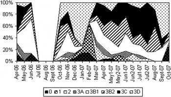
In our analysis, we, therefore, considered both follicular sexes together (Figure 11). The sexual rest (stage 0) was observed only in a small number of individuals in spring (April 2006, April 2007 and the first half of May 2007), in summer 2006 (June 2006), in autumn (October 2007) and in winter (December, January and February 2007). The highest percentage of individuals at the sexual rest was recorded in January 2007 and has not exceeded 17.24%. Initiation of gametogenesis (stage 1) took place in the same periods and during the following months. The maximum frequency was 50% and was recorded in February 2007. Advanced gametogenesis (stage 2) occurred in spring (March and April 2006, and April 2007) and summer (late June and July 2007) in a minority of individuals. Indeed, the largest percentage was reached in March 2007 (24%). Maturation (stage 3A), also observed in a small number of individuals was recorded in spring (in May 2006 and during the period March–May 2007), the early summer of 2007 (first half of June) and autumn 2006 (November) but the percentage of individuals at this gametogenic stage did not exceed 13%. Spawning (stage 3B1 and stage 3B2) was continuous throughout the year; we noted spring peaks (May 2006 and the end of the first half of May 2007), summer peaks (June 2006, first half of June 2007 and first half of August 2007) and autumn peaks (November 2006). In winter, gametic emissions were few and concerned just a minority of individuals; the lowest percentages were recorded in January and February 2007. Gametogenic restoration (stage 3C) appeared throughout the year except in winter. The highest proportions were recorded during spring (April 2006 and April 2007) and summer (June and July 2007). Finally, the spent stage (3D) took place after the peak spawning periods in summer (August 2007), in winter (December 2006 and January and February 2007) with a peak in January (79.62%) and autumn (October 2007).
Temporal distribution of Fulvia fragilis gametogenic stages (April 2006–October 2007).

Relationship between environmental parameters and gametogenesis
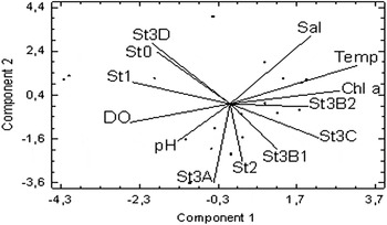
In order to highlight a possible correlation between the gametogenic stages and environmental variables, we applied a principal component analysis (PCA). Four principal components were retained explaining 72.99% of the total inertia. We noted that the first two components explained 52.54% of the total inertia. The PCA indicated that the three most significant environmental parameters relative to the axis of the component 1 were the sea surface temperature, the chlorophyll-a concentration and the concentration of dissolved oxygen (Figure 12). Stages 0, 1 and 3D were positively correlated with the concentration of dissolved oxygen in the environment and negatively correlated to temperature and chlorophyll-a. This happened during the cold months of the year: December, January and February. Stages 3B2 and 3C (with a peak in July 2007) were strongly positively correlated with sea surface temperature and chlorophyll-a. Moreover, stages 2 and 3B1 were positively correlated with these parameters.
Principal component analysis (PCA) (environmental parameters and gametogenic stages). Chl a, chlorophyll-a; DO, dissolved oxygen; Sal, salinity; St, stage; Temp, sea surface temperature.

DISCUSSION
This paper constitutes the first work to study the reproduction biology of F. fragilis. In literature, the most studied species of Cardiidae are Cerastoderma glaucum and Cerastoderma edule (e.g. Zaouali, Reference Zaouali1974; Guillou et al., Reference Guillou, Bachelet, Desprez, Ducotoy, Madani, Rybarczyk, Sauriau, Sylvand, Elkaim and Glemarc1990, Reference Guillou, Bachelet, Rybarczyk and Glemarc1992; Derbali et al., Reference Derbali, Jarboui and Ghorbel2009). These two species are strictly gonochoric. In addition, certain species of the family of Cardiidae, such as Clinocardium nuttalli (Gallucci & Gallucci, Reference Gallucci and Gallucci1982), Fulvia mutica (Liu et al., Reference Liu, Li, Yuan and Zhang2008) and Acrosterigma (Vasticardium) burchardi (Limpanont et al., Reference Limpanont, Yang, Won, Han, Lee, Lee and Choi2010), present a simultaneous hermaphroditism. Generally, bivalves show a wide variation of the sexual reproduction mode from strictly gonochoric species to the simultaneous hermaphroditic species. According to Heller (Reference Heller1993), the simultaneous or sequential hermaphroditism has been observed in 11 of 45 super families of bivalves and mainly in freshwater bivalves.
We established during our histological study of the reproduction of F. fragilis that this bivalve is hermaphrodite. Referring to the report of Coe (Reference Coe1943), F. fragilis, producing simultaneously two types of gametes, is a normal hermaphroditic functional bivalve. According to the theory of Ghiselin (Reference Ghiselin1969), this mode of reproduction is linked to low densities, low mobility of the animal and the possibilities of depopulations associated with the unstable environments. Fulvia fragilis answers well to these criteria. Indeed, in the Bay of Tunis, we found this bivalve living in the intertidal zone at low bathymetries influenced by the extreme climatic conditions. Ansell et al. (Reference Ansell, Bodoy and Massé1979) noted that the geographical distribution of littoral bivalves would be highly influenced by environmental conditions including a higher specific thermal range corresponding to the maximum lethal temperature compatible with the survival of 50% of the population (LT50). During our monitoring, we observed high summer mortalities when the sea surface temperatures reached values exceeding 30°C and probably close to sublethal and lethal levels, especially in July 2006 (30.9°C) and in September 2007 (30.2°C). We note that in Lake Timsah (Suez Canal, Egypt) where this species occurs in large densities, the maximum summer temperatures, during the years 1996 and 1997, did not exceed 29°C (Mohammad et al., Reference Mohammad, Mohallal, Mohammed and Attia2006). In addition, F. fragilis lives at the surface or just below a few centimetres of the sediment which facilitates its predation. We collected, during our samplings, several shells of dead perforated individuals due to the predation of Hexaplex trunculus. However, F. fragilis presents a muscular foot which enables it to move by saltatory movements; this potential mobility does not contribute to the evolution of its reproduction mode, as in the case of Clinocardium nuttalli (Bivalvia: Cardiidae) (Gallucci & Gallucci, Reference Gallucci and Gallucci1982). According to these authors, ‘…sessileness in bivalves would not appear to be contributory factor in the evolution of the mode of reproduction’.
The gametogenic scale of F. fragilis was determined for the first time. The eight stages (sexual rest, initiation of gametogenesis, advanced gametogenesis, ripe, partial spawning, advanced spawning, restoration and spent) are well-defined when referring to the germinal cells.
Our study revealed that F. fragilis presents an asynchronous gametogenic cycle. We detected this asynchrony not only between the individuals which do not always present the same stage of maturation but also between the follicles of the same individual that present several stages in various surfaces of the gonad. The coexistence of various stages is current in the bivalves in temperate areas, which explains the phenomenon of successive individual spawns (Ansell, Reference Ansell1961; Gaspar et al., Reference Gaspar, Ferreira and Monteiro1999; Tirado et al., Reference Tirado, Salas and Márquez2003). In addition, it is important to note that during all the stages of the sexual maturation, haemocytes are present in inter-follicular spaces. They are abundant during stages 3B2, 3D and 0. The haemocytes could have a role of macrophages ensuring the transfer of nutrients to the oocytes and the spermatozoa in maturation following the example described for Pecten maximus (Le Pennec et al., Reference Le Pennec, Dorange, Beninger, Donval and Widowati1992). According to Darriba et al. (Reference Darriba, San Juan and Guerra2004), the presence of haemocytes during all the reproduction cycle is the proof that these cells have a double function: (1) a macrophage function during the gametic lysis; and (2) a nutrition function, providing nutrients for the gametes in formation. The haemocytes can thus be assimilated, in the gonad, as transfer nutrients cells and may be reserve cells in F. fragilis. This last point could be supported by the rapid turnover between the time when the gametes are emitted and the beginning of the later gametogenesis.
Several studies showed that the reproduction of the marine bivalves is mainly affected by the environmental conditions including particularly temperature and the availability of the nutrients (e.g. Ansell, Reference Ansell1974; Mann, Reference Mann1979; Lubet, Reference Lubet1981; Guillou et al., Reference Guillou, Bachelet, Rybarczyk and Glemarc1992; Brey, Reference Brey1995; Darriba et al., Reference Darriba, San Juan and Guerra2004; Enríquez-Díaz et al., Reference Enríquez-Díaz, Pouvreau, Chávez-Villaba and Le Pennec2009). The gametogenic cycles depend on external factors (environmental parameters) and on internal factors (endocrine regulations, like the sex steroids in Ruditapes decussatus (Ketata et al., Reference Ketata, Guermazi, Rebai and Hamza-Chaffai2007), and energy competitions between somatic and germinal compartments (Lubet & Mann, Reference Lubet and Mann1987)). The temporal distribution of the gametogenesis stages of this species enabled us to deduce that F. fragilis has no real period of sexual rest. Indeed, we noted a continuous spawning activity. The initiation of gametogenesis reached its peak in February 2007, a period corresponding to the lowest temperatures (15.1°C), low concentrations of chlorophyll-a (0.94µg/l) and high levels of dissolved oxygen (7.36mg/l). In some bivalve species, the initiation of gametogenesis could be primarily due to the low temperature, which is the case for Mesoderma mactroides (Hermann et al., Reference Hermann, Alfaya, Lepore, Penchaszadech and Laudien2009). In other bivalves, it is due to the low nutrients levels in the environment which is the case for Ensis arcuatus using its reserves (Darriba et al., Reference Darriba, San Juan and Guerra2004) and Pecten maximus using mainly its glycogen and lipids reserves present in the digestive gland (Le Pennec & Le Pennec, Reference Le Pennec and Le Pennec2002). In F. fragilis, this gametogenic stage would be simultaneously stimulated by a concomitant drop in the sea temperature and the amount of nutrients available in the environment and by high concentrations of dissolved oxygen. Also the statistical method of the PCA confirmed this phenomenon. The stages (2) and (3A) represented only small proportions of both male and female follicles. Indeed, the maximum proportion of stage (2) did not exceed 24% in March 2007. This explains the appearance of stage (3A) during the following months and in part the following gametic emission.
The rarity of the mature individuals is due to the speed of the spawning. During our observations, we noticed that the maturation stage coexists systematically with the partial spawning stage. However, we noted spring and summer successive spawns during the period from March 2007 until the end of August 2007. These spawns were mainly due to the gonadic restorations. Indeed, in this period, the stage 3C is represented in very important proportions and the rest stage was almost absent. The spawning of F. fragilis is continuous; nevertheless rarer in winter (December, January and February 2007) and marking seasonal peaks during the rest of the year. We recorded summer spawning peaks in June 2006 and June 2007. The first peak corresponds to the month of the most intense spawns during all the study period. At this period, we recorded high value of chlorophyll-a concentration (6.21µg/l) and a rise of temperature from 18.9°C to 28.2°C. The second peak also corresponds to rises in temperature (23°C to 27.2°C) as well as in nutrients availability in the environment (1.01µg/l to 3.18µg/l). The autumnal peak was recorded in October 2006 following a rise in the chlorophyll-a concentration (2.38µg/l to 5.49µg/l) and a high temperature of 27.2°C. The spring peak took place during the first half of May 2007 stimulated by a rise in temperature from 19.4°C to 22.1°C. We conclude that F. fragilis has an unspecified spawning temperature. Rises in temperatures might stimulate the massive spawning. The high values in the chlorophyll-a concentration, during spawning periods, indicate some relationship between levels of phytoplankton and spawning; this phenomenon was noticed in other bivalves such as Crassostrea gigas (Ruiz et al., Reference Ruiz, Abad, Sedano, Garcia-Martin and López Sández1992), Venus verrucosa (Tirado et al., Reference Tirado, Salas and Márquez2003) and Ensis arcuatus (Darriba et al., Reference Darriba, San Juan and Guerra2004). Moreover, during the period from April 2007 to September 2007 we observed that successive spawning values of temperature increased continuously. However, when chlorophyll-a concentration reached 8.09µg/l, the majority of the female follicles and a large number of male follicles were in gametogenic restoration. We conclude that in F. fragilis, the high quantities of nutrients available maintain the gonadic activity and ensure the gametic regeneration and in consequence the successive spawning. In other bivalves such as Ensis arcuatus, the successive spawning due to the gonadic restoration takes place in the periods when the nutrients are not very abundant in the environment (Darriba et al., Reference Darriba, San Juan and Guerra2004). The correlations between the spawning and the gametic restoration stages with the sea surface temperature and the nutrient availability in the environment, in F. fragilis, were also affirmed by the PCA. The majority of the follicles reached the spent stage during December 2006 and January 2007. The gonadic spent stage reached the maximum values of 81% and 77% respectively for the male and female follicles in January, a month which corresponds to the minimal recorded value of the chlorophyll-a and to low temperatures.
Fulvia fragilis, as we have already noted, has a diffuse gonad and it shows male, female and some hermaphroditic follicles intermingled. The presence of hermaphroditic follicles has already been reported in another simultaneous hermaphrodite Cardiidae Acrosterigma (Vasticardium) burchardi (Limpanont et al., Reference Limpanont, Yang, Won, Han, Lee, Lee and Choi2010).
The simultaneous maturation of the male and female gametes, the presence of mature oocytes surrounded by spermatozoa in the hermaphroditic follicle and in the male gonoducts (Figure 10), and the intermingling arrangement of the different types of follicles, enable us to conclude that the assumptions of internal-fertilization and probably self-fertilization in this species are not excluded. According to Mackie (Reference Mackie and Wilbur1984), in most bivalves, the cross-fertilization is typical, but in the hermaphrodite simultaneous species, self-fertilization could be a common process. Coe (Reference Coe1943) indicated that at the hermaphrodite bivalves which present spermatogonia and oogonia simultaneously activated; some were capable of self-fertilization. In the literature, several studies suggest the possibility of self-fertilization in certain species of bivalves. Furthermore, several authors (Woods, Reference Woods1931; Okada, Reference Okada1935; Coe, Reference Coe1943; Meier-Brook, Reference Meier-Brook1970 in Petinelli & Bicchierai, 2009) reported evidence of fertilization in gonoducts. Generally, self-fertilization was described in larviparous simultaneous hermaphroditic bivalves because embryonic development usually occurs in the gill chambers. We mention the example of the Sphaeriidae; the self-fertilization was described following the observation of oocytes surrounded by spermatozoa in the gonoducts, by Stepanoff (Reference Stepanoff1865) and by Okada (Reference Okada1935). Indeed, Pisidium henslowanum (Sphaeriidae) presents mature oocytes surrounded by spermatozoa in the lumen of the female follicle (Pettinelli & Bicchierai, Reference Pettinelli and Bicchierai2009). These authors concluded that fertilization could be intra-follicular in this species. This same phenomenon was interpreted as an evidence of self-fertilization by Araujo & Ramos (Reference Araujo and Ramos1997, Reference Araujo and Ramos1999). Furthermore, a recent study of the reproduction of the Asian clam Corbicula fluminea (Park & Chung, Reference Park and Chung2004) suggested that this hermaphrodite bivalve presents a self-fertilization: ‘At a glance, some intermingling of a number of spermatozoa and a few oocytes in the same follicle suggest that self-fertilization may occur in the hermaphroditic follicle’. The self-fertilization is recognized as being a natural process in hermaphrodite simultaneous pectinids (Beaumont, Reference Beaumont1986; Beninger & Le Pennec, Reference Beninger, Le Pennec and Shumway1991; Ibarra et al., Reference Ibarra, Cruz and Romero1995 in Martínez et al., Reference Martínez, Mettifogo, Pérez and Callejas2007). However, in studying the reproduction of the protandric oviparous species Bankia setacea, Coe (Reference Coe1941) concluded that under the functionally hermaphroditic condition, self-fertilization occurs occasionally.
In conclusion, F. fragilis may be considered as a functional simultaneous hermaphrodite bivalve which presents a continuous gonadic activity. Self-fertilization seems possible. Even in the absence of self-fertilization, the simultaneous hermaphroditism has an advantage: in the case of low densities, the contact of two individuals always ends by a successful fertilization, which is not the case in gonochoric individuals (Tomlinson, Reference Tomlinson1966). Yet, the simultaneous hermaphroditism has some disadvantages. Indeed, this mode of reproduction requires high energy costs for the maintenance of two reproductive structures compared to a gonochoric individual (Heath, Reference Heath1977; Charnov, Reference Charnov1979). Indeed, Heath (Reference Heath1977) explains that gonochorists have more energy available for gamete production because ‘they have a lower expenditure on reproductive structures’. In addition, the probable self-fertilization in F. fragilis would imply a reduced viability of the resulting offspring (Heath, Reference Heath1977). The mode of reproduction of F. fragilis ensures the success of its installation but the high energy costs due to the hermaphroditism, the effort of reproduction following the continuous spawning, the addictive predators consuming it, as in the case of Hexaplex trunculus, the probably low viabilities caused by self-fertilization and the high temperatures can explain summer mortalities and its scarcity at the Bay of Tunis, during our study. This vulnerability could also be owing to its ‘progressive adaptation’ to the Tunisian waters. However, we noted that this species is again present in relatively high densities since May 2010 and this proves the adaptation of the species to its new ‘receiver’ habitat.
ACKNOWLEDGEMENTS
The authors thank Professor Mohamed Samir Boubaker, consultant of the Anatomo-Pathology Department in the ‘Institut Pasteur de Tunis’, for hosting us in his laboratory. We are grateful to Salma Feriani and Aouatef Souissi for guidance in histological techniques for this study. We also thank Lamia Gargouri for his assistance with photographs. We express our special thanks to Mourad Ben Slama for her help in statistical treatments. We acknowledge Fabio Crocetta and Ermanno Quaggiotto for helping in literature research. We thank Roberto Pettinelli for useful comments. The authors wish also to thank the anonymous referees for their useful and helpful comments on the manuscript.