





Introduction
Successful embryo selection remains a major challenge in assisted reproductive technologies (ART). Although advances in ovarian stimulation, embryo culture systems and laboratory practice have improved IVF outcomes, implantation rates remain limited. A substantial proportion of morphologically good-quality blastocysts fail to implant or result in a live birth, indicating that embryo morphology alone does not fully reflect developmental competence (Cimadomo et al., Reference Cimadomo, Rienzi, Conforti, Forman, Canosa, Innocenti, Poli, Hynes, Gemmell, Vaiarelli, Alviggi, Ubaldi and Capalbo2023).
Morphological assessment has been the basis of embryo selection for more than two decades. The Gardner and Schoolcraft grading system, together with the recommendations of the Istanbul Consensus and its recent update, provides a standardised framework that is widely used in clinical practice (Gardner and Schoolcraft, Reference Gardner and Schoolcraft1999; ALPHA Scientists and ESHRE Embryology, Reference Balaban, Brison, Calderón, Catt, Conaghan, Cowan, Ebner, Gardner, Hardarson, Lundin, Magli, Mortimer, Mortimer, Munné, Royere, Scott, Smitz, Thornhill, van Blerkom and Van den Abbeel2011; Coticchio et al., Reference Coticchio, Ahlström, Arroyo, Balaban, Campbell, De Los Santos, Ebner, Gardner, Kovačič, Lundin, Magli, Mcheik, Morbeck, Rienzi, Sfontouris, Vermeulen and Alikani2025). Nevertheless, embryos with similar morphological characteristics may differ considerably in chromosomal status, metabolic activity, gene expression and implantation potential. These observations have stimulated the search for additional, preferably non-invasive, markers of embryo competence (Cimadomo et al., Reference Cimadomo, Rienzi, Conforti, Forman, Canosa, Innocenti, Poli, Hynes, Gemmell, Vaiarelli, Alviggi, Ubaldi and Capalbo2023; Coticchio et al., Reference Coticchio, Ahlström, Arroyo, Balaban, Campbell, De Los Santos, Ebner, Gardner, Kovačič, Lundin, Magli, Mcheik, Morbeck, Rienzi, Sfontouris, Vermeulen and Alikani2025).
During the last decade, several complementary approaches have been investigated. Time-lapse monitoring (TLM) enables continuous assessment of embryo development under stable culture conditions, whereas artificial intelligence (AI) has been introduced to improve the consistency of image interpretation. At the same time, metabolomic, proteomic, transcriptomic and cell-free DNA-based analyses of spent culture media have provided new opportunities to investigate embryo-derived molecular signals without embryo biopsy (Meseguer et al., Reference Meseguer, Herrero, Tejera, Hilligsøe, Ramsing and Remohí2011; Cimadomo et al., Reference Cimadomo, Rienzi, Conforti, Forman, Canosa, Innocenti, Poli, Hynes, Gemmell, Vaiarelli, Alviggi, Ubaldi and Capalbo2023; Coticchio et al., Reference Coticchio, Ahlström, Arroyo, Balaban, Campbell, De Los Santos, Ebner, Gardner, Kovačič, Lundin, Magli, Mcheik, Morbeck, Rienzi, Sfontouris, Vermeulen and Alikani2025).
Although many of these approaches have produced encouraging results, only a few have been adopted into routine clinical practice, while most remain investigational. Differences in study design, laboratory protocols, analytical methods and clinical endpoints make comparison between studies difficult. Moreover, biomarkers that perform well in single-centre studies often show lower reproducibility in external validation studies. Consequently, the clinical value of many proposed biomarkers remains uncertain (Kim et al., Reference Kim, Lee and Jun2022; Cimadomo et al., Reference Cimadomo, Rienzi, Conforti, Forman, Canosa, Innocenti, Poli, Hynes, Gemmell, Vaiarelli, Alviggi, Ubaldi and Capalbo2023; Jiang and Bormann, Reference Jiang and Bormann2023).
Early studies suggested that spent culture media could identify embryos with high implantation potential independently of conventional morphology (Seli et al., Reference Seli, Sakkas, Scott, Kwok, Rosendahl and Burns2007). However, subsequent studies failed to confirm these findings consistently, and methodological as well as technical limitations prevented widespread clinical adoption (Uyar and Seli, Reference Uyar and Seli2014). Similar challenges have been encountered with several other promising biomarkers proposed for embryo selection.
In this review, we summarise the current evidence on conventional morphology, TLM, AI and emerging omics-based approaches for embryo selection. Rather than considering these technologies in isolation, we discuss their biological rationale, current evidence, clinical applicability and limitations and highlight the challenges that continue to limit their integration into routine IVF practice.
Conventional morphological evaluation of IVF embryos
Conventional morphological evaluation remains the cornerstone of embryo selection in routine IVF practice and primarily relies on embryo grading based on cell number, blastomere size and symmetry and the degree of fragmentation at the cleavage stage (Veeck Gosden, Reference Veeck Gosden1999) as well as blastocoel expansion, inner cell mass (ICM) and trophectoderm (TE) morphology at the blastocyst stage, as originally described by Gardner and Schoolcraft (Gardner and Schoolcraft, Reference Gardner and Schoolcraft1999). To reduce inter-and intra-laboratory variability, standardised evaluation criteria were established during the Istanbul Consensus Workshop on Embryo Assessment (ALPHA Scientists and ESHRE Embryology, Reference Balaban, Brison, Calderón, Catt, Conaghan, Cowan, Ebner, Gardner, Hardarson, Lundin, Magli, Mortimer, Mortimer, Munné, Royere, Scott, Smitz, Thornhill, van Blerkom and Van den Abbeel2011). These recommendations were recently updated, refining definitions for cleavage-stage, morula and blastocyst assessment, while incorporating guidance on abnormal cleavage patterns identified through extended culture and TLM (Coticchio et al., Reference Coticchio, Ahlström, Arroyo, Balaban, Campbell, De Los Santos, Ebner, Gardner, Kovačič, Lundin, Magli, Mcheik, Morbeck, Rienzi, Sfontouris, Vermeulen and Alikani2025). Despite its widespread use, conventional morphology remains a simple, inexpensive and standardised method for embryo assessment; however, its ability to predict embryo competence is limited (Minasi et al., Reference Minasi, Colasante, Riccio, Ruberti, Casciani, Scarselli, Spinella, Fiorentino, Varricchio and Greco2016; Majumdar et al., Reference Majumdar, Majumdar, Verma and Upadhyaya2017).
According to the updated consensus, high-quality cleavage-stage embryos are characterised by appropriate cell number, symmetrical blastomeres, minimal fragmentation and the absence of multinucleation. The revised recommendations also recognise abnormal cleavage patterns, such as direct, reverse and irregular cleavage and provide guidance for their interpretation during extended embryo culture. For morula-stage embryos, standardised criteria for evaluating compaction and developmental progression have likewise been introduced. Blastocyst assessment has also been refined through standardised grading of blastocoel expansion and morphological evaluation of the ICM and TE, with the updated consensus replacing the previous numerical grading system with alphabetical categories (A–D) (Coticchio et al., Reference Coticchio, Ahlström, Arroyo, Balaban, Campbell, De Los Santos, Ebner, Gardner, Kovačič, Lundin, Magli, Mcheik, Morbeck, Rienzi, Sfontouris, Vermeulen and Alikani2025).
The updated consensus further incorporates findings from an international survey of 529 embryologists representing 82 countries, highlighting considerable variation in embryo grading practices, timing of embryo assessment and the implementation of adjunctive technologies such as TLM and computational image analysis. These findings emphasise the continuing need for internationally harmonised embryo assessment criteria (Coticchio et al., Reference Coticchio, Ahlström, Arroyo, Balaban, Campbell, De Los Santos, Ebner, Gardner, Kovačič, Lundin, Magli, Mcheik, Morbeck, Rienzi, Sfontouris, Vermeulen and Alikani2025).
In parallel, the Spanish Society for the Study of Reproductive Biology (ASEBIR) has introduced a simplified and more clinically oriented embryo assessment system. Analyses based on the updated ASEBIR framework demonstrated that ICM grading was not significantly associated with implantation or live birth, whereas blastocyst expansion and TE morphology were independent predictors of both outcomes. Furthermore, cleavage-stage morphology contributed little additional prognostic information once embryos had successfully developed to the blastocyst stage, supporting the growing preference for blastocyst-stage assessment when extended embryo culture is feasible (Pons et al., Reference Pons, Carrasco, Rives, Delgado, Martínez-Moro, Martínez-Granados, Rodriguez, Cairó and Cuevas-Saiz2023).
Despite continuous efforts toward standardisation, morphology alone remains insufficient for accurately predicting embryo euploidy and implantation potential (Minasi et al., Reference Minasi, Colasante, Riccio, Ruberti, Casciani, Scarselli, Spinella, Fiorentino, Varricchio and Greco2016; Majumdar et al., Reference Majumdar, Majumdar, Verma and Upadhyaya2017; Singh et al., Reference Singh, Pathak, Singh and Desai2022). Moreover, conventional morphology provides only a static snapshot of embryo development and cannot capture the dynamic biological processes underlying embryo competence. These limitations have driven the development of complementary approaches, including dynamic imaging, AI and omics-based technologies, aimed at improving the accuracy and objectivity of embryo selection.
Time-lapse monitoring (TLM) and morphokinetics
TLM has emerged as one of the most widely adopted adjuncts to conventional morphological assessment in IVF laboratories. These systems enable uninterrupted embryo culture by capturing images at regular intervals, typically every 10 minutes, without removing embryos from the incubator. This approach preserves stable culture conditions while providing continuous information on embryo development (Lundin and Park, Reference Lundin and Park2020). In several commercially available systems, the acquired images can be further processed using built-in computational algorithms to generate embryo-specific morphokinetic scores that support embryo ranking during selection (Giménez et al., Reference Giménez, Conversa, Murria and Meseguer2023).
Ongoing research has focused on identifying morphokinetic parameters that may facilitate embryo (de)selection, as well as on developing robust predictive algorithms. Particular attention has been given to the synchrony of embryonic cell divisions and the timing of key developmental milestones, including cleavage, compaction, blastulation and blastocyst expansion (Meseguer et al., Reference Meseguer, Herrero, Tejera, Hilligsøe, Ramsing and Remohí2011, Reference Meseguer, Rubio, Cruz, Basile, Marcos and Requena2012; Campbell et al., Reference Campbell, Fishel, Duffy and Montgomery2013; Cetinkaya et al., Reference Cetinkaya, Pirkevi, Yelke, Colakoglu, Atayurt and Kahraman2015; Braga et al., Reference Braga, Setti, Guilherme, Morishima, Iaconelli and Borges2023). In addition, previous studies have demonstrated that embryo morphokinetics may be influenced by maternal and treatment-related factors, including maternal age, body mass index (BMI), ovarian reserve, ovarian stimulation protocols and endometriosis, emphasising the importance of interpreting morphokinetic parameters within their clinical context (Gurbuz et al., Reference Gurbuz, Gode, Uzman, Ince, Kaya, Ozcimen, Ozcimen and Acar2016; Boynukalin et al., Reference Boynukalin, Serdarogullari, Gultomruk, Coban, Findikli and Bahceci2019; Boucret et al., Reference Boucret, Tramon, Riou, Ferré-L’Hôtellier, Bouet and May-Panloup2022; Yuksel et al., Reference Yuksel, Ozer, Duzguner, Akca, Bio, Bio, Kahraman, Liperis and Serdarogullari2024; Bashiri et al., Reference Bashiri, Akbarzadeh, Hosseini, Amjadi, Afzali, Khadivi, Taheri and Zandieh2025; Sezer et al., Reference Sezer, Yazici, Ozer, Yelke and Ozkara2025).
Despite these advances, evidence regarding the clinical benefit of TLM remains inconsistent. A randomised study comparing embryo selection based on conventional morphology and TLM reported no consistent clinical advantage of TLM over conventional assessment, with higher pregnancy and live birth rates observed in the conventional morphology group for cleavage-stage embryo transfers (Meng et al., Reference Meng, Xu, Zheng, Li, Ding, Xu, Pu, Wang and Wu2022). More recently, two large multicentre, double-blind randomised controlled trials have provided high-quality evidence regarding the clinical utility of time-lapse systems. The SelecTIMO trial demonstrated that neither uninterrupted embryo culture nor time-lapse-based embryo selection improved cumulative ongoing pregnancy rates compared with conventional embryo culture and morphology-based selection (Kieslinger et al., Reference Kieslinger, Vergouw, Ramos, Arends, Curfs, Slappendel, Kostelijk, Pieters, Consten, Verhoeven, Besselink, Broekmans, Cohlen, Smeenk, Mastenbroek, de Koning, van Kasteren, Moll, van Disseldorp, Brinkhuis, Kuijper, van Baal, van Weering, van der Linden, Gerards, Bossuyt, van Wely and Lambalk2023). Similarly, the subsequent TILT trial reported no significant improvement in live birth rates with either time-lapse-based embryo selection or uninterrupted culture compared with standard IVF laboratory practice (Bhide et al., Reference Bhide, Chan, Lanz, Alqawasmeh, Barry, Baxter, Gonzalez Carreras, Choudhury, Cheong, Chung, Collins, Cong, Doidge, Heighway, Patel, Pardo, Rattos, Wright, Dodds, Perez, Khan and Thangaratinam2024). These findings suggest that the additional morphokinetic information provided by TLM does not necessarily translate into improved clinical outcomes when used as a standalone embryo selection strategy.
Beyond its potential role in embryo selection, TLM has also been investigated as a non-invasive tool for predicting embryo ploidy. Serrano-Novillo et al. evaluated 374 blastocysts from PGT-A cycles and reported that euploid embryos reached several developmental milestones earlier than aneuploid embryos. The authors also identified a novel cytoplasmic movement parameter (St2) associated with embryo ploidy. Nevertheless, the overall predictive performance remained modest (Area Under Curve (AUC) ∼ 0.69), indicating that morphokinetic analysis alone cannot reliably replace invasive genetic testing for non-invasive prediction of embryo ploidy (Serrano-Novillo et al., Reference Serrano-Novillo, Uroz and Márquez2023).
Overall, TLM provides valuable dynamic information on embryo development while maintaining uninterrupted culture conditions. These findings indicate that the principal value of TLM currently lies in continuous embryo observation and objective developmental annotation rather than improved embryo selection when used in isolation. Future improvements are likely to arise from integrating TLM-derived features with AI, metabolomics and other multi-omic approaches rather than relying on morphokinetic parameters alone (Munné et al., Reference Munné, Horcajadas, Seth-Smith, Perugini and Griffin2025).
Computational and data-driven (AI) embryo evaluation
While conventional morphokinetic analysis and TLM have provided valuable insights into embryo development, their predictive performance remains limited. These limitations have stimulated the application of AI to embryo assessment, enabling automated analysis of complex developmental patterns beyond human visual interpretation.
The increasing availability of large time-lapse datasets has accelerated the development of AI-based embryo assessment systems aimed at improving the objectivity, consistency and reproducibility of embryo evaluation. Most AI models are based on deep learning algorithms trained using static embryo images or, more commonly, time-lapse videos annotated with known clinical outcomes, including implantation, fetal heartbeat, live birth or PGT-A results (Tran et al., Reference Tran, Cooke, Illingworth and Gardner2019; Chavez-Badiola et al., Reference Chavez-Badiola, Flores-Saiffe-Farías, Mendizabal-Ruiz, Drakeley and Cohen2020). Compared with conventional morphological assessment, AI-assisted systems have demonstrated high concordance with experienced embryologists while substantially reducing inter-observer variability, thereby supporting a more standardised approach to embryo evaluation (Storr et al., Reference Storr, Venetis, Cooke, Kilani and Ledger2017; Dimitriadis et al., Reference Dimitriadis, Zaninovic, Badiola and Bormann2022).
Although AI models have been developed using different imaging modalities, ranging from static embryo images to time-lapse videos, substantial heterogeneity in study design, imaging platforms and validation strategies continues to limit their generalisability and direct comparison across studies.
One of the most actively investigated applications of AI is the non-invasive prediction of embryo ploidy. Recent studies have reported encouraging predictive performance, with AUC values generally ranging from approximately 0.65 to 0.87, depending on the algorithms, imaging data and validation methods employed (Jiang and Bormann, Reference Jiang and Bormann2023). Although these findings demonstrate the potential of AI-assisted ploidy prediction, substantial variability in predictive accuracy and study methodology currently precludes replacement of invasive genetic testing such as PGT-A. Accordingly, AI-based ploidy assessment should presently be regarded as a promising adjunctive technology requiring further prospective, multicentre validation before routine clinical implementation.
Importantly, recent prospective clinical evidence has begun to evaluate whether AI-assisted embryo selection translates into improved reproductive outcomes. In a multicentre, randomised, double-blind, non-inferiority trial, Illingworth et al. compared deep learning-assisted embryo selection with conventional morphology-based assessment in IVF. The study demonstrated that AI-assisted embryo selection was non-inferior to standard embryologist assessment with respect to ongoing pregnancy and other key reproductive outcomes but did not establish clear clinical superiority. These findings suggest that, although AI has the potential to standardise embryo evaluation and reduce observer variability, current evidence remains insufficient to support replacement of experienced embryologists or routine implementation of AI as a standalone embryo selection strategy (Illingworth et al., Reference Illingworth, Venetis, Gardner, Nelson, Berntsen, Larman, Agresta, Ahitan, Ahlström, Cattrall, Cooke, Demmers, Gabrielsen, Hindkjær, Kelley, Knight, Lee, Lahoud, Mangat, Park, Price, Trew, Troest, Vincent, Wennerström, Zujovic and Hardarson2024).
Overall, current evidence suggests that AI should be viewed as a clinical decision-support tool rather than an autonomous embryo selection system. Although recent randomised clinical evidence supports the feasibility and safety of AI-assisted embryo selection, convincing evidence demonstrating superiority over expert embryologist assessment or consistent improvements in live birth outcomes is still lacking. Moreover, limited prospective validation, poor cross-centre generalisability and variability among AI algorithms continue to restrict routine clinical implementation (Illingworth et al., Reference Illingworth, Venetis, Gardner, Nelson, Berntsen, Larman, Agresta, Ahitan, Ahlström, Cattrall, Cooke, Demmers, Gabrielsen, Hindkjær, Kelley, Knight, Lee, Lahoud, Mangat, Park, Price, Trew, Troest, Vincent, Wennerström, Zujovic and Hardarson2024; Cimadomo et al., Reference Cimadomo, Coticchio and Rienzi2026).
Non-invasive genomics in IVF
Non-invasive preimplantation genetic testing (ni-PGT-A)
Non-invasive preimplantation genetic testing for aneuploidy (ni-PGT-A) is based on the analysis of cell-free DNA (cfDNA) released into spent embryo culture (SC) medium, offering the possibility of assessing embryo ploidy without the need for TE biopsy. Initial multicentre studies demonstrated the feasibility of this approach and reported moderate to high concordance between cfDNA analysis and conventional TE biopsy, although considerable variability was observed among participating centres, likely reflecting differences in embryo quality and laboratory protocols. Additional analyses comparing cfDNA obtained from spent culture medium with both TE and ICM samples also showed encouraging concordance, supporting the biological potential of this approach (Rubio et al., Reference Rubio, Navarro-Sánchez, García-Pascual, Ocali, Cimadomo, Venier, Barroso, Kopcow, Bahçeci, Kulmann, López, De la Fuente, Navarro, Valbuena, Sakkas, Rienzi and Simón2020).
Since these initial reports, considerable efforts have been directed toward optimising laboratory protocols and improving the analytical performance of ni-PGT-A. A recent clinical study comparing conventional TE biopsy with ni-PGT-A reported comparable ongoing pregnancy and miscarriage rates, suggesting that non-invasive approaches may become clinically feasible under optimised conditions (Sakkas et al., Reference Sakkas, Navarro-Sánchez, Ardestani, Barroso, Bisioli, Boynukalin, Cimadomo, Frantz, Kopcow, Andrade, Ozturk, Rienzi, Weiser, Valbuena, Simón and Rubio2024). Nevertheless, several studies have identified important technical limitations, including maternal DNA contamination, embryonic mosaicism, low cfDNA yield and amplification errors, all of which may compromise diagnostic accuracy (Hanson et al., Reference Hanson, Tao, Hong, Comito, Pangasnan, Seli, Jalas and Scott2021; Rubio et al., Reference Rubio, Racowsky, Barad, Scott and Simon2021).
Accordingly, recent systematic reviews, expert consensus statements and society recommendations have concluded that, although niPGT-A represents one of the most promising non-invasive approaches to embryo assessment, current evidence remains insufficient to support its replacement of conventional trophectoderm biopsy-based PGT-A in routine clinical practice (Volovsky et al., Reference Volovsky, Scott and Seli2024, de Albornoz et al., Reference de Albornoz, Arroyo, Iriarte, Vendrell, Vidal and Roig2025; Leigh et al., Reference Leigh, Scott, Rechitsky, Handyside, Munne, Wells, Vermeesch, Kahraman, Gianaroli, Rubio, Katz-Jaffe, Cram, Xu, Chen, Gao, Madjunkova, Harton, Gordon, Shulman, Simpson and Kuliev2026). Future improvements in cfDNA isolation, contamination control and sequencing technologies may enhance the clinical utility of ni-PGT-A. However, robust prospective multicentre studies and standardised laboratory protocols remain essential before its widespread clinical implementation.
Future advances in cfDNA analysis may eventually enable non-invasive embryo testing to extend beyond aneuploidy screening toward more comprehensive genomic applications, including whole-genome sequencing and downstream applications such as polygenic risk assessment. However, these applications remain highly exploratory, as current limitations in cfDNA quantity, quality, contamination and analytical accuracy preclude their reliable implementation in routine clinical practice.
Transcriptomics and non-coding RNA studies in IVF
Transcriptomic analyses investigate gene expression patterns that reflect the functional activity of embryonic cells. In reproductive medicine, transcriptomic profiling has emerged as a promising non-invasive approach for assessing embryo competence by analysing messenger RNAs (mRNAs), microRNAs (miRNAs) and other extracellular RNAs released into SCM. These analyses are commonly performed using quantitative PCR or high-throughput RNA sequencing, followed by bioinformatic analyses to identify biological pathways associated with embryo development and implantation.
Among transcriptomic biomarkers, miRNAs have received the greatest attention because of their remarkable stability in extracellular fluids and their regulatory role in early embryonic development. Initial studies demonstrated that specific miRNAs detected in spent culture medium were associated with embryo ploidy and pregnancy outcome, suggesting their potential as non-invasive biomarkers of embryo competence (Rosenbluth et al., Reference Rosenbluth, Shelton, Wells, Sparks and Van Voorhis2014). Subsequent investigations identified additional implantation-associated miRNAs, including miR-20a and miR-30c, and reported predictive accuracies of approximately 77–78% for implantation potential (Capalbo et al., Reference Capalbo, Ubaldi, Cimadomo, Noli, Khalaf, Farcomeni, Ilic and Rienzi2016). More recently, RNA sequencing studies have identified broader extracellular RNA signatures associated with successful pregnancy, leading to predictive models with reported accuracies approaching 80% (Kamijo et al., Reference Kamijo, Hamatani, Sasaki, Suzuki, Abe, Inoue, Iwai, Ogawa, Odawara, Tanaka, Mikashima, Suzuki, Miyado, Matoba, Odawara and Tanaka2022).
Recent advances have expanded transcriptomic profiling beyond individual miRNAs. Large-scale extracellular RNA sequencing has demonstrated dynamic changes in embryo-derived RNAs throughout preimplantation development and has identified stage-specific transcriptomic signatures associated with embryo quality and developmental arrest (Wu et al., Reference Wu, Zhou, Yan, Connel, Garzo, Yeo, Zhang, Su and Zhong2024). Similarly, transcriptomic profiling has revealed distinct gene expression patterns between euploid and aneuploid embryos, reflecting differences in cellular metabolism, cell-cycle regulation and developmental competence (Licciardi et al., Reference Licciardi, Lhakhang, Kramer, Zhang, Heguy and Tsigiros2018). More recently, RNA sequencing has also been explored as a potential adjunct to genetic testing, demonstrating encouraging concordance with PGT-A while identifying implantation-associated gene expression profiles in extended embryo culture models (Ortega-Jaén et al., Reference Ortega-Jaén, Mora-Martinez, Capalbo, Mifsud, Boluda-Navarro, Mercader, Martín, Pardiñas, Gil and de Los Santos2025).
Despite these promising findings, considerable heterogeneity remains a major challenge. Embryonic transcriptomic profiles are influenced by multiple biological and technical variables, including culture media composition, fertilisation technique, embryo cryopreservation, chromosomal status, parental characteristics and infertility diagnosis (Bonetti et al., Reference Bonetti, Haddad, Domingues, Alegretti, Motta, Seeley and Silva2019; Martinez et al., Reference Martinez, Baccarelli, Liang, Dioni, Mansur, Adir, Bollati, Racowsky, Hauser and Machtinger2019; Ducreux et al., Reference Ducreux, Barberet, Guilleman, Pérez-Palacios, Teissandier, Bourc’his and Fauque2023; Pérez-Sánchez et al., Reference Pérez-Sánchez, Pardiñas, Díez-Juan, Quiñonero, Domínguez, Martin, Vidal, Beltrán, Mifsud, Mercader, Pellicer, Cobo and de Los Santos2023; Mendola et al., Reference Mendola, Biswas, Schindler, Walmsley, Russell, Angle and Garrisi2024; Trigg and Conine Reference Trigg and Conine2024; Bazrgar et al., Reference Bazrgar, Khajehoseini, Eftekhari-Yazdi, Bakhtiarizadeh, Gourabi, Saei, Pazhoomand, Hosseinishenatal and Mohammadi2025). Consistent with these observations, a recent meta-analysis reported only moderate overall diagnostic performance of non-coding RNAs and highlighted substantial inter-study variability, limiting their immediate clinical applicability (Huang et al., Reference Huang, Chen, Ng, Yeung and Lee2023).
Consequently, transcriptomic profiling represents a rapidly evolving strategy for non-invasive embryo assessment by providing functional information beyond embryo morphology. However, methodological variability, limited external validation and the influence of multiple confounding factors currently preclude its routine clinical implementation. Future studies integrating transcriptomic data with complementary omics technologies, such as proteomics and metabolomics, may improve the accuracy and clinical utility of transcriptomics-based embryo selection.
Metabolomics and secretomics in IVF
Metabolomic and secretomic profiling of SCM has emerged as one of the most promising non-invasive approaches for assessing embryo competence. This strategy is based on the premise that developing embryos continuously consume and release metabolites, proteins and signalling molecules into the surrounding culture environment, thereby generating a biochemical fingerprint that reflects their metabolic activity, mitochondrial function and developmental potential. Advances in analytical platforms, including mass spectrometry (MS), nuclear magnetic resonance (NMR) spectroscopy and Raman spectroscopy, have substantially expanded the ability to characterise these embryo-derived molecular signatures (Huang et al., Reference Huang, Sun, Wang, Du, Li, Lin, Feng, Lei, Lin, Chen and Zeng2013; Wallace et al., Reference Wallace, Cottell, Cullinane, McAuliffe, Wingfield and Brennan2014; Eldarov et al., Reference Eldarov, Gamisonia, Chagovets, Ibragimova, Yarigina, Smolnikova, Kalinina, Makarova, Zgoda, Sukhikh and Bobrov2022).
Numerous studies have demonstrated associations between metabolomic profiles and embryo quality, implantation potential and chromosomal status. Altered amino acid turnover, energy metabolism and lipid metabolism have consistently been associated with embryo competence, while specific metabolites related to lysine, tryptophan, glutamine, arginine and proline metabolism have been proposed as potential biomarkers of implantation or euploidy (Botros et al., Reference Botros, Sakkas and Seli2008; Eldarov et al., Reference Eldarov, Gamisonia, Chagovets, Ibragimova, Yarigina, Smolnikova, Kalinina, Makarova, Zgoda, Sukhikh and Bobrov2022; Miao et al., Reference Miao, Feng, Wang, Lian, Meng, Song, Yuan, Geng and Wu2022; Liang et al., Reference Liang, Duan, Fu, Chen, Wang, Fan, Meng, Chen and Shi2023; Cabello-Pinedo et al., Reference Cabello-Pinedo, Abdulla, Mas, Fraire, Maroto, Seth-Smith, Escriba, Teruel, Crespo, Munné and Horcajadas2024). More recently, predictive models integrating metabolomic signatures with machine learning approaches have reported encouraging diagnostic performance for implantation and aneuploidy prediction, further supporting the potential clinical utility of metabolomic profiling (Meng et al., Reference Meng, Huang, Diao, Gao, Zhang, Kong, Gao, Jiang, Qin, Chen, Xu, Gao, Liang and Hu2023; Cabello-Pinedo et al., Reference Cabello-Pinedo, Abdulla, Mas, Fraire, Maroto, Seth-Smith, Escriba, Teruel, Crespo, Munné and Horcajadas2024).
In parallel, secretomic analyses have identified several embryo-derived proteins and cytokines associated with implantation competence, including HLA-G, leukaemia inhibitory factor (LIF), interleukins and extracellular matrix proteins (Bahrami-Asl et al., Reference Bahrami-Asl, Hajipour, Rastgar Rezaei, Novinbahador, Latifi, Nejabati, Farzadi, Fattahi, Nouri and Dominguez2021; Palomar et al., Reference Palomar, Yagüe-Serrano, Martínez-Sanchis, Iniesta, Quiñonero, Fernández-Colom, Monzó, Rubio, Molina and Domínguez2025). Increasing evidence also suggests that combining secretomic biomarkers with morphometric features or TLM parameters may improve predictive performance compared with individual biomarkers alone (Dominguez et al., Reference Dominguez, Meseguer, Aparicio-Ruiz, Piqueras, Quiñonero and Simón2015; Pais et al., Reference Pais, Sharara, Zmuidinaite, Butler, Keshavarz and Iles2020; Palomar et al., Reference Palomar, Yagüe-Serrano, Martínez-Sanchis, Iniesta, Quiñonero, Fernández-Colom, Monzó, Rubio, Molina and Domínguez2025).
Early enthusiasm for metabolomic embryo assessment also illustrates the importance of rigorous external validation before clinical implementation. Initial studies using near-infrared (NIR) spectroscopy to analyse SCM suggested that metabolomic profiling could improve embryo selection beyond conventional morphology, leading to the commercialisation of the Molecular Biometrics (Norwood, MA, USA) platform (Sfontouris et al., Reference Sfontouris, Lainas, Sakkas, Zorzovilis, Petsas and Lainas2013). However, subsequent prospective randomised studies and an individual patient data meta-analysis failed to demonstrate improvements in live birth rates, providing insufficient evidence to support NIR-based embryo selection in addition to conventional morphological assessment (Vergouw et al., Reference Vergouw, Heymans, Hardarson, Sfontouris, Economou, Ahlström, Rogberg, Lainas, Sakkas, Kieslinger, Kostelijk, Hompes, Schats and Lambalk2014). Other spectroscopic approaches, including Raman spectroscopy, also generated encouraging early results for the non-invasive assessment of embryo reproductive potential (Scott et al., Reference Scott, Seli, Miller, Sakkas, Scott and Burns2008). However, these findings did not lead to an independently validated and routinely adopted embryo selection strategy, and robust evidence demonstrating improvement in clinically meaningful outcomes remains lacking. Similar barriers remain relevant to current metabolomic and secretomic approaches. Low sample volumes, variability in embryo culture conditions, differences in analytical platforms and bioinformatic pipelines, together with the lack of standardised protocols and prospective multicentre validation, continue to limit reproducibility and clinical translation (Munné et al., Reference Munné, Horcajadas, Seth-Smith, Perugini and Griffin2025). Consequently, despite decades of research, no metabolomic or secretomic biomarker has yet been incorporated into routine clinical embryo selection as a standalone tool.
Future perspectives
The future of embryo assessment is likely to move beyond the evaluation of isolated biomarkers toward the integration of complementary sources of biological information. Rather than representing competing strategies, TLM, AI and omics-based technologies should be regarded as complementary components of a multimodal embryo assessment strategy. Collectively, these advances are shifting embryo assessment from subjective morphological grading toward a more comprehensive framework in which imaging, molecular biomarkers and computational analyses are combined to characterise embryo competence more objectively (Figure 1).
Current landscape of non-invasive embryo assessment. AI-assisted figure preparation: ChatGPT (GPT-5.6 Sol, OpenAI).

Figure 1. Long description
A central blastocyst and embryo culture dish connect three complementary sources of non-invasive information. Morphological information includes cell number and symmetry, fragmentation, and blastocyst grade and expansion. The molecular profile includes metabolomics, proteomics, transcriptomics using cell-free RNA, and secretomics using exosomes. The genetic profile includes cell-free DNA analysis for niPGT-A, whole-genome sequencing, and polygenic risk assessment. Arrows from these three domains converge on AI-assisted multimodal integration, which combines complementary data to predict embryo competence. The integrated assessment then leads to clinical embryo prioritization and personalized transfer, defined as selection of the embryo or embryos with the highest predicted probability of live birth. The figure presents these approaches as non-invasive, dynamic, and complementary rather than as independent replacement tests.
One of the most promising future directions is the integration of imaging-based and molecular data within unified computational frameworks. A major strength of AI is its ability to integrate conventional morphology, morphokinetic parameters, extracellular transcriptomic profiles, metabolomic signatures and secreted protein biomarkers into comprehensive predictive models that better reflect embryo competence than any individual modality alone. Such multimodal approaches may improve the prediction of implantation, ongoing pregnancy and live birth while simultaneously reducing reliance on invasive procedures.
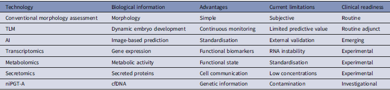
However, the successful translation of these technologies into routine clinical practice will require substantially greater methodological standardisation. Variability in embryo culture conditions, incubation systems, media composition, image acquisition protocols, analytical platforms and bioinformatic pipelines continues to limit reproducibility across IVF laboratories. Future multicentre prospective studies should therefore prioritise standardised protocols, external validation using independent datasets and clinically meaningful outcome measures, particularly cumulative live birth rather than surrogate endpoints alone. These principles are consistent with recent expert recommendations emphasising that emerging embryo assessment technologies should be adopted into clinical practice only after rigorous validation, standardised methodologies and demonstration of meaningful improvements in clinically relevant outcomes (Leigh et al., Reference Leigh, Scott, Rechitsky, Handyside, Munne, Wells, Vermeesch, Kahraman, Gianaroli, Rubio, Katz-Jaffe, Cram, Xu, Chen, Gao, Madjunkova, Harton, Gordon, Shulman, Simpson and Kuliev2026). The principal advantages and current limitations of the major non-invasive embryo assessment technologies are summarised in Table 1.
Comparison of current non-invasive embryo assessment technologies

Table 1. Long description
The table compares seven embryo assessment technologies. Conventional morphology assesses morphology, is simple, is limited by subjectivity, and is classified as routine. Time-lapse monitoring assesses dynamic embryo development through continuous monitoring, has limited predictive value, and is a routine adjunct. Artificial intelligence provides image-based prediction and standardization but requires external validation and is classified as emerging. Transcriptomics assesses gene expression and functional biomarkers but is limited by RNA instability and remains experimental. Metabolomics assesses metabolic activity and functional state but lacks standardization and remains experimental. Secretomics assesses secreted proteins and cell communication but is limited by low concentrations and remains experimental. Non-invasive PGT-A assesses cell-free DNA for genetic information, is limited by contamination, and remains investigational.
Another important consideration is the biological complexity of embryo development. Embryo competence cannot be fully explained by a single molecular layer, as genomic, transcriptomic, proteomic and metabolomic processes continuously interact throughout preimplantation development. Importantly, even a morphologically high-quality, morphokinetically appropriate and chromosomally euploid blastocyst may fail to implant or progress despite transfer into a receptive endometrium (Cimadomo et al., Reference Cimadomo, Rienzi, Conforti, Forman, Canosa, Innocenti, Poli, Hynes, Gemmell, Vaiarelli, Alviggi, Ubaldi and Capalbo2023). This observation underscores that embryo competence reflects the outcome of highly complex molecular, metabolic and embryo-endometrial interactions that remain only partially understood. Consequently, future research should increasingly adopt systems biology approaches capable of integrating multiple omics layers to better characterise the dynamic regulatory networks underlying embryo viability and implantation potential. Recent opinion papers have also questioned the long-standing assumption that embryo selection technologies alone, regardless of their increasing sophistication, can substantially improve IVF outcomes, emphasising the biological complexity of embryo competence and implantation (Gleicher et al., Reference Gleicher, Gayete-Lafuente, Barad, Patrizio and Albertini2025). Accordingly, future advances are likely to depend less on identifying a single ‘best biomarker than on integrating complementary imaging, molecular and computational information within robust, clinically validated frameworks.
Equally important is the need to improve the interpretability and transparency of AI systems. While highly accurate prediction models are desirable, clinical decision-making in reproductive medicine requires algorithms that are explainable, reproducible and generalisable across different patient populations and laboratory settings. AI should therefore be developed as a clinical decision-support tool that complements, rather than replaces, the expertise of embryologists. Evolution and clinical readiness of embryo assessment technologies were summarised in Figure 2.
Evolution and clinical readiness of embryo assessment Technologies. AI-assisted figure preparation: ChatGPT (GPT-5.6 Sol, OpenAI).

Figure 2. Long description
A left-to-right timeline summarizes the development and present clinical readiness of embryo assessment technologies in IVF. Morphology in the 1990s and blastocyst grading in the 2000s are classified as routine clinical practice. Time-lapse monitoring, introduced in the late 2000s, is classified as limited adjunctive use. Omics technologies in the early 2010s are experimental and investigational. Artificial intelligence in the late 2010s is classified as limited adjunctive use. Non-invasive PGT-A using cell-free DNA, introduced around 2015 to 2017, and whole-genome sequencing in the late 2010s are classified as experimental and investigational. Integrated multimodal assessment is placed in the future and is not yet in clinical use. An arrow beneath the timeline indicates increasing technological complexity and biological information from left to right.
Consequently, future innovations should be judged not only by their technological sophistication but also by their ability to improve outcomes that matter to patients. Ultimately, the goal of embryo assessment should not be the identification of the perfect embryo’, but the development of reliable, evidence-based and patient-centred strategies that improve the likelihood of a healthy live birth while minimising unnecessary interventions. The biological complexity of embryo competence was represented in Figure 3.
The biological complexity of embryo competence. AI-assisted figure preparation: ChatGPT (GPT-5.6 Sol, OpenAI).

Figure 3. Long description
A blastocyst labelled embryo competence is positioned at the center of a circular diagram. Ten domains point toward embryo competence: embryo morphology, including cell number, fragmentation, blastocyst grade and expansion; morphokinetics, including cell-cycle timings and developmental dynamics; genetics, including aneuploidy, copy-number variations and monogenic variants; epigenetics, including DNA methylation, histone modifications and imprinting; transcriptomics, including gene expression, alternative splicing and non-coding RNAs; proteomics, including protein abundance and post-translational modifications; metabolomics, including energy metabolism, amino acids, lipids and redox status; the maternal immune system, including immune tolerance, cytokines and immune-cell subsets; endometrial receptivity, including the hormonal environment and decidualization; and placental development, including trophoblast invasion, angiogenesis and placental function. Question marks around the outer circle denote unknown biological interactions. The figure indicates that no single current technology captures all determinants of implantation and live birth.
Conclusion
Embryo assessment has evolved from conventional morphology-based evaluation toward integrative, non-invasive and data-driven strategies. Approaches such as TLM, AI, metabolomic and secretomic profiling and non-invasive genomic testing provide complementary insights into embryo viability and implantation potential. Methodological heterogeneity, variability in sample characteristics and limited generalisability remain important challenges across studies. At the same time, increasing evidence suggests that integrating complementary technologies, rather than relying on any single biomarker, represents the most realistic path toward improving embryo selection. Importantly, previous experience with several promising technologies also demonstrates that encouraging preliminary findings alone are insufficient for clinical adoption without rigorous external validation and evidence of improved live birth outcomes. The future of embryo assessment is unlikely to depend on a single transformative technology but rather on the thoughtful integration of complementary imaging, molecular and computational approaches supported by rigorous clinical validation. Such an evidence-based strategy is more likely to improve embryo selection while preserving the central role of experienced embryologists in clinical decision-making and ultimately improving patient outcomes.
Acknowledgements
None.
Author contributions
Gulcin Ozkara: Conceptualisation-Equal, Data curation-Equal, Investigation-Equal, Methodology-Equal, Writing – original draft-Equal, Writing – review & editing-Equal.
Competing interests
The author declare that no conflict of interest exist.
Funding information
Not applicable.
Declaration of generative AI use
During the preparation and revision of this manuscript, the author used ChatGPT (GPT-5.6 Sol, OpenAI) to assist with English-language editing and the preparation and editing of Figures 1–3. All AI-assisted text and visual content was subsequently reviewed and edited by the author, who takes full responsibility for the scientific content and final version of the manuscript.



 Open access

