Introduction
Laryngeal haemangioma is a comparatively rare benign tumour of the larynx. The first larynx haemangioma was described by Mackenzie in 1871.Reference MacKenzie1 In 1921, Sweetser classified haemangiomas as infantile or adult.Reference Sweetser2 Adult laryngeal haemangiomas are clearly defined, with a bluish red colour, appearing most often in the region of the glottis and supraglottis. They occur more frequently in males. The principal symptom is hoarseness, occasional haemoptysis, and, in advanced cases, dysphagia and difficulty breathing.Reference Berkes and Sente3, Reference Prasad, Prasad and Bhat4 Compared to the infantile type, adult haemangiomas do not show the tendency of spontaneous regression.
Adult haemangiomas are mostly treated surgically. Various methods are utilised, including surgical removal,Reference Lucioni, Marioni, Della Libera and Rizzotto5 carbon dioxide (CO2)Reference Lucioni, Marioni, Della Libera and Rizzotto5 or potassium titanyl phosphate (KTP)Reference Kawakami, Hayashi, Yoshimura, Ichihara, Nishikawa and Ichihara6 laser therapy, cryosurgery,Reference O'Neill, Snyder and Toomey7 and sclerotherapy.Reference Orlando, Caldas, Campos, Nishinari, Krutman and Wolosker8 The treatment employed depends on: the patient's age; the tumour type, size and location; and the patient's complaints.Reference Berkes and Sente3
In this report, we present a patient with a cavernous haemangioma of the right supraglottic mucosa of the aryepiglottic fold, which was successfully excised by transoral robotic surgery without complications. To our knowledge, it is the first case reported in the medical literature in which transoral robotic surgery was utilised. The ethics committee of our hospital approved the study.
Case report
A 58-year-old woman presented to the otolaryngology department having experienced a lump-in-the-throat sensation over a period of 1 year. There was nothing significant regarding her medical history. She had no history of smoking or alcohol abuse.
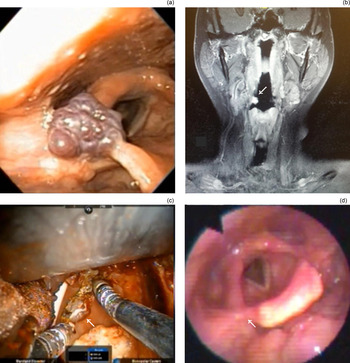
Otolaryngological physical examination findings were normal apart from a laryngeal lesion. On video-laryngoscopic examination, a lobulated, dark red mass was observed in the right supraglottic region. The lesion originated from the aryepiglottic fold and extended to the laryngeal side of the epiglottis (Figure 1a). A T1-hyperintense lesion was observed at the level of the right supraglottis on contrast-enhanced magnetic resonance imaging (MRI) (Figure 1b). As the patient was concerned about the risk of spontaneous rupture or airway compromise if the laryngeal haemangioma was left untreated, surgery was asked.
(a) Pre-operative image of supraglottic haemangioma at diagnosis: a lobulated, dark red tumour was seen at the level of the right aryepiglottic fold. (b) A hyperintense lesion was seen on coronal, T1-weighted, contrast-enhanced magnetic resonance imaging at the level of the right supraglottis (white arrow). (c) Intra-operative image of transoral robotic surgery excision of supraglottic haemangioma (white arrow). (d) Post-operative laryngoscopic view showing minimal scarring with preservation of the mucosal tissue surrounding the right aryepiglottic fold (white arrow).

Transoral robotic surgery was performed and the haemangioma was completely removed under general anaesthesia (Figure 1c). The tumour was grasped using Maryland 5 mm forceps to expose its wide-based stump. It was coagulated around its stump with monopolar cautery, using a spatula-tipped electrode. The minimal oozing that remained was easily and quickly controlled by cauterisation. No difficulty was encountered during dissection of the lesion.
The patient was stable during the post-operative period. She was discharged from hospital on the 2nd post-operative day, with no discomfort. There was improvement in the lump-in-the-throat sensation experienced by the patient post-operatively.
On video-laryngoscopic examination two months after the operation, minimal post-operative scarring was observed, with preservation of the mucosal tissue surrounding the right aryepiglottic fold (Figure 1d). The patient was followed up regularly thereafter, and no recurrence was noted over the subsequent six months.
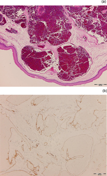
Histological examination of the vascular lesion showed clustered irregular-shaped, dilated, blood-filled, thin-walled vascular spaces, lined by a single layer of endothelial cells, within the oedematous subepithelial stroma. The lining cells were positive for cluster of differentiation 31 and negative for D2-40 in immunohistochemical staining (Figure 2).
(a) Histological examination of vascular lesion showed clustered irregular-shaped, dilated, blood-filled, thin-walled vascular spaces, lined by a single layer of endothelial cells, within the oedematous subepithelial stroma (H&E; ×40). (b) The lining cells were positive for cluster of differentiation 31 in immunohistochemical staining (×100).

Discussion
Haemangiomas of the larynx are generally classified as adult or infantile types. Infantile haemangiomas are usually subglottic and may cause fluctuating respiratory distress and biphasic stridor.Reference Ferguson9 Conversely, adult haemangiomas are rare and usually seen in the glottic or supraglottic region. They are more often of cavernous form and cause vague symptoms,Reference Lucioni, Marioni, Della Libera and Rizzotto5, Reference Huang, Lee and Shen10 such as hoarseness, cough, haemoptysis, dyspnoea and a lump sensation (as in our patient). Sixty-five per cent of haemangiomas occur in the head and neck region.Reference Shpitzer, Noyek, Witterick, Kassel, Ichise and Gullane11 The causative or predisposing factors are not well understood and have not been convincingly enumerated. The aetiological factors are thought to be vocal abuse, cigarette smoking and laryngeal trauma.Reference Kimmelman, Sugar and Lowry12 Malignant transformation of such tumours is very rare, with few case reports.Reference McRae, Gatland, McNab Jones and Khan13 Our patient was not a cigarette smoker and she had no history of vocal abuse, intubation or laryngeal trauma. Respiratory distress is almost never seen in adults.Reference Kimmelman, Sugar and Lowry12
Histologically, haemangiomas are composed of large, irregular, blood-filled channels lined with a single layer of endothelial cells between loose fibrous tissue septa of varying thickness. The lesions are generally of the cavernous type. Cavernous haemangiomas differ from capillary haemangiomas: the former have vascular channels that are less well circumscribed, and they are larger and usually located deeper in submucosal tissues. Sluggish blood flow may result in organised or dystrophically calcified thrombi within dilated vessels.Reference Kimmelman, Sugar and Lowry12
Laryngeal haemangiomas are diagnosed primarily by physical examination and history. Endoscopy is almost always sufficient for the diagnosis of a haemangioma. Doppler ultrasound, computed tomography, technetium imaging and plain radiographs can play a role in determining the dimensions and extent of haemangiomas.Reference Stal, Hamilton and Spira14 If the lesion is extensive, angiography and MRI with contrast may be useful in confirming the vascular nature of an adult laryngeal haemangioma and in determining its extent. Biopsies are not indicated because of the risk of severe bleeding.Reference Van Aalst, Bhuller and Sadove15, Reference Rahbar, Nicollas, Roger, Triglia, Garabedian and McGill16 Our patient underwent a contrast-enhanced MRI scan, which showed a T1-hyperintense lesion that was isolated and appeared to be restricted to the level of the right supraglottis (Figure 1b).
No active treatment is advised for adult laryngeal haemangiomas unless the lesions are symptomatic or show a tendency to involve other parts. There is no consensus and no well-established treatment protocol for adult laryngeal haemangiomas, as only anecdotal case reports or very limited case series are available in the medical literature. Factors influencing the choice of therapy include patient age, and tumour type, size and location. Adult haemangiomas do not show a tendency to spontaneously regress; however, they are not very progressive tumours. Therefore, clinical observation is the best therapy in most cases.
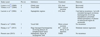
If bleeding, respiratory tract stenosis or cosmetic problems occur, aggressive therapies are needed. Corticosteroid injection,Reference Lucioni, Marioni, Della Libera and Rizzotto5, Reference Kawakami, Hayashi, Yoshimura, Ichihara, Nishikawa and Ichihara6 laser ablation with CO2 or KTP lasers,Reference Lucioni, Marioni, Della Libera and Rizzotto5, Reference Kawakami, Hayashi, Yoshimura, Ichihara, Nishikawa and Ichihara6 cryosurgery,Reference O'Neill, Snyder and Toomey7 and radiation therapyReference Huang, Lee and Huang17 have all been utilised. For small lesions, excision with microlaryngoscopic techniques or laser ablation can be used.Reference Yilmaz, Aktepe and Altuntaş18, Reference Yan, Olszewski, Hoffman, Zhuang, Ford and Dailey19 For large lesions, tracheostomy and an open surgical approach may be required. Table I summarises the treatments for adult laryngeal haemangioma.
Summary of treatments for adult laryngeal haemangioma

Pt = patient; CO2 = carbon dioxide; RT = radiotherapy; TORS = transoral robotic surgery
Laser surgery is thought to be relatively effective and less invasive than surgical removal. Lesion excision with a CO2 laser is generally accepted.Reference Lucioni, Marioni, Della Libera and Rizzotto5, Reference Lomeo, McDonald and Finneman20 In limited or pedunculated supraglottic cavernous haemangiomas, CO2 laser vaporisation of the lesion with super-pulse modality at 4–8 W is advised.Reference Lucioni, Marioni, Della Libera and Rizzotto5 However, CO2 laser cauterisation is ineffective in extended lesions and in large vessels with significant bleeding. In addition, several reports indicate that use of the CO2 laser when treating subglottic haemangiomas is associated with the increased risk of damage to adjacent mucosa and of developing subglottic stenosis post-operatively.Reference Cotton and Tewfik21
• Excision of adult laryngeal haemangiomas with transoral robotic surgery can be very successful, with a low incidence of complications
• Patients with small laryngeal haemangiomas should consider transoral robotic excision to avoid excessive removal of normal tissues and enable easier control of bleeding
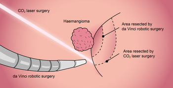
The literature on the use of robotic surgery for managing laryngeal haemangiomas is lacking. We recommend utilising a transoral robotic system to ablate this vascular tumour because of its several advantages (Figure 3). For instance, the robotic system can be directed to the relevant tissues by using a spatula-tipped monopolar cautery instrument; thus, it is very adaptable and precise, thereby avoiding excessive removal of normal tissues and shortening the recovery time after surgery. Furthermore, as transoral robotic surgery assisted excision is a minimally invasive approach, blood loss is minimal and it is easy to control bleeding during the operation. Therefore, we recommend robotic surgery as a first choice treatment, especially for small or medium-sized laryngeal haemangiomas.
Illustration comparing excision using a carbon dioxide (CO2) laser with transoral robotic surgery; in the latter method, the robotic arm can easily bypass the tumour, cutting through the back of the tumour from attached mucosa. This adaptable, precise technique enables preservation of more of the surrounding tissue, with easier-to-control bleeding during the operation.