Introduction
Until the discovery of the life cycle of Toxoplasma in 1970, intestinal coccidia of cats and dogs were classified under the genus Isospora and considered to have a direct fecal–oral cycle (Dubey, Reference Dubey2018). Frenkel and Dubey (Reference Frenkel and Dubey1972) found encysted extra-intestinal stage of Isospora felis and I. rivolta in rodents. Based on these observations, Frenkel (Reference Frenkel1977) proposed new genus, Cystoisospora, for Isospora species of cats and dogs. Barta et al. (Reference Barta, Schrenzel, Carreno and Rideout2005) supported the validity of Cystoisospora based on molecular phylogeny and morphological differences between avian and mammalian Isospora species; Isospora species in mammals lack Stieda bodies that are present in avian Isospora species. Therefore, Isospora species in cats and dogs are now classified as Cystoisospora (Dubey, Reference Dubey2018).
Three or more species of Cystoisospora, C. canis (Nemeséri, Reference Nemeséri1960; Lepp and Todd, Reference Lepp and Todd1974; Mitchell et al., Reference Mitchell, Zajac, Charles, Duncan and Lindsay2007), C. ohioensis (Dubey, Reference Dubey1975, Reference Dubey1978a, Reference Dubey1978b), C. neorivolta (Dubey and Mahrt, Reference Dubey and Mahrt1978) and C. burrowsi (Trayser and Todd, Reference Trayser and Todd1978; Rommel and Zielasko, Reference Rommel and Zielasko1981) have been reported as intestinal coccidia of dogs. Of these, full asexual and sexual life cycle stages at the light microscope level are known only for C. ohioensis (Dubey, Reference Dubey1978a, Reference Dubey1978b). Only partial stages are known for C. canis (Lepp and Todd, Reference Lepp and Todd1974), C. neorivolta and C. burrowsi (Dubey and Mahrt, Reference Dubey and Mahrt1978; Trayser and Todd, Reference Trayser and Todd1978). The oocyst dimensions for C. neorivolta are unknown and endogenous stages for C. burrowsi were not described in detail leaving several uncertainties. It is not clear based on their descriptions, whether C. neorivolta and C. burrowsi are the same or different parasites and there is no way to determine their identity now because there are no archived specimens (Dubey and Mahrt, Reference Dubey and Mahrt1978; Trayser and Todd, Reference Trayser and Todd1978). However, C. neorivolta and C. burrowsi are structurally distinct from C. ohioensis, based on location of endogenous stages; C. ohioensis stages are in the surface epithelial enterocytes whereas C. burrowsi and C. neorivolta stages are in sub-epithelial tissue. Because of the difficulty of distinguishing C. ohioensis, C. neorivolta and C. burrowsi without details of development, these organisms are referred to as C. ohioensis-like (Dubey et al., Reference Dubey, Weisbrode and Rogers1978).
Dubey et al. (Reference Dubey, Weisbrode and Rogers1978) reported clinical coccidiosis in a naturally infected dog associated with a C. ohioensis-like organism. In the present report, I re-evaluated electron micrographs and histological preparations from this dog to determine if differences in structure were present that could help determine differential diagnosis of C. ohioensis-like organisms. The results provide new information on ultrastructure of meronts and gamonts.
Materials and methods
The dog had died of acute enteritis and intestines were initially fixed in 10% buffered formalin and subsequently processed for transmission electron microscopy (TEM) as described (Dubey et al., Reference Dubey, Weisbrode and Rogers1978). Smears of intestines were fixed in methanol and stained with Giemsa (Dubey et al., Reference Dubey, Weisbrode and Rogers1978). After 40 years, paraffin blocks and TEM grids from this case had been discarded and were not available for further studies. Therefore, for the present study, two sections that had been stained in 1978 with haematoxylin and eosin were re-stained with periodic acid Schiff (PAS) reaction. TEM negatives were scanned digitally and re-evaluated here.
Results
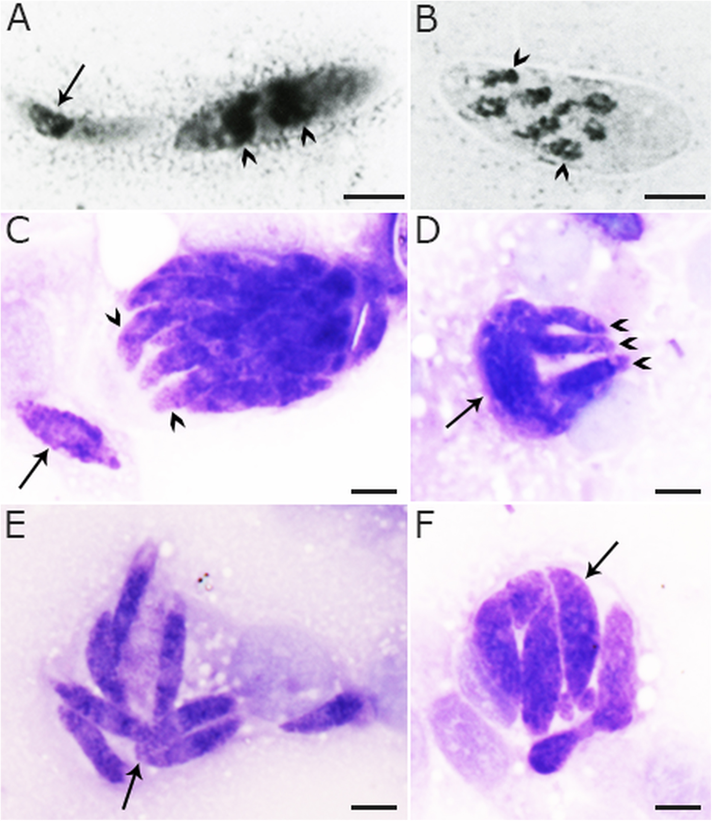
In sections stained with haematoxylin and eosin or PAS, endogenous stages were present in surface epithelium and in the lamina propria of both small and large intestine. In smears of intestines stained with Giemsa, merozoites and crescent/merozoite-shaped meronts were seen containing up to eight nuclei (Fig. 1A and B). Multinucleated round meronts were not seen. Merozoites of different shapes and sizes were seen. Mode of division was not clear but binucleated merozoites, and division into two were indicative of endyogeny. In some preparations merozoites two protruded from an undivided mass (Fig. 1C and D).
Endogenous stages of a Cystoisospora ohioensis-like organism in intestinal smears of a naturally infected dog. Giemsa stain. Bars = 5 µm. (A) Slender merozoite with one nucleus (arrow) and a multinucleate (arrowheads) meront. (B) Meront with eight nuclei (arrowheads). (C) A trinucleate meront-like structure (arrow) and a meront with protruding merozoites (arrowheads). (D) Merozoites (arrowheads) budding from a multinucleated mass (arrow). (E) Mature merozoites, with two merozoites apparently attached to each other (arrow). (F) An intracellular meront with mature merozoites (arrow).

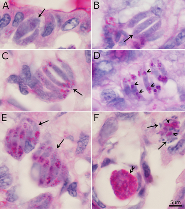
In sections stained with PAS, merozoites of different shapes and sizes were seen (Fig. 2A–F). Slender, uninucleate merozoites were 5–12 µm long (Fig. 2C and D). All stages of merogony were PAS-positive. The PAS-positivity seems to increase with maturation of merozoites. The intensity of staining, the sizes of PAS-positive granules and their location in merozoites varied with plane of section of merozoites (Fig. 2F). Immature microgamonts with centrally located nuclei were PAS-negative. The microgametes were PAS-negative but the residual bodies were PAS-positive (Fig. 2H). Both immature and mature macrogamonts were PAS-positive (Fig. 2F).
Asexual and sexual stages of a Cystoisospora ohioensis-like organism in sections of intestines of a naturally infected dog. Periodic acid Schiff reaction (PAS) counter stained with haematoxylin. The scale bar is 5 µm and applies to all parts. (A–C) Developing meronts/merozoites (arrows) with relatively few PAS-positive granules. (D) Slender merozoites with two areas of PAS-positive granules (arrowheads). (E) Two groups with tightly packed merozoites (arrows). (F) An intensely PAS-positive macrogamont (double arrowheads) and a microgamont with peripherally arranged microgametes (arrows) and few PAS-positive residual bodies (arrowheads).

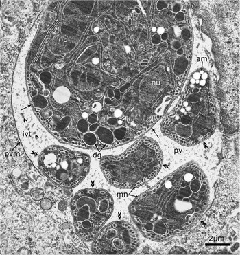
By TEM, mature merozoites were identified in meronts. Figure 3 shows two groups of merozoites within one parasitophorous vacuole (pv). Parts of six free merozoites are enclosed in the same pv; intravacuolar tubules are visible among free merozoites. The merozoites have numerous micronemes, dense granules and amylopectin granules (am). The large membrane body is interpreted as undivided meront with more than three nuclei and amylopectin granules.
Transmission electron micrograph of a Cystoisospora ohioensis-like organism in ileum of a naturally infected dog. Two groups of merozoites are enclosed in parasitophorous vacuolar membrane (pvm) in an enterocyte. Note one large meront (arrows) and six free merozoites (double arrowheads) outside the main group. The merozoites in the main group are twisted and individual boundaries are not clearly demarcated. Note numerous intravacuolar tubules (ivt) in the parasitophorous vacuole (pv). Also, note nucleus (nu), micronemes, amylopectin granules (am) and numerous dense granules (dg).

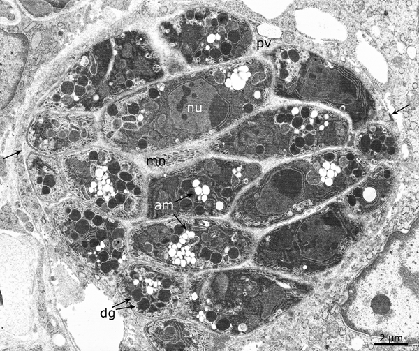
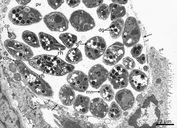
Figure 4 shows a meront with eight or more tightly packed merozoites. The merozoites are twisted; therefore, their number is uncertain. Figure 5 shows a meront with 12 or more mature loosely arranged merozoites within 1 parasitophorous vacuole (pv). One of the salient features of the merozoites is the presence of many amylopectin granules (Figs 3–5). Merozoites contained several micronemes that were arranged haphazardly. The nucleus was located centrally or in the posterior part of the merozoite. Only few rhoptries could be identified definitively (Fig. 5). There were many dense granules, and it was difficult to distinguish rhoptries from dense granules unless neck or blind ends of rhoptries were visible. A residual body was not seen in meronts.
Transmission electron micrograph of a Cystoisospora ohioensis-like organism in ileum of a naturally infected dog. A mature meront (arrows) with more than eight merozoites packed in parasitophorous vacuole (pv). The merozoites are twisted, thus their number is uncertain. Note prominent amylopectin granules (am), numerous dense granules (dg) and micronemes (nm).

Transmission electron micrograph of a Cystoisospora ohioensis-like organism in ileum of a naturally infected dog. A mature meront (arrows) with several merozoites loosely arranged in parasitophorous vacuole (pv). Note prominent amylopectin granules (am), conoid (co), dense granules (dg), few rhoptries (rh) and several micronemes (nm) in merozoites.

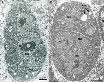
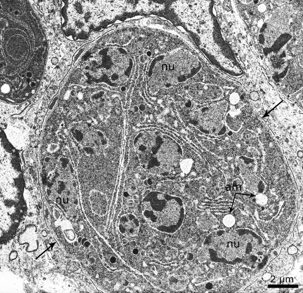
Early microgamont/meront-like structures contained 2–5 nuclei with few or no amylopectin granules (Figs. 6 and 7). Later stages of microgamonts could be distinguished from meronts by the presence of peripherally arranged nuclei (Fig. 8).
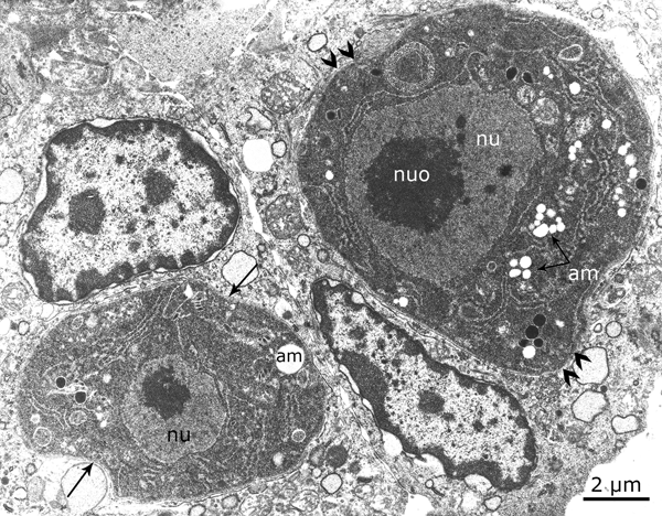
Transmission electron micrograph of a Cystoisospora ohioensis-like organism in ileum of a naturally infected dog. An uninucleated undifferentiated organism (arrows) and a macrogamont (double arrowheads). The macrogamont has a large nucleus (nu) and with a large nucleolus (nuo), and contains several amylopectin granules (am).

Transmission electron micrograph of a Cystoisospora ohioensis-like organism in ileum of a naturally infected dog. Two microgamont/meront-like organisms with two (A) or four (B) nuclei. Note only a few amylopectin granules (am).

Transmission electron micrograph of a Cystoisospora ohioensis-like organism in ileum of a naturally infected dog. A microgamont enclosed in a thin parasitophorous vacuolar membrane (arrows) with several nuclei (nu), some of them are at the periphery and few amylopectin granules (am). Note that the chromatin in nuclei is condensed.

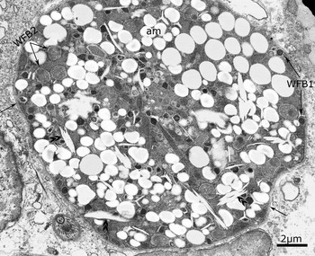
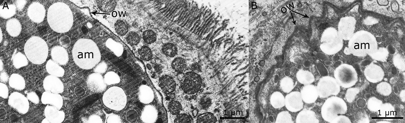
Earliest macrogamonts were distinguished from microgamonts by the presence of a single nucleus with a large nucleolus, and many amylopectin granules that were PAS-positive (Fig. 6). Mature macrogamonts contained a central nucleus, electron-dense wall-forming bodies (WFB) type 1 and spongy WFB type 2, numerous amylopectin granules and canaliculi (Fig. 9). WFB were absent in oocysts (Fig. 10).
Transmission electron micrograph of a macrogamont of Cystoisospora ohioensis-like organism in ileum of a naturally infected dog. Note electron dense wall-forming bodies type 1 (WFB 1) and spongy WFB 2, numerous amylopectin (am) granules, and canaliculi (double arrowheads).

Transmission electron micrograph of developing oocysts of a Cystoisospora ohioensis-like organism in ileum of a naturally infected dog. (A and B) The oocysts are intracellular in enterocytes and contain numerous amylopectin granules (am) but no wall-forming bodies. Note oocysts wall (ow).

Discussion
Review of literature indicates that feline Cystoisospora species (C. felis, C. rivolta) merozoites undergo more than one asexual generation in the original host cell without leaving it to enter new host cells (Shah, Reference Shah1971; Dubey, Reference Dubey1979; Dubey, Reference Dubey2018). By light microscopy, the meronts contained up to 10 nuclei. Ferguson et al. (Reference Ferguson, Birch-Andersen, Hutchison and Siim1980a) using the TEM demonstrated that C. felis merozoites divided by endodyogeny in enterocytes of a cat 6 days after feeding oocysts. They further indicated that the daughters undergo a further cycle of endodyogeny within the same pv (Ferguson et al., Reference Ferguson, Birch-Andersen, Hutchison and Siim1980a). Even large meronts seen were the product of endodyogeny. Boch et al. (Reference Boch, Göbel, Heine and Erber1981) confirmed multiplication of meronts by endodyogeny.
The mode of division of the canine Cystoisospora species is not fully understood. Lepp and Todd (Reference Lepp and Todd1974) reported three generations of meronts in C. canis; they schematically depicted as many as 20 separate nuclei in meronts and as many as 72 merozoites in a group but the details are not clear in their illustrations and it is likely that multinucleated microgamonts were misinterpreted as schizonts (Dubey and Lindsay, Reference Dubey and Lindsay2019a). In a preliminary report, Hilali et al. (Reference Hilali, Ghaffar and Scholtyseck1979) described the ultrastructure of meronts and merozoites of C. canis in the intestines of dogs. Merozoites divided by endodyogeny; no multinucleated stage was found. However, details are missing. Thus, mode of division of C. canis schizonts is not clear.
Only partial details are available concerning asexual development of C. neorivolta and C. burrowsi (Dubey and Mahrt, Reference Dubey and Mahrt1978; Trayser and Todd, Reference Trayser and Todd1978). In the present study, details of asexual development of a C. ohioensis-like organism are presented. A group of merozoites shown in Fig. 3 here contained free merozoites and developing merozoites in the same pv; these were misinterpreted by Dubey et al. (Reference Dubey, Weisbrode and Rogers1978) as an immature meront with four nuclei. Up to eight nuclei were seen in meronts in preparations stained with Giemsa but the mode of division was not clear and multinucleated round or oval-shaped meronts commonly seen in Eimeria species (Levine, Reference Levine1973) were not detected. A review of literature and re-review of histological sections of enteric stages of C. felis, C. rivolta, C. canis, C. ohioensis, of dogs and cats revealed that there is lack of confirmation of schizony in their asexual stages (Dubey, Reference Dubey2018; Dubey and Lindsay, Reference Dubey and Lindsay2019a, Reference Dubey2019b). Therefore, asexual stages were termed meronts.
In the present study, mature merozoites were present along with immature meronts in the same vacuole (Fig. 3). The undivided mass was considered multinucleated meront and not a residual body. This is normal pattern in asexual stages of cat and dog coccidia (Dubey, Reference Dubey2018; Dubey and Lindsay, Reference Dubey2019b, Reference Dubey and Lindsay2019c). However, ultrastructural studies are needed to resolve mode of multiplication of all canine Cystoisospora species. The finding of many amylopectin granules in merozoites in the present study is unusual for coccidian merozoites. The amylopectin granules stain red with PAS, and thus can be distinguished by light microscopy. Unlike Cystoisospora merozoites, Eimeria merozoites are generally PAS-negative.
Dubey (Reference Dubey1979, 2014) who studied the life cycle of C. felis and C. rivolta of cats and C. ohioensis of dogs proposed the term ‘types’ instead of asexual generations because of numerous generations within a given host cell. This kind of division is different than the fixed numbers of generations of schizonts in Eimeria species (Levine, Reference Levine1973). Shah (Reference Shah1971), who studied the life cycle of C. felis called the groups with more than one generation of schizonts as ‘cysts’. However, cyst is not a good term because cyst is a term best used for an orally infective stage of coccidia and monozoic tissue cysts of C. canis can occur in paratenic hosts has been used for encysted stages in extra-intestinal organs (Lindsay et al., Reference Lindsay, Houk, Mitchell and Dubey2014). Dubey and Frenkel (Reference Dubey and Frenkel1972) first proposed the term ‘type’ for intestinal stages of Toxoplasma gondii because there were many generations of the parasite in feline enterocytes in the intestine. In conclusion, ‘type’ is more applicable than generations with respect to Cystoisospora spp. (Dubey and Lindsay, Reference Dubey and Lindsay2019a).
The development of Cystoisospora spp. oocyst wall is largely unknown. In C. felis, two types of WFB, type 1 and type 2, have been described (Ferguson et al., Reference Ferguson, Birch-Andersen, Hutchison and Siim1980b). The WFB1 were electron dense and smaller in size than WFB2 which were spongy (Ferguson et al., Reference Ferguson, Birch-Andersen, Hutchison and Siim1980b). The WFB arise during the maturation of the macrogamont and form the oocyst wall. In the present study, WFB were similar in structure as in C. felis. TEM of optimally preserved samples will be needed for full characterization of WFB and oocyst wall formation of canine Cystoisospora species. Nevertheless, the present study is the first for TEM of gamonts of a canine Cystoisospora.
Author ORCIDs
J. P. Dubey 0000-0003-4455-2870
Acknowledgements
I would like to thank Camila Cezar, Fernando Murata, Meghan Sadler, and Oliver Kwok for assistance and Prof Steven E. Weisbrode for his advice and past collaborations.
Financial support
This research received no specific grant from any funding agency, commercial or not-for-profit sectors.
Conflict of interest
None.
Ethical standards
No experiments were performed. Archived specimens were studied.