Introduction
In many near-shore temperate reef ecosystems, rock lobsters are a dominant predator effecting a top-down control of benthic community structure (Tarr et al., Reference Tarr, Williams and MacKenzie1996; Mayfield & Branch, Reference Mayfield and Branch2000; Shears & Babcock, Reference Shears and Babcock2003). However, detailed knowledge of rock lobster diets and spatial and temporal variability in diet is unknown. This information is crucial to informed management at an ecosystem level of rocky reefs and the important fisheries they support. The establishment of no-take marine protected areas (MPAs), where commercial and recreational fishing is prohibited, has provided a useful resource for monitoring of key temperate reef organisms (Edgar et al., Reference Edgar, Moverley, Barrett, Peters and Reed1997). This has recently led to speculation that southern rock lobster (Jasus edwardsii) predation may underpin an apparent decrease in densities of young abalone within the Tasmanian MPAs (Edgar & Barrett, Reference Edgar and Barrett1999; Barrett et al., Reference Barrett, Edgar, Morton and Buxton2003). Currently, there is no method to test this idea or to evaluate the predation patterns of southern rock lobsters in the wild. Furthermore, these are both high value fishery species and, thus, an interesting trophic interaction to monitor.
Previous research on predator-prey relationships of crustaceans has largely involved gut dissection and relied on visual analysis to identify prey remains (Hickman, Reference Hickman1945; Fielder, Reference Fielder1965; Ennis, Reference Ennis1973; Jernakoff et al., Reference Jernakoff, Phillips and Fitzpatrick1993; Mayfield et al., Reference Mayfield, Lopata and Branch2000c). This approach has shown that the diet of lobsters varies widely with season and that lobsters are selective feeders (Joll & Phillips, Reference Joll and Phillips1984; Barkai et al., Reference Barkai, Davis and Tugwell1996; Mayfield et al., Reference Mayfield, Atkinson, Branch and Cockcroft2000a,Reference Mayfield, Branch and Cockcroftb). The physical process of digestion, however, makes visual assessment of gut contents difficult, and gut content analysis has many biases due to varying rates of gut retention, digestion and erosion of prey material. The collection of foregut contents for inspection also necessitates the mortality of the studied lobster (Hickman, Reference Hickman1945; Williams, Reference Williams1981; Joll & Phillips, Reference Joll and Phillips1984; Jernakoff et al., Reference Jernakoff, Phillips and Fitzpatrick1993; Mayfield et al., Reference Mayfield, Atkinson, Branch and Cockcroft2000a,Reference Mayfield, Lopata and Branchc) so that large scale surveys are problematic. Killing animals to identify prey is also unlikely to be acceptable if the study population resides in an MPA (Ward et al., Reference Ward, Heinemann and Evans2001). Visual inspection of gut contents is known to miss soft-bodied prey organisms, such as abalone and other gastropod molluscs, and may result in up to 90% of the gut contents being unidentifiable (Mayfield et al., Reference Mayfield, Lopata and Branch2000c). Non-lethal techniques to determine dietary intake are essential because of the high value of live rock lobsters in the commercial fishery and to enable ongoing work inside MPAs.
For most marine predators, unbiased identification of prey is problematic, but new protocols are emerging that allow the unambiguous detection of prey species in predator diets. Recent advances in molecular biology have been used to elucidate diets of animals in a wide range of taxonomic groups, such as fish (Rosel & Kocher, Reference Rosel and Kocher2002), giant squid (Deagle et al., Reference Deagle, Jarman, Pemberton and Gales2005a), wasps (Kasper et al., Reference Kasper, Reeson, Cooper, Perry and Austin2004), spiders (Agusti et al., Reference Agusti, Shayler, Harwood, Vaughan, Sunderland and Symondson2003) and even introduced insects capable of foraging in native forests (Sheppard et al., Reference Sheppard, Henneman, Memmott and Symondson2004). Molecular techniques take advantage of unique DNA sequences in species or in groups of organisms and can provide detailed and precise information about predator-prey relationships (Agusti et al., Reference Agusti, Shayler, Harwood, Vaughan, Sunderland and Symondson2003; Nejstgaard et al., Reference Nejstgaard, Frischer, Raule, Gruebel, Kohlberg and Verity2003; Jarman & Wilson, Reference Jarman and Wilson2004; Deagle et al., Reference Deagle, Jarman, Pemberton and Gales2005a; Juen & Traugott, Reference Juen and Traugott2005).
In the marine environment, protocols have been developed successfully to isolate prey DNA from the faeces of predators (Nejstgaard et al., Reference Nejstgaard, Frischer, Raule, Gruebel, Kohlberg and Verity2003; Jarman et al., Reference Jarman, Deagle and Gales2004; Deagle et al., Reference Deagle, Tollit, Jarman, Hindell, Trites and Gales2005b; Vestheim et al., Reference Vestheim, Edvardsen and Kaartvedt2005). The DNA is amplified using polymerase chain reaction (PCR) with specific primers that target gene regions of prey species (Jarman et al., Reference Jarman, Deagle and Gales2004; Sheppard & Harwood, Reference Sheppard and Harwood2005). The techniques developed have shown that the identity of prey species can be unambiguously detected in this manner (Symondson, Reference Symondson2002). A major incentive for development of non-invasive techniques as an alternative to direct stomach content analysis has been to allow the non-lethal study of endangered or protected animal species (Farrell et al., Reference Farrell, Roman and Sunquist2000; Jarman et al., Reference Jarman, Gales, Tierney, Gill and Elliott2002; Symondson, Reference Symondson2002; Jarman et al., Reference Jarman, Deagle and Gales2004; Jarman & Wilson, Reference Jarman and Wilson2004; Deagle et al., Reference Deagle, Tollit, Jarman, Hindell, Trites and Gales2005b).
In Tasmania, lobsters are the basis for a lucrative commercial fishery where fishers land and sell their lobsters live to discerning domestic and international markets. For fishers to support our research, it was necessary to obtain dietary information while still leaving the lobsters in perfect condition for subsequent sale. The capacity to use animals caught in traps was also important to enable researchers to obtain samples aboard commercial rock lobster fishing vessels, to access remote areas of lobster habitat and to obtain animals from deep-water reefs outside standard scuba diving depths.
The aims of this paper were threefold. Firstly, we demonstrated a non-lethal approach to obtaining dietary samples; secondly, we determined if faecal material could be used to identify prey using molecular methods; and, thirdly, we determined if the longevity of the DNA signal was sufficient to include prey items from lobsters that would be retained in traps for up to 24 h.
Materials and methods
Feeding trials
Rock lobsters for feeding trials were captured by scuba diving in the Crayfish Point Marine Reserve at Taroona, Tasmania (42.95°S, 147.34°E) in April 2004 and May 2005. Lobsters were collected opportunistically, ensuring an even distribution of sexes and a wide range of sizes. All captured lobsters were measured and carapace length (CL) was recorded to the nearest millimeter.
Captured lobsters were immediately taken to the laboratory and kept in aerated, flow-through seawater tanks. For the duration of the feeding trials, lobsters were maintained under ambient light conditions and water temperatures in outdoor aquaria at the Marine Research Laboratories, Taroona, Tasmania.
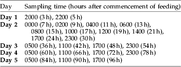
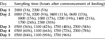
For each trial, the lobsters were placed in one section of a 450 l tank separated into three sections with plastic mesh and dividers. Each lobster was provided with a 400 mm×200 mm concrete block as a shelter. All lobsters were starved for 72 h prior to each feeding trial to facilitate gut evacuation and to remove any remaining prey DNA from the digestive tract. For each trial, approximately 15 g of fresh food material was given to each lobster. This material was prepared by shucking live abalone (Haliotis rubra) and cutting the foot tissue into approximately 15 g portions. Common sea urchins (Heliocidaris erythrogramma) and long-spined sea urchins (Centrostephanus rodgersii) were shucked alive and approximately 15 g portions of roe and viscera were used as food. Food was introduced to each lobster at 1700 h and individual lobsters were monitored for feeding activity. Only lobsters that actively fed and consumed the entire food sample within the first hour were used in the feeding trials. No additional food was provided to lobsters for the duration of the trial and each lobster was sampled once per feeding trial. Lobsters were selected for faecal collection over the next five days at the times (hours after commencement of feeding) given in table 1. Lobsters were selected randomly to eliminate any tank effect. For each sampling time, faecal material was collected from a minimum of three separate lobsters. For each of the individual faecal samples, PCR assays were performed twice to guarantee the consistency of the result. These feeding experiments were repeated over a period of 65 days with 116 faecal samples collected in total. A total of 61 lobsters were used in these trials, comprising 24 females and 37 males. Lobsters ranged in size from 53 to 161 mm carapace length.
Feeding trial schedule over five days, covering 96 h after initiation of feeding episodes. Each time indicates when samples were collected and the hours after the commencement of feeding.
Faecal material collection
A ‘cradle’ device was designed and built for the purpose of restraining lobsters while samples could be taken from the hindgut region. Lobsters were placed in the cradle, which holds the animal upside-down and immobilizes the posterior four pairs of walking legs. The anterior telson was held firmly by the researcher to stabilize the abdominal region and tail while collecting faeces.
Lobster faeces were collected using a 100–1000 μl pipette with disposable tips. For each faecal sample, a new sterile tip was used to prevent contamination between samples. The tip was inserted directly into the anal pore of the lobster to remove faeces from the hindgut region. The collected material was immediately pipetted into a 1.5 ml micro centrifuge tube containing 500 μl of 70% ethanol. Ethanol has been shown to be an effective preservative for field samples and does not require freezing or any special handling (Jarman et al., Reference Jarman, Deagle and Gales2004). The volume collected varied from approximately 10 μl to 1 ml depending on the size of lobster and fullness of hindgut at the time of sampling. Ethanol was removed from samples before DNA extraction by centrifuging at 10,000 g for 30 s. Excess ethanol was poured off and the sample tubes centrifuged again. Any remaining ethanol was then removed by pipette.
DNA extraction
There were no previously published studies using rock lobster faecal material as a source of prey DNA. Tissue samples from predator and prey species were collected to test PCR primer specificity and to use as both positive and negative DNA controls in later experiments. Tissue samples were taken from fresh animals and a small (~0.25 ml) portion was used for total cellular DNA extraction. Another portion (~2 ml) of the tissue was stored in 70% ethanol as a voucher specimen. The DNeasy® Tissue Kit (QIAGEN) was used to extract genomic DNA from predator and prey tissues, and the manufacturer's animal tissue protocol was followed.
The Ultra Clean™ Fecal DNA Kit (Mo Bio Laboratories, Inc.) was used for DNA extractions on rock lobster faecal samples following the manufacturer's protocols with the supplied proprietary buffers and reagents. All DNA extracted from faecal samples using this kit was ready for PCR, and the manufacturer's protocol appeared to remove any potential PCR inhibitors.
PCR amplification
Precautions were taken during preparation of PCR reactions to minimize the possibility of contamination by extraneous DNA. Aerosol-resistant barrier pipette tips were used for preparing all PCR reactions, and pipette tips were either sterile and pre-packaged or autoclaved prior to use. All PCR reactions were prepared in a dedicated hood where PCR tubes, pipettes and pipette tips were subjected to UV light for a minimum of 10 min prior to setting up each PCR reaction.
The components of the 20 μl PCRs were 50 mM KCL, 15 mM Tris.HCl pH 3.0, 5.0 mM MgCl2, 100 μg ml−1 bovine serum albumin, 0.5 mM of each dNTP, 2 mM each primer (Geneworks), 1 unit AmpliTaq Gold® DNA polymerase (Sigma), and ~20 ng template DNA. Both positive and negative controls were run with each batch of PCRs. For negative controls, 2 μl MilliQ H2O was used as template; and, for positive controls, ~20 ng template DNA from Haliotis rubra, Centrostephanus rodgersii and Heliocidaris erythrogramma was used to confirm reaction success. PCR replication was performed by running reactions a minimum of two times to ensure a consistent result.
The PCR primer sets used in this experiment (table 2) were developed previously to distinguish Haliotis rubra tissue from that of other Haliotis species for forensic purposes (Elliott et al., Reference Elliott, Bartlett, Evans, Officer and Haddon2002) and for identification of echinoderms as prey items in marine ecosystems (Jarman et al., Reference Jarman, Redd and Giles2006). Primers were obtained from GeneWorks Pty Ltd Custom Oligonucleotide service and diluted to 10 μM for use in setting up PCR reactions.
PCR primers used, including sequence of each primer, target organism or group, and DNA region amplified. DNA regions are mitochondrial cytochrome oxidase subunit I (mt COI) and the nuclear large subunit ribosomal RNA gene (18s rDNA).

PCR thermal cycling conditions for HalCO2GENA and HalCO2GENB primers were: denaturation and DNA polymerase activation at 95°C for 10 min followed by 10 initial amplification cycles: 94°C for 30 s, annealing at 60–55°C for 30 s, 72°C for 1 min with a decrease in the annealing temperature by 0.5°C for each of the 10 cycles. Twenty-five further amplification cycles were carried out: 94°C for 30 s, 55°C for 30 s and 72°C for 1 min. The final extension step was 72°C for 5 min and the reaction was held at 10°C until removed from the MJ Research PTC-2001 Thermal Cycler.
PCR thermal cycling conditions for EchinNSS18sf/EchinNSS18sr primers were: denaturation and DNA polymerase activation at 95°C for 10 min followed by 35 amplification cycles: 95°C for 5 s, annealing temperature at 51°C for 30 s, 72°C for 30 s. The final extension step was 72°C for 5 min and the reaction was held at 10°C until removed from the MJ Research PTC-2001 Thermal Cycler.
PCR products were visualized by electrophoresis on 1.5% agarose gels stained with ethidium bromide (5 μl ethidium bromide per 100 ml agarose). Each gel was loaded with 7 μl of PCR product, and a 100 bp ladder was used on every gel to determine fragment size and to confirm the results of each PCR. All agarose gels ran for 20 min at 80 V.
Results
Feeding trials
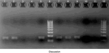
Using HalCO2GENA and HalCO2GENB primers, fragments of 193 bp were successfully amplified from both black-lipped abalone tissue DNA and faecal samples from lobsters fed a diet of black-lipped abalone (fig. 1). Using EchinNSSf1 and EchinNSSr1 primers, fragments of 160 bp were successfully amplified from Centrostephanus rodgersii and Heliocidaris erythrogramma tissue DNA, and from faecal samples from lobsters fed a diet of both urchin species (fig. 1). Prey was initially detected in all samples at 7 h after feeding and was consistently detected in all samples until 60 h after the feeding episode. There were no detections in any of the samples taken at 3 and 5 h or for the six sampling times from 66 h to 96 h after commencement of feeding.
Specificity of HalCO2GENA/HalCO2GENB and EchinNSSf1/EchinNSSr1 PCR primers. Agarose gel showing PCR products amplified from rock lobster feeding experiments using group and species-specific PCR primers. Lanes 1–5 show DNA amplification using PCR primers specific to Haliotis rubra and lanes 7–11 show DNA amplification using PCR primers specific to Echinodermata. Lanes 6 and 12 are 100 bp DNA reference ladders indicating relative size of DNA fragments in sample lanes. Lanes 1, 2 and 3 indicate PCR amplified rock lobster faecal samples from 8, 24 and 96 h after feeding on black-lipped abalone. Lane 5 shows DNA amplification using Haliotis rubra tissue. Lanes 7, 8, 9 and 10 show PCR amplified rock lobster faecal samples from 12, 36, 48 and 72 h after feeding on Centrostephanus rodgersii. Lanes 4 and 11 are negative controls with water instead of DNA sample template.

Discussion
The faecal collection technique developed met the important requirement of being non-lethal. During the feeding trials, several individual lobsters were sampled ten times over four months using the non-destructive faecal collection technique with no obvious deleterious impacts. The ability to repeatedly sample the same individual lobster confirms the low impact nature of this method and allows for a greater range of experimentation on lobster diet and feeding behaviour. Faecal material was easily and successfully extracted from both sexes of lobsters and a wide range of size classes (53 mm CL-161 mm CL). This indicates that faecal material is useful to study ontogenetic dietary shifts and dietary changes associated with maturity and reproduction.
Molecular prey detection depends upon the ability of DNA to resist digestion in the predator gut and on the power of PCR to amplify a prey specific region of DNA from semi-digested material (Jarman et al., Reference Jarman, Gales, Tierney, Gill and Elliott2002; Nejstgaard et al., Reference Nejstgaard, Frischer, Raule, Gruebel, Kohlberg and Verity2003; Deagle et al., Reference Deagle, Tollit, Jarman, Hindell, Trites and Gales2005b; Parsons et al., Reference Parsons, Piertney, Middlemas, Hammond and Armstrong2005). The extent of DNA breakdown after digestion by rock lobsters was previously unknown, although Mayfield et al. (Reference Mayfield, Lopata and Branch2000c) found that digestion rendered the use of serological methods unsuitable. This study shows, for the first time, that prey DNA survives digestion, can be isolated from lobster faecal material and can be successfully amplified using PCR.
The longevity of the molecular signal indicates that samples obtained from traps during routine fishing operations would retain prey DNA consumed prior to entering the trap. In Jasus edwardsii fisheries, traps are set for a maximum of 24 h. These results suggest that dietary information should still be available from faecal material for at least the 24 h prior to entering the trap as the dietary signal was recorded in all samples up to 60 h after the commencement of feeding.
For animals such as rock lobsters where previous dietary studies have been inconclusive, DNA holds considerable promise for assessing specific prey variability across spatial and temporal scales. Although rock lobsters and abalone are common on temperate reefs around the world (e.g. South Africa, New Zealand, Australia), there are no records of the presence of abalone in rock lobster dietary studies (Hickman, Reference Hickman1945; Tarr et al., Reference Tarr, Williams and MacKenzie1996; Mayfield & Branch, Reference Mayfield and Branch2000; Mayfield et al., Reference Mayfield, Lopata and Branch2000c). Commercial abalone divers often report empty abalone shells in front of lobster dens. The failure of previous studies to identify abalone in lobster guts is possibly due to the ‘foot’ tissue being the only part consumed by rock lobsters and without any shell fragments, this tissue would be unidentifiable. Understanding the interaction between these two valuable recreational and commercial species is one example of the potential of DNA dietary studies for improving our understanding of marine ecosystems. Importantly, the ability to obtain dietary information non-destructively enables comparisons between fished and non-fished (e.g. MPAs) regions, to better understand the trophic impacts of harvesting marine resources.
Acknowledgements
This research was a component of grant 2004/013 ‘Towards integrated multi-species management of Australia's SE reef fisheries: A Tasmanian example’, which is supported by funding from the Fisheries Research and Development Corporation on behalf of the Australian Government. Funding was also provided by the ANZ Charitable Trust Australia's Holsworth Wildlife Research Endowment. We would additionally like to thank all the individuals at the Marine Research Laboratories who assisted with rock lobster husbandry and the collection of lobsters and prey species. All experiments were carried out in accordance with the Australian Code of Practice for the Care and Use of Animals for Scientific Purposes 7th Edition 2004.