Introduction
Bipolar disorder (BD) is related to a clinically relevant cognitive impairment that affects multiple domains, such as executive function, speed of processing, attention, working memory, verbal memory or visual memory (Robinson and Ferrier, Reference Robinson and Ferrier2006; Mann-Wrobel et al., Reference Mann-Wrobel, Carreno and Dickinson2011; Bora and Pantelis, Reference Bora and Pantelis2015).
Several cross-sectional studies have reported a relationship between number of episodes and severity of neurocognitive dysfunction (Martínez-Arán et al., Reference Martínez-Arán, Vieta, Reinares, Colom, Torrent, Sánchez-Moreno, Benabarre, Goikolea, Comes and Salamero2004b; Cardoso et al., Reference Cardoso, Bauer, Meyer, Kapczinski and Soares2015; Passos et al., Reference Passos, Mwangi, Vieta, Berk and Kapczinski2016). Patients who experience more than three manic episodes may have a more severe impairment (López-Jaramillo et al., Reference López-Jaramillo, Lopera-Vásquez, Gallo, Ospina-Duque, Bell, Torrent, Martínez-Arán and Vieta2010). Furthermore, a systematic review has reported an association between number of manic episodes and verbal memory deficit (Robinson and Ferrier, Reference Robinson and Ferrier2006), although it has been argued that the opposite hypothesis could also be true, meaning that more frequent episodes could be a consequence of greater neurocognitive impairment (Martino et al., Reference Martino, Igoa, Marengo, Scápola and Strejilevich2018). In fact, most longitudinal studies do not support a progressive course of neurocognitive dysfunction (Balanzá-Martínez et al., Reference Balanzá-Martínez, Tabarés-Seisdedos, Selva-Vera, Martínez-Arán, Torrent, Salazar-Fraile, Leal-Cercós, Vieta and Gómez-Beneyto2005; Tabarés-Seisdedos et al., Reference Tabarés-Seisdedos, Balanzá-Martínez, Sánchez-Moreno, Martinez-Aran, Salazar-Fraile, Selva-Vera, Rubio, Mata, Gómez-Beneyto and Vieta2008; Braw et al., Reference Braw, Erez, Sela, Gvirts, Hare, Bloch and Levkovitz2013; Santos et al., Reference Santos, Aparicio, Bagney, Sánchez-Morla, Rodríguez-Jiménez, Mateo and Jiménez-Arriero2014; Mora et al., Reference Mora, Portella, Forcada, Vieta and Mur2016; Schouws et al., Reference Schouws, Comijs, Dols, Beekman and Stek2016). Indeed, executive function, attention and speed of processing deficits do not seem to worsen over time (Mur et al., Reference Mur, Portella, Martínez-Arán, Pifarré and Vieta2008), but memory impairment could be progressive and it can be related to both the course of the disorder (Robinson and Ferrier, Reference Robinson and Ferrier2006) and the use of polypharmacy (Balanzá-Martínez et al., Reference Balanzá-Martínez, Selva, Martínez-Arán, Prickaerts, Salazar, González-Pinto, Vieta and Tabarés-Seisdedos2010). Similarly, recent meta-analyses do not support the notion of progressive cognitive decline in most patients with BD (Samamé et al., Reference Samamé, Martino and Strejilevich2014; Bora and Özerdem, Reference Bora and Özerdem2017), although the possibility of a progressive neurocognitive impairment in a subset of patients having frequent episodes cannot be ruled out (Bora and Özerdem, Reference Bora and Özerdem2017).
Besides the course of illness and the number of episodes, another factor that may allegedly impact cognitive performance is the history of psychosis, which is modestly associated with greater severity of cognitive deficits (Jiménez-López et al., Reference Jiménez-López, Aparicio, Sánchez-Morla, Rodriguez-Jimenez, Vieta and Santos2017; Bora, Reference Bora2018). Neuroimaging studies show that patients with a history of psychosis have larger reductions in grey matter volume (Maggioni et al., Reference Maggioni, Altamura and Brambilla2017), and reduced cortical thickness as compared with patients without a history of psychosis. Structural alterations have been related to both duration of illness (Hibar et al., Reference Hibar, Westlye, Doan, Jahanshad, Cheung, Ching, Versace, Bilderbeck, Uhlmann, Mwangi, Krämer, Overs, Hartberg, Abé, Dima, Grotegerd, Sprooten, Bøen, Jimenez, Howells, Delvecchio, Temmingh, Starke, Almeida, Goikolea, Houenou, Beard, Rauer, Abramovic, Bonnin, Ponteduro, Keil, Rive, Yao, Yalin, Najt, Rosa, Redlich, Trost, Hagenaars, Fears, Alonso-Lana, van Erp, Nickson, Chaim-Avancini, Meier, Elvsåshagen, Haukvik, Lee, Schene, Lloyd, Young, Nugent, Dale, Pfennig, McIntosh, Lafer, Baune, Ekman, Zarate, Bearden, Henry, Simhandl, McDonald, Bourne, Stein, Wolf, Cannon, Glahn, Veltman, Pomarol-Clotet, Vieta, Canales-Rodriguez, Nery, Duran, Busatto, Roberts, Pearlson, Goodwin, Kugel, Whalley, Ruhe, Soares, Fullerton, Rybakowski, Savitz, Chaim, Fatjó-Vilas, Soeiro-de-Souza, Boks, Zanetti, Otaduy, Schaufelberger, Alda, Ingvar, Phillips, Kempton, Bauer, Landén, Lawrence, van Haren, Horn, Freimer, Gruber, Schofield, Mitchell, Kahn, Lenroot, Machado-Vieira, Ophoff, Sarró, Frangou, Satterthwaite, Hajek, Dannlowski, Malt, Arolt, Gattaz, Drevets, Caseras, Agartz, Thompson and Andreassen2018) and neurocognitive impairment (Cao et al., Reference Cao, Passos, Mwangi, Bauer, Zunta-Soares, Kapczinski and Soares2016). These findings could suggest that impairment of neurocognitive function in patients with psychotic symptoms has a progressive course.
Aims of study. (1) To examine the trajectory of neurocognitive deficit in patients with BD in comparison with healthy controls; (2) to examine if there is a subgroup of patients with a progressive course of neurocognitive dysfunction; (3) to examine if the course of the disorder during the follow-up is related to change on neurocognitive functioning.
Methods
Participants were enrolled in the Cuenca Bipolar Disorder Follow-up Study, a prospective study carried out at the Department of Psychiatry of the Hospital Virgen de la Luz de Cuenca (Castilla-La Mancha, Spain). The study was approved by the Clinical Research Ethics Committee of the Cuenca Health Area. Written informed consent was obtained from all participants after the procedures had been fully explained and prior to enrolment in the study.
Ninety-nine outpatients with a diagnosis of BD according to the Diagnostic and Statistical Manual of Mental Disorders, Fourth Edition (DSM-IV) criteria were included in the present study. Data from 80 out of the 99 patients had been included in a previous study (Santos et al., Reference Santos, Aparicio, Bagney, Sánchez-Morla, Rodríguez-Jiménez, Mateo and Jiménez-Arriero2014). Diagnosis was confirmed for each participant using the Structured Clinical Interview for DSM-IV (SCID-I). At the time of inclusion in the study, all patients met the following criteria for euthymia: lower than seven points on the Hamilton Depression Rating Scale (HDRS) and less than six points on the Young Mania Rating Scale (YMRS), for at least three consecutive monthly evaluations (Van Gorp et al., Reference Van Gorp, Altshuler, Theberge, Wilkins and Dixon1998; Sánchez-Morla et al., Reference Sánchez-Morla, García-Jiménez, Barabash, Martínez-Vizcaíno, Mena, Cabranes-Díaz, Baca-Baldomero and Santos2008). Moreover, patients were classified as psychotic (BD-P) or non-psychotic (BD-NP) depending on the presence or absence of a history of psychotic symptoms (delusions and/or hallucinations), which was determined according to both the SCID-I and a comprehensive review of the psychiatric chart. Exclusion criteria were: (1) presence of medical disease or neurological illness which can cause cognitive impairment; (2) substance dependence or abuse in the last year, except nicotine; (3) a history of electroconvulsive therapy; (4) a history of brain trauma or concussion with loss of consciousness; (5) current intellectual quotient (IQ) score lower than 70; (6) <6 years of education; (7) age under 18 or over 55 years at the time of inclusion; (8) having been included in a structured cognitive rehabilitation programme; (9) refusal to sign written informed consent.
The control group consisted of 40 healthy volunteers (HC), aged between 18 and 55, which had been included in a prior study of our team (Santos et al., Reference Santos, Aparicio, Bagney, Sánchez-Morla, Rodríguez-Jiménez, Mateo and Jiménez-Arriero2014). The SCID-I clinical interview was used to rule out the presence of psychiatric disorders in this group. Control subjects met the same exclusion criteria as patients. In addition, control subjects with a family history of severe mental disorder (psychosis or major affective disorder) in first-degree relatives were excluded.
Both patients and healthy controls were assessed at two time points: at the start of the study (T1), and after 5 years (T2). The same inclusion and exclusion criteria were used at T1 and at T2.
Neurocognitive assessment was performed with a battery of 12 neurocognitive tests (17 neurocognitive measures), which were assigned to the six neurocognitive domains identified by the MATRICS initiative (Nuechterlein et al., Reference Nuechterlein, Barch, Gold, Goldberg, Green and Heaton2004) based on the criteria described in the literature (Heinrichs and Zakzanis, Reference Heinrichs and Zakzanis1998; Lezak et al., Reference Lezak, Howieson, Bigler and Tranel2012), and previously used by our group (Santos et al., Reference Santos, Aparicio, Bagney, Sánchez-Morla, Rodríguez-Jiménez, Mateo and Jiménez-Arriero2014; Jiménez-López et al., Reference Jiménez-López, Aparicio, Sánchez-Morla, Rodriguez-Jimenez, Vieta and Santos2017): (1) speed of processing: Trail Making Test – Part A (TMT-A), Wechsler Adult Intelligence Scale (WAIS-III) digit-symbol coding subtest, Category Fluency Test (animal naming); (2) working memory: WAIS-III digit-span backward subtest, letter–number sequencing subtest of the Wechsler Memory Scale (WMS-III); (3) attention/vigilance: Degraded Stimulus Continuous Performance Test (DS-CPT); (4) verbal learning and memory: California Verbal Learning Test (CVLT); (5) visual memory: Rey–Osterrieth Complex Figure Test (ROCFT); (6) executive functions: Wisconsin Card Sorting Test (WCST), Stroop Test interference, Trail Making Test – Part B (TMT-B), FAS test. Cronbach's α coefficients for each neurocognitive domain were the following: speed of processing = 0.721, working memory = 0.586, executive function = 0.828, visual memory = 0.937, verbal memory = 0.826 and attention = 0.854.
Additionally, the vocabulary subtest of the WAIS-III was used to or estimate premorbid IQ. Similarly, a short form of the WAIS-III was used to assess the current IQ (Fuentes Durá et al., Reference Fuentes Durá, Romero Peris, Dasí Vivó and Ruiz Ruiz2010).
Patients were classified at T1 considering their performance in the neurocognitive assessment, in two categories: patients with cognitive impairment (CI+) and patients without cognitive impairment (CI−), according to the criteria used in the previous studies (Kremen et al., Reference Kremen, Seidman, Faraone, Toomey and Tsuang2000; Jiménez-López et al., Reference Jiménez-López, Aparicio, Sánchez-Morla, Rodriguez-Jimenez, Vieta and Santos2017). Following these criteria, patients are classified as CI+: (1) when at least two neurocognitive domains were more than 2 s.d.s below the mean obtained in the control group; or (2) when performance in one neurocognitive domain was more than 3 s.d.s below the mean of the control group.
For clinical assessment, the Spanish version of both the Hamilton Depression Rating Scale (HAM-D) (Ramos-Brieva and Cordero-Villafafila, Reference Ramos-Brieva and Cordero-Villafafila1988) and the YMRS (Colom et al., Reference Colom, Vieta, Martínez-Arán, Garcia-Garcia, Reinares, Torrent, Goikolea, Banús and Salamero2002) were used.
All patients included in the study were clinically assessed at least once every 3 months throughout the follow-up, using the HAM-D and the YMRS. The presence of depressive and hypomanic/manic episodes was established based on the DSM-IV criteria.
Both the neurocognitive and clinical evaluations were administered by experienced members of the research team.
Statistical analysis
Statistics were conducted using the statistical package SPSS 24.0 for Windows (SPSS Inc., Chicago, IL, USA). Initially, we examined whether the different study variables were normally distributed. Natural logarithm, square root, square or cubic transformations were performed to normalise the distribution of scores when necessary. Non-parametric tests (Mann–Whitney U test) were used when a variable could not be normalised. Differences in demographic characteristics between groups were verified using single-factor analysis of variance (ANOVA) or Mann–Whitney U test when a variable could not be normalised. The χ2 test or Fisher's exact test were used to examine associations in categorical data.
The neurocognitive measures were transformed into z-scores based on controls’ scores. Additionally, data were recoded in order that higher scores always indicated better performance. Scores for each neurocognitive domain were obtained by averaging the z-scores of all neurocognitive measures within each of the six domains. Neurocognitive composite index (NCI) was calculated averaging the scores of each neurocognitive domain. Internal consistency for each neurocognitive domain was examined by calculating Cronbach's α coefficients. To assess longitudinal performance on neurocognitive tasks, repeated-measures ANOVA was performed for NCI, each neurocognitive domain and also for each neurocognitive measure within these domains. Symptom scales (HDRS and YMRS) and premorbid IQ were used as covariates. Effects of group, time and group-by-time interactions were examined.
In order to analyse changes on neurocognitive functioning throughout the 5 years of follow-up, we also calculated the NCI change (Duff, Reference Duff2012), as the difference between NCI score at T2 and the NCI score at T1, in a way that, when neurocognitive functioning had worsened, negative scores were obtained. The same procedure was used to calculate changes for each neurocognitive domain. Multivariate analysis of variance (MANOVA) was used to examine differences in NCI change between CI+ and CI−, and between BD-P and BD-NP, using HAM-D and YMRS scores as covariates. Relationships between demographic, clinical and neurocognitive measures, and NCI-change were assessed using Pearson's correlation coefficients. We then performed a multiple regression analysis (stepwise regression) to establish the variables that predict neurocognitive change, using the variable NCI change as the dependent variable, and clinical and sociodemographic measures significantly associated with NCI change as predictor variables.
Results
As Fig. 1 shows, 99 patients with BD were initially assessed, of whom 76 were reassessed at the end of the follow-up period. Six patients decided to dropout and four patients died (two patients committed suicide). Eleven patients who were being followed up could not be reassessed at T2 because they did not meet the criteria for euthymia at that point. Other two patients refused to be reassessed. There were no differences in the measures of psychopathology, course prior to inclusion in the study and neurocognition at T1 between patients who completed (n = 76) and those who did not complete the follow-up for any reason (n = 23). Of the final sample, 54 patients (71%) had a history of psychotic symptoms (BDP) and 63 patients had BD type 1 (BD-1), and 13 had BD type 2 (BD-2).
Flow chart of 5-year follow-up of patients with bipolar disorder.

The time elapsed between the first and second assessments was 63.6 months (s.d.: 5.9 months; range: 55–73 months) for patients and 64.1 months (s.d. = 5.4 months; range: 59–69 months) for the control group (p = 0.458; F = 0.788).
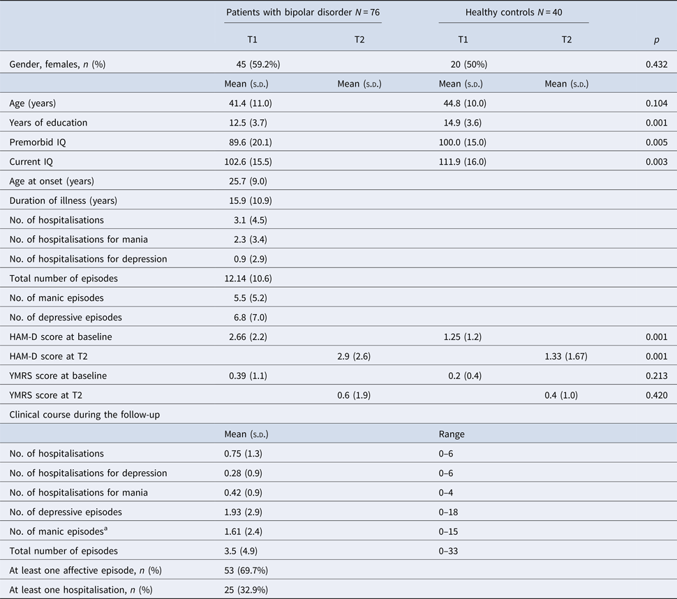
Table 1 shows demographic and clinical characteristics of patients with BD and HC. There were no differences in age and gender distributions between the two groups. However, HC had more years of education, higher premorbid and current IQ, and lower HAM-D scores at T1 and T2, than patients with BD. In addition, a repeated-measures ANOVA was performed for the two psychopathological measures. There was no effect of time or group × time interaction for both HAM-D (effect of time: Pillai's trace: 0.003, F (1,114): 0.294, p = 0.589; group × time interaction: Pillai's trace: 0.001, F (1,114): 0.066, p = 0.797) and YMRS (effect of time: Pillai's trace: 0.010, F (1,114): 1.173, p = 0.28; group × time interaction: Pillai's trace: 0.001, F (1,114): 0.010, p = 0.921).
Demographic and clinical characteristics of the patient and control group at baseline (T1) and at 5-year follow-up (T2)

s.d., standard deviation; T1, at baseline; T2, at follow-up of 5 years; IQ, intellectual quotient; HAM-D, Hamilton Depression Rating Scale; YMRS, Young Mania Rating Scale.
a Number of manic + hypomanic episodes.
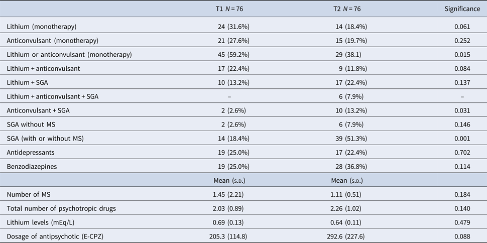
In addition, Table 1 shows the clinical course of patients during the follow-up. Pharmacological treatment of patients at T1 and T2 is provided in Table 2.
Pharmacological treatment in patients with bipolar disorder

T1, at baseline; T2, at follow-up of 5 years; SGA, second-generation antipsychotics; MS, mood stabilisers; E-CPZ, mg equivalents of chlorpromazine.
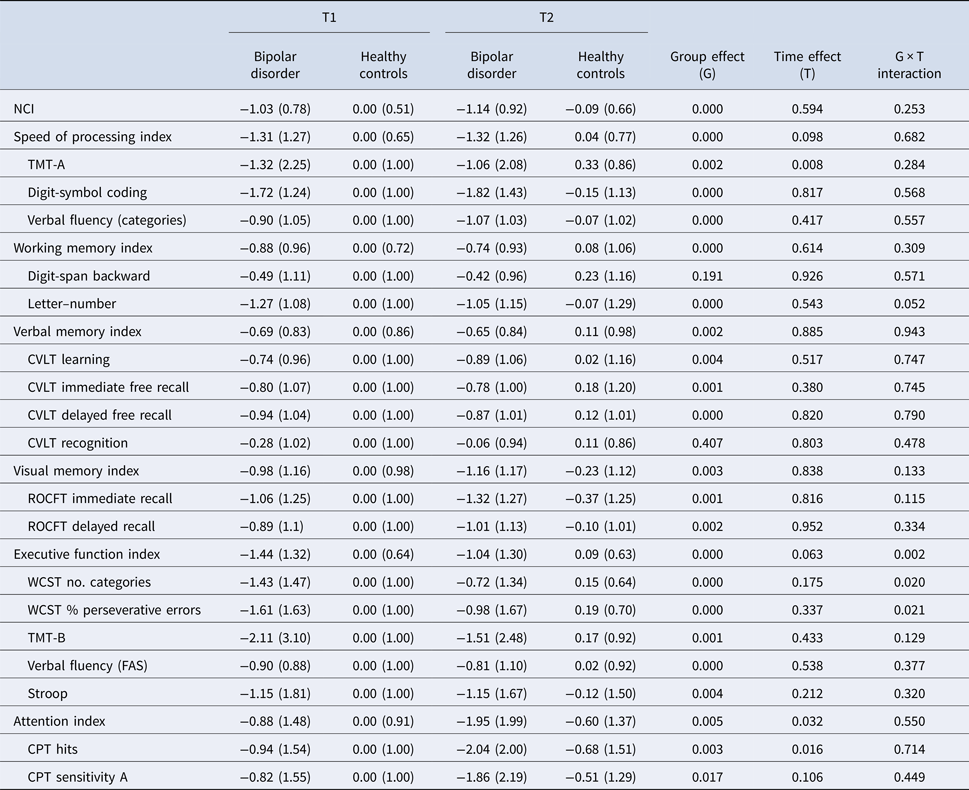
Table 3 shows the z-scores for each neurocognitive measure and NCI, as well as the results of the repeated-measures ANOVA. There was no effect of time or group × time interaction either for NCI or for any measure included in the domains working memory, verbal memory and visual memory (Table 3). However, an effect of time was observed for a measure of speed of processing (TMT-A) and for the attention domain (attention index and CPT hits). In the executive function domain score and in the two measures of the WCST, a group × time interaction was observed in a way that there was a greater improvement on performance in the group of patients than in the control group. However, when measures of WCST were not included into the executive function domain, both the effect of time and the group × time interaction were not maintained (effect of time: Pillai's trace: 0.019, F (1,109) = 2.083, p = 0.152; group × time interaction: Pillai's trace: 0.033, F (1,109): 3.708, p = 0.057). In the domain of attention, an effect of time was also observed (worse performance at T2 than at T1), although there was no group × time interaction.
Repeated-measures ANOVA z-scores (covariates: intellectual quotient, Hamilton Depression Rating Scale and Young mania Rating Scale at T1 and T2)

T1, at baseline; T2, at follow-up of 5 years; NCI, neurocognitive composite index; TMT-A, Trail Making Test – Part A; CVLT, California Verbal Learning Test; ROCFT, Rey–Osterrieth Complex Figure Test; WCST, Wisconsin Card Sorting Test; TMT-B, Trail Making Test – Part B; CPT, Continuous Performance Test.
Values are presented as mean (standard deviation).
Change in neurocognitive function
Differences between BD and HC were not observed either in the NCI change (BD: mean: −0.14, s.d.: 0.46; HC: mean: −0.10, s.d.: 0.39; F (1): 0.451; p = 0.503) or when the six neurocognitive domains were analysed (MANOVA: Pillai's trace: 0.071; F (6,109): 1.390; p = 0.225). In addition, differences in NCI change between BD-P (N = 54) and BD-NP (N = 22) were not observed (BD-P: mean: −0.19, s.d.: 0.49; BD-NP: mean: −0.02, s.d.: 0.39; F (1): 1.464; p = 0.230). When the six neurocognitive domains were analysed (MANOVA: Pillai's trace: 0.120; F (5,66): 1.805; p = 0.124), differences between the two subgroups of patients were not found either after controlling for psychopathology.
At T1, 28 patients were classified as patients with neurocognitive impairment (CI+), and 48 without neurocognitive impairment (CI−). Differences in NCI change were not found between CI+ and CI− (CI+: mean: −0.15, s.d.: 0.48; CI−: mean: −0.14, s.d.: 0.45; F (1): 0.045; p = 0.833). No effect of group was observed when the six domains were considered (MANOVA: Pillai's trace: 0.044; F (5,66): 0.607; p = 0.695). There were no differences between CI+ and CI− for the different measures of the course of the disorder during the follow-up: number of hospitalisations: 0.6 (s.d.: 1.3) v. 0.8 (s.d.: 1.4), F: 0.502, p = 0.481; number of hospitalisations for mania: 0.5 (s.d.: 0.9) v. 0.4 (s.d.: 0.9), F: 0.109, p = 0.743; number of hospitalisations for depression: 0.1 (s.d.: 0.4) v. 0.4 (s.d.: 1.1), F: 1.567, p = 0.215; total number of episodes: 2.7 (s.d.: 2.6) v. 4.0 (s.d.: 5.9), F: 1.105, p = 0.297; number of manic episodes: 1.4 (s.d.: 1.6) v. 1.7 (s.d.: 2.8), F: 0.338, p = 0.563; number of depressive episodes: 1.4 (s.d.: 1.5) v. 2.3 (s.d.: 3.5), F: 1.517, p = 0.222.
A correlation between NCI change and the number of manic and hypomanic episodes observed during the follow-up period (r = −0.314; p = 0.006), number of hospitalisations for mania (r = −0.231; p = 0.045), total number of episodes (r = −0.241; p = 0.036) and number of drugs at T2 (r = −0.308; p = 0.007) was found. However, no correlation was found between NCI change and age (r = −0.164; p = 0.156), duration of illness (r = −0.209; p = 0.07), number of depressive episodes (r = −0.161; p = 0.165), total number of hospitalisations (r = −0.159; p = 0.171), number of hospitalisations for depression (r = 0.04; p = 0.972), dose of antipsychotics (mEq chlorpromazine/day) at T1 (r = −0.165; p = 0.298) and at T2 (r = −0.185; p = 0247) or number of psychotropic drugs at T1 (r = −0.05; p = 0.519). Additionally, a difference in NCI change between males and females has been observed in the group of patients with BD: males: −0.27 (s.d.: 0.47); females: −0.05 (s.d.: 0.44); F (1,74): 4.11; p = 0.046.
Stepwise regression analysis
When NCI change was used as a dependent variable, only three of the measures were included into the model (Table 4): number of manic episodes (including number of manic and hypomanic episodes), gender and total number of psychotropic drugs. Together, the three variables explained the 23.4% of the variance of the NCI change. Number of manic and hypomanic episodes was the variable that explained a higher percentage of the variance of NCI change (12%).
Stepwise regression analysis for dependent variable change of NCI

a Includes number of manic and hypomanic episodes; T2 = follow-up of 5 years.
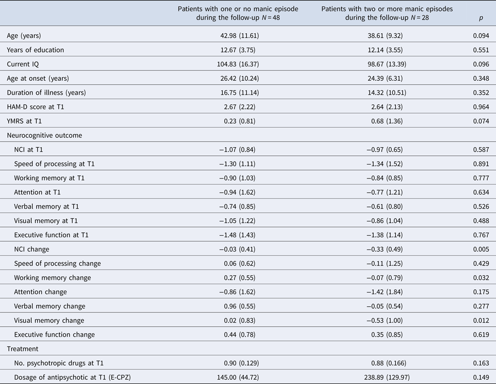
Then, comparisons between patients with two or more manic episodes during the follow-up period and patients that had one or no manic relapse were performed. The ANOVA showed that patients with more than one manic episode have a greater decrease in NCI and in working memory and visual memory domains. No differences were observed in clinical or neurocognitive variables at T1 between both groups (Table 5).
Comparisons between demographic, clinical, pharmacological and neurocognitive measures amongst patients with bipolar disorder depending on the number of manic episodes during the follow-up

IQ, intellectual quotient; HAM-D, Hamilton Depression Rating Scale; YMRS, Young Mania Rating Scale; T1, baseline; T2, follow-up of 5 years; NCI, neurocognitive composite index; E-CPZ, mg equivalents of chlorpromazine.
Analysis of variance (ANOVA). Values are presented as mean (standard deviation).
Discussion
In this study, we have examined the trajectory of neurocognitive functioning in a sample of euthymic BD patients during a 5-year follow-up period. We have not found evidence of progression in neurocognitive dysfunction either in the cognitive composite index or in any of the neurocognitive domains for the whole cohort, confirming the results of other longitudinal studies (Mur et al., Reference Mur, Portella, Martínez-Arán, Pifarré and Vieta2008; Tabarés-Seisdedos et al., Reference Tabarés-Seisdedos, Balanzá-Martínez, Sánchez-Moreno, Martinez-Aran, Salazar-Fraile, Selva-Vera, Rubio, Mata, Gómez-Beneyto and Vieta2008; Mora et al., Reference Mora, Portella, Forcada, Vieta and Mur2013; Ryan et al., Reference Ryan, Assari, Pester, Hinrichs, Angers, Baker, Marshall, Stringer, Saunders, Kamali, McInnis and Langenecker2016), even when older adults (Gildengers et al., Reference Gildengers, Chisholm, Butters, Anderson, Begley, Holm, Rogers, Reynolds and Mulsant2013; Schouws et al., Reference Schouws, Comijs, Dols, Beekman and Stek2016), adolescents (Bombin et al., Reference Bombin, Mayoral, Castro-Fornieles, Gonzalez-Pinto, de la Serna, Rapado-Castro, Barbeito, Parellada, Baeza, Graell, Payá and Arango2013) or newly diagnosed (Torres et al., Reference Torres, Kozicky, Popuri, Bond, Honer, Lam and Yatham2014) with BD were examined. Furthermore, in contrast to previous studies (Mur et al., Reference Mur, Portella, Martínez-Arán, Pifarré and Vieta2008; Santos et al., Reference Santos, Aparicio, Bagney, Sánchez-Morla, Rodríguez-Jiménez, Mateo and Jiménez-Arriero2014), we did not find a worsening in the verbal memory domain during the follow-up. The improvement found on the WCST should be interpreted as a consequence of practice effect (Szöke et al., Reference Szöke, Trandafir, Dupont, Méary, Schürhoff and Leboyer2008). It has been pointed out that the improvement in performance on the WCST due to practice effects is quite large, even after long periods between assessments (Basso et al., Reference Basso, Bornstein and Lang1999). Moreover, the capacity of several measures of the WCST to reflect changes is limited by ceiling effect (Basso et al., Reference Basso, Bornstein and Lang1999, Reference Basso, Lowery, Bornstein and Ghormley2001), what could explain the difference between patients and control subjects.
The second objective of this study was to examine if there is a subgroup of patients with a progressive course of neurocognitive dysfunction. We have not found significant differences in the trajectory of neurocognitive functioning between patients with and without a history of psychotic symptoms. These results are in line with those from Lera-Miguel et al. (Reference Lera-Miguel, Andrés-Perpiñá, Fatjó-Vilas, Fañanás and Lázaro2015), who did not find differences between patients with BD-P and BD-NP with early onset, in practically the totality of neurocognitive measures. In addition, the trajectory of neurocognitive performance in the subgroup of patients classified as cognitively impaired at T1 did not differ from that of patients without cognitive dysfunction. Moreover, we have not found significant differences between CI+ and CI− BD patients in measures of the illness course during the follow-up. This result is important because it does not support the notion that BD patients with more severe cognitive impairment are also those who suffer from more frequent episodes (Martino et al., Reference Martino, Igoa, Marengo, Scápola and Strejilevich2018).
The most important finding of the present study is the confirmation of a negative correlation between number of manic and hypomanic episodes and number of hospitalisations due to manic episodes and neurocognitive change during the follow-up. Other course-related variables such as number of depressive episodes, total number of episodes, total number of hospitalisations or illness duration were not associated with neurocognitive change. The number of manic episodes, number of psychotropic drugs at T2 and gender explain 23.4% of the variance in cognitive change. The number of manic and hypomanic episodes is the variable that explains a greater percentage of the variance (around 12%). Hence, our results confirm, for the first time in a longitudinal design, that BD patients who have a greater number of manic or hypomanic episodes may constitute a subgroup characterised by the progression of neurocognitive impairment. These findings are consistent with the results of some cross-sectional studies (Martínez-Arán et al., Reference Martínez-Arán, Vieta, Reinares, Colom, Torrent, Sánchez-Moreno, Benabarre, Goikolea, Comes and Salamero2004b, Reference Martínez-Arán, Vieta, Torrent, Sanchez-Moreno, Goikolea, Salamero, Malhi, Gonzalez-Pinto, Daban, Alvarez-Grandi, Fountoulakis, Kaprinis, Tabares-Seisdedos and Ayuso-Mateos2007; Robinson and Ferrier, Reference Robinson and Ferrier2006; Post et al., Reference Post, Fleming and Kapczinski2012) suggesting that manic episodes have a negative impact on neurocognition and the course of BD, and supporting the concept of high heterogeneity amongst BD cohorts as regards to neurocognitive performance and functioning (Burdick et al., Reference Burdick, Russo, Frangou, Mahon, Braga, Shanahan and Malhotra2014; Solé et al., Reference Solé, Jiménez, Torrent, del Mar Bonnin, Torres, Reinares, Priego, Salamero, Colom, Varo, Vieta and Martínez-Arán2016, Reference Solé, Bonnin, Jiménez, Torrent, Torres, Varo, Valls, Montejo, Gómez-Ocaña, Tomioka, Vieta, Martinez-Aran and Reinares2018; Russo et al., Reference Russo, Van Rheenen, Shanahan, Mahon, Perez-Rodriguez, Cuesta-Diaz, Larsen, Malhotra and Burdick2017). In this line, neuroimaging studies indicate that structural abnormalities observed in BD patients may be progressive (Hibar et al., Reference Hibar, Westlye, Doan, Jahanshad, Cheung, Ching, Versace, Bilderbeck, Uhlmann, Mwangi, Krämer, Overs, Hartberg, Abé, Dima, Grotegerd, Sprooten, Bøen, Jimenez, Howells, Delvecchio, Temmingh, Starke, Almeida, Goikolea, Houenou, Beard, Rauer, Abramovic, Bonnin, Ponteduro, Keil, Rive, Yao, Yalin, Najt, Rosa, Redlich, Trost, Hagenaars, Fears, Alonso-Lana, van Erp, Nickson, Chaim-Avancini, Meier, Elvsåshagen, Haukvik, Lee, Schene, Lloyd, Young, Nugent, Dale, Pfennig, McIntosh, Lafer, Baune, Ekman, Zarate, Bearden, Henry, Simhandl, McDonald, Bourne, Stein, Wolf, Cannon, Glahn, Veltman, Pomarol-Clotet, Vieta, Canales-Rodriguez, Nery, Duran, Busatto, Roberts, Pearlson, Goodwin, Kugel, Whalley, Ruhe, Soares, Fullerton, Rybakowski, Savitz, Chaim, Fatjó-Vilas, Soeiro-de-Souza, Boks, Zanetti, Otaduy, Schaufelberger, Alda, Ingvar, Phillips, Kempton, Bauer, Landén, Lawrence, van Haren, Horn, Freimer, Gruber, Schofield, Mitchell, Kahn, Lenroot, Machado-Vieira, Ophoff, Sarró, Frangou, Satterthwaite, Hajek, Dannlowski, Malt, Arolt, Gattaz, Drevets, Caseras, Agartz, Thompson and Andreassen2018) and are related to number of manic episodes (Strakowski et al., Reference Strakowski, DelBello, Zimmerman, Getz, Mills, Ret, Shear and Adler2002; Abé et al., Reference Abé, Ekman, Sellgren, Petrovic, Ingvar and Landén2015; Cao et al., Reference Cao, Passos, Mwangi, Bauer, Zunta-Soares, Kapczinski and Soares2016). The deleterious effect associated with manic episodes has been related to an increase in inflammatory and oxidative activity that would be present during manic episodes (Kapczinski et al., Reference Kapczinski, Mwangi, Cassidy, Librenza-Garcia, Bermudez, Kauer-Sant'anna, Kapczinski and Passos2017). Likewise, in line with previous studies (Martínez-Arán et al., Reference Martínez-Arán, Vieta, Colom, Torrent, Sanchez-Moreno, Reinares, Benabarre, Goikolea, Brugue, Daban and Salamero2004a), our results indicate that polypharmacy also has a negative impact on cognition in BD.
Different results have been found in a recent longitudinal study (Martino et al., Reference Martino, Igoa, Marengo, Scápola and Strejilevich2018), which concludes that the trajectory of cognitive deficits in BD is relatively independent of number of episodes. However, there are important differences in the characteristics of the samples which could explain the discrepancies. In contrast with the study of Martino et al. (Reference Martino, Igoa, Marengo, Scápola and Strejilevich2018), in our sample, type 1 BD patients and BD patients with a history of psychotic symptoms are predominant. Moreover, patients included in our study had an earlier age at onset and greater number of episodes during the follow-up, which may have given enough power to find the association in a subgroup of patients.
This study has several strengths, amongst which stands out that patients were euthymic in both assessments. In addition, the sample was relatively large and included patients without a history of substance abuse/dependence or psychiatric comorbidity. Likewise, all subjects were evaluated using a comprehensive neuropsychological battery, which includes the six neurocognitive domains defined by the MATRICS initiative. The inclusion of a healthy control group is another important strength, especially considering that both patients and controls were followed-up and assessed after 5 years.
The current study also has some limitations. First, the interval of 5 years between the two evaluations could be too short to detect a possible progression in cognitive decline for the overall sample. Longer follow-up periods may be more appropriate and give more statistical power (Schouws et al., Reference Schouws, Comijs, Dols, Beekman and Stek2016). Second, all patients were receiving pharmacological treatment, including second-generation antipsychotics (SGA), especially at T2. Although we have not found a correlation between the neurocognitive change and the dose of SGA at T1 and T2, and the doses of SGA did not enter into the regression model, we cannot rule out that antipsychotic treatment might have a negative impact on neurocognitive function (Balanzá-Martínez et al., Reference Balanzá-Martínez, Selva, Martínez-Arán, Prickaerts, Salazar, González-Pinto, Vieta and Tabarés-Seisdedos2010). Third, the sample included in the current study is principally composed by patients with a long course of the illness. The possibility that progression of cognitive deterioration is more evident in early stages of the disease should be considered (Vieta et al., Reference Vieta, Salagre, Grande, Carvalho, Fernandes, Berk, Birmaher, Tohen and Suppes2018). However, Torres et al. (Reference Torres, Kozicky, Popuri, Bond, Honer, Lam and Yatham2014) analysed the 1-year trajectory of cognitive deficits in recently diagnosed bipolar patients and no progression of neurocognitive impairment was found, although again, 1 year might be too short time to show any effects.
Our data suggest that the trajectory of cognitive deterioration in patients with BD is variable, although this variability is not related to any clinical subtype. However, the neurocognitive trajectory may be mediated by clinical course, in a way that neurocognitive decline could be present in a subgroup of patients characterised by more recurrent hypomanic and manic episodes. These results may have important clinical implications since they suggest that the prevention of both manic and hypomanic episodes can have a positive impact on the trajectory of cognitive function. Further longitudinal studies with larger sample sizes and longer periods of follow-up are necessary to confirm the effect of the recurrence of manic episodes on cognitive function in patients with BD.
Acknowledgements
None.
Financial support
This research was supported in part by the grant PI16/00359 (Fondo de Investigaciones Sanitarias, FIS), by Madrid's Regional Government (B2017/BMD-3740 AGES CM 2-CM) and European Union Structural Funds, and by the Centro de Investigación Biomédica en Red de Salud Mental (CIBERSAM) of the Instituto de Salud Carlos III.
Conflict of interest
Dr Vieta has received grants and served as a consultant, advisor or CME speaker for the following entities: AB-Biotics, Aequus, Adamed, Alexza, Allergan, Almirall, AstraZeneca, Bial, Bristol-Myers Squibb, Dainippon Sumitomo Pharma, Elan, Eli Lilly, Esteve, Ferrer, Forest Research Institute, Gedeon Richter, Glaxo-Smith-Kline, Janssen-Cilag, Jazz, Johnson & Johnson, Lundbeck, Merck, Novartis, Organon, Otsuka, Pfizer, Pierre-Fabre, Qualigen, Roche, Rovi, Sanofi-Aventis, Servier, Shering-Plough, Shire, Solvay, Sunovion, Takeda, Telefónica, Teva, the Spanish Ministry of Science and Innovation (CIBERSAM), the Seventh European Framework Programme (ENBREC), the Stanley Medical Research Institute, United Biosource Corporation and Wyeth. Dr R. Rodriguez-Jimenez has been a consultant for, spoken in activities of, or received grants from: Instituto de Salud Carlos III, Fondo de Investigación Sanitaria (FIS), Centro de Investigación Biomédica en Red de Salud Mental (CIBERSAM), Madrid Regional Government (S2010/BMD-2422 AGES; B2017/BMD-3740 AGES CM 2-CM), JanssenCilag, Lundbeck, Otsuka, Pfizer, Ferrer, Juste. All other authors declare that they have no conflicts of interest.