Background
Anomalous left coronary artery from the pulmonary artery is a rare congenital anomaly with an incidence of 1/300,000 live births, accounting for 0.25–0.5% of CHD.Reference Keith 1 In its most common form, the left coronary artery arises from the main pulmonary artery, and the right coronary artery arises normally from the right aortic cusp. The right coronary artery typically becomes enlarged and ectatic with advancing age. Without surgical repair, 90% of patients with anomalous left coronary artery from the pulmonary artery syndrome die within the 1st year of life.Reference Wesselhoeft, Fawcett and Johnson 2
Anomalous left coronary artery from the pulmonary artery does not present as a haemodynamic problem in utero. Relatively similar oxygen concentrations in the main pulmonary artery and aorta due to parallel circulations in the fetus result in normal myocardial perfusion, and therefore there is no stimulus for collateral vessel formation between the right and left coronary artery systems.
In the neonatal period, however, as pulmonary artery pressures and resistance decrease, retrograde flow into the pulmonary artery develops. This results in coronary steal, in which a left-to-right shunt leads to myocardial hypoperfusion, ischaemia, and possibly infarction in the territory of the anomalous vessel. The ensuing left ventricular dysfunction and mitral valve insufficiency can lead to congestive heart failure. If survival beyond infancy occurs without surgical repair, prominent collateral vessels develop between the right and the left coronary circulations, with coronary artery dilation due to volume overload. Unrepaired anomalous left coronary artery from the pulmonary artery in adults is characterised by chronic ischaemia and risk of sudden death.
Definitive surgical repair is the treatment of choice for anomalous left coronary artery from the pulmonary artery, including direct re-implantation of the anomalous coronary artery to the aorta or creation of an intrapulmonary aortocoronary tunnel as described by Takeuchi et al.Reference Takeuchi, Imamura and Katsumoto 3 The role of re-vascularisation in an asymptomatic patient with prominent collateral vessels and years of subclinical myocardial ischaemia, however, is somewhat contentious, as myocardium supplied by long-standing ischaemic vasculature may be permanently scarred and fibrotic, and unable to regain function despite intervention.Reference Shapiro, Mergo, Austin, Kantor and Gerber 4
Myocardial fibrosis has been described in patients with anomalous left coronary artery from the pulmonary artery on the basis of histological evaluationReference Shivalkar, Borgers, Daenen, Gewillig and Flameng 5 and late gadolinium-enhancement cardiac MRI.Reference Secinaro, Ntsinjana and Tann 6 To our knowledge, however, no previous studies have reported cardiac MRI T1 mapping in the anomalous left coronary artery from the pulmonary artery. The aim of this case report was to describe multi-modality imaging in an adult patient with unrepaired anomalous left coronary artery from the pulmonary artery, including cardiac MRI T1 mapping.
Case presentation
A 29-year-old gravida1 para1 woman with increasing fatigue was referred for evaluation after a transthoracic echocardiogram demonstrated a “coronary fistula”. She denied exertional chest pain, palpitations, pre-syncope, syncope, and breathlessness with exertion. She was contemplating a second pregnancy. Her resting electrocardiogram was normal. Further investigations included stress echocardiography, coronary CT angiography, nuclear stress testing, and cardiac MRI. Imaging was carried out over a period of 2 years during baseline and follow-up assessments because of ongoing reluctance of the patient to undergo surgery. The patient led a relatively sedentary life and remained asymptomatic in her daily activities.
Transthoracic echocardiography demonstrated the left coronary artery arising from the main pulmonary artery with large collateral vessels in the interventricular septum. The right coronary artery was dilated and tortuous (Supplementary Fig 1). Stress exercise echocardiography demonstrated reversible ischaemia with mild hypokinaesia of the septum at peak exercise (Supplementary video 1). The patient exercised on a treadmill for 9 minutes, achieving a workload of 10 metabolic equivalents. Ischaemic electrocardiogram changes were seen at peak exercise including 1.5-mm horizontal ST depression in leads II, III, aVF. No chest pain was reported.
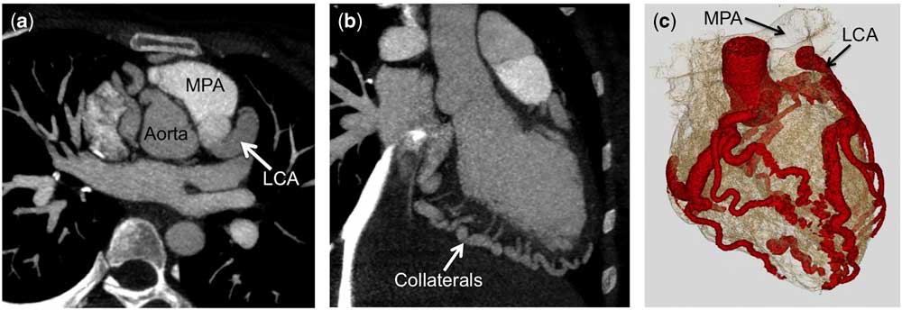
Coronary CT confirmed anomalous origin of the left coronary artery from the left inferolateral aspect of the main pulmonary artery (Fig 1). The anomalous left coronary artery was dilated measuring up to 11 mm in diameter. The right coronary artery had a normal origin arising from the right coronary sinus and was enlarged and tortuous. There were multiple collateral vessels along the right ventricular wall and interventricular septum, communicating with the left coronary artery.
Coronary CT demonstrates ( a ) the left coronary artery (LCA) arising from the main pulmonary artery (MPA) and ( b and c ) multiple prominent collateral vessels linking the right and the left coronary circulations.

Persantine sestamibi myocardial perfusion confirmed the stress echocardiographic findings, and revealed a moderate defect within the left anterior descending coronary artery territory with near-complete reversibility.
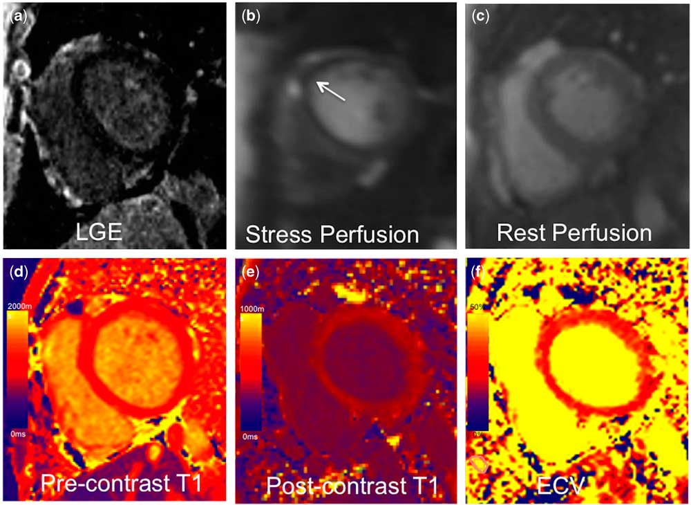
Cardiac dipyridamole stress MRI at 1.5 T revealed a reversible stress-induced subendocardial perfusion defect in the basal-to-mid anterior and anteroseptal wall with no associated defect on rest perfusion or late gadolinium-enhanced images, in keeping with coronary steal phenomenon (Fig 2).
Cardiac MRI images demonstrate ( a ) no late gadolinium enhancement (LGE), ( b ) a stress-induced subendocardial perfusion defect at the basal-to-mid anterior wall (arrow), ( c ) no rest perfusion defect, ( d ) global non-contrast myocardial T1 value of 1117 ms, ( e ) post-contrast T1 value of 495 ms, and ( f ) myocardial extracellular volume value of 32.4%.

The left ventricle was mildly dilated (indexed end-diastolic volume 118 ml/m2) with normal global systolic function (ejection fraction 65%). The calculated Qp:Qs based on phase-contrast analysis was 1.4:1, in keeping with left-to-right shunt flow.
Pre-contrast and 15-minute post-contrast (0.15 mmol/kg gadobutrol) T1 maps using a modified Look–Locker inversion recovery sequenceReference Kellman, Arai and Xue 7 at 1.5 T showed global myocardial T1 values of 1117 ms and 495 ms, respectively (Fig 2). Care was taken to avoid obvious myocardial collaterals on contouring. The left ventricular endocardial and epicardial borders were manually contoured. Values from the American Heart Association 16-segment model were averaged to represent global T1 values.Reference Cerqueira, Weissman and Dilsizian 8 Myocardial extracellular volume fraction was calculated on the basis of the approach proposed by Arheden et al.Reference Arheden, Saeed and Higgins 9 Global myocardial extracellular volume fraction was 32.4%, with no significant difference between coronary artery territories (left anterior descending 32.6±3.6%, right coronary artery 33.1±3.2%, and circumflex 32.4±3.2%, p=0.767). The post-contrast T1 mapping acquisition was performed more than 30 minutes after the administration of adenosine, which is an important consideration, as T1 and extracellular volume values are affected by the intramyocardial blood flow increase during and immediately following vasodilator stress.Reference Mahmod, Piechnik and Levelt 10 Non-contrast T1 and extracellular volume fraction values were higher than normal reference ranges, suggesting the presence of diffuse interstitial myocardial fibrosis.Reference Hanneman, Nguyen and Thavendiranathan 11 Alternatively, such global changes could reflect elevated myocardial blood volume related to extensive collateralisation.Reference Liu, Wijesurendra and Francis 12
Conclusion
To the best of our knowledge, this is the first report of myocardial T1 mapping in a patient with anomalous left coronary artery from the pulmonary artery. Globally elevated non-contrast T1 and extracellular volume fraction values in this patient suggest the possible presence of diffuse interstitial myocardial fibrosis, not evident on other imaging studies, which may reflect ongoing subclinical ischaemia due to the anomalous left coronary system and the extensively collateralised right coronary system. Cardiac MRI T1 mapping allows for a new dimension of myocardial characterisation, which may provide insight into subtle, diffuse abnormalities at the tissue level.
Acknowledgement
None.
Financial Support
This study received no specific grant from any funding agency or from commercial or not-for-profit sectors.
Conflicts of Interest
None.
Ethical Standards
The authors assert that all procedures contributing to this study comply with the ethical standards of the relevant national guidelines on human experimentation and with the Helsinki Declaration of 1975, as revised in 2008, and has been approved by the institutional committees.
Supplementary materials
To view supplementary material for this article, please visit https:/doi.org/10.1017/S1047951117000932