Introduction
Bacterial spread from the tonsil into the potential space between tonsillar capsule and pharyngeal muscle bed may cause a peritonsillar abscess or ‘quinsy’.Reference Gleeson1 It is the foremost complication of bacterial tonsillitis, and is the most common deep space infection of the head and neck. Standard treatment is surgical drainage by needle or knife in the supratonsillar fold, alongside medical and antimicrobial therapy.Reference Windfuhr, Nematian and Ziogou2 Drainage is performed by a solo practitioner. This treatment provides immediate relief for patients and prevents complications; hence, prompt and safe surgical treatment is a priority.Reference Khayr and Taepke3 This task often falls to the most junior ‘on-call’ otolaryngologist, and causes trepidation in the novice because of the dexterity needed to operate on an anxious, unwell patient with trismus.
Simulation in ENT has been growing;Reference Thone, Winter, García-Matte and González4 the opportunity to practise new skills in a controlled setting has clear benefits for patient safety and soothes operator anxiety. Peritonsillar abscess simulation has been attempted in a number of innovative ways, including: inserting water balloons in the oropharynx region of a resuscitation mannikin;Reference Murphy, Murphy and Sama5 constructing a model oropharynx from latex moulage;Reference Taylor and Chang6 creating an oropharynx in a coffee cup made from gelatine and a balloon, placed under a Resusci Anne mask;Reference Bunting, Wilson, Malloy and Malekzadeh7 and a cod liver oil capsule in a custom-made mould, secured to a Laryngotech intubation simulator.Reference Giblett and Hari8
The challenge in developing any simulation model is finding the balance between ease of use, affordability, replicability and educational value. Although all of the aforementioned simulators offer trainees an undeniable educational boon, they suffer from inherent qualities that limit their widespread use: some rely on expensive specialist equipment from other departments, others are complex or time-consuming in their assembly, requiring specialist technicians for their construction.
In view of this, we developed an educationally useful, simple, low-cost peritonsillar abscess simulator. It is easily constructed from basic equipment present on any ENT ward, and a packet of party balloons. The following article introduces the Newport Quinsy Simulator and describes a formal evaluation.
Materials and methods
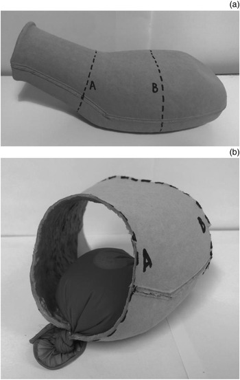
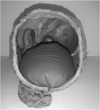
The Newport Quinsy Simulator was created using common equipment found on any ward. A cardboard urinary bottle is cut at both ends to create an oropharynx, with a disposable glove filled with water attached to the floor of the model as a tongue (Figure 1). One author (LG) painted an oropharynx with a peritonsillar abscess, and a paper image of this was secured to the rear of the model (Figure 2).
Initial construction of the model oropharynx: (a) lines A and B mark areas to be cut; and (b) ‘tongue’ is inserted into model.

Trainee's view of the model oropharynx and tongue.

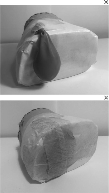
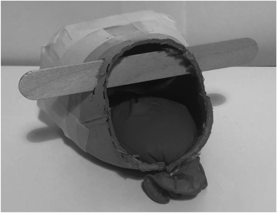
A balloon containing water or a purulent-appearing material, like custard, is secured directly behind the image of the abscess (Figure 3a), taking care not to overlap the infected tonsil. The balloon should be full but not tense, to avoid bursting. Tape is then applied over the balloon and the edges of the urine bottle, which gently presses the balloon inwards, producing the three-dimensional ‘bulge’ of peritonsillar swelling (Figure 3b). An optional wooden tongue depressor is secured at the opening to simulate trismus (Figure 4).
Securing the balloon (abscess) to the model: (a) balloon correctly positioned behind the abscess in the image; and (b) tape applied to secure balloon.

A fully assembled Newport Quinsy Simulator model.

The total assembly time is approximately 10 minutes. Each model can be used for around five attempts for needle aspiration of approximately 1 ml before the balloon requires re-filling or replacement.
Evaluation of the model was undertaken by seeking multi-stakeholder feedback following use of the simulator. The ‘expert’ group comprised ENT consultants, and middle-grade and advanced nurse practitioners. The ‘novice’ group consisted of ENT senior house officers (SHOs), SHOs who cross-cover ENT when ‘on-call’ and medical students.
Five-point Likert scales were used by the expert group to assess the accuracy of the synthesised anatomy and the technical skills needed to use the simulator. The novice group quantified their level of experience and then rated their confidence in draining a peritonsillar abscess before using the simulator; they then rated any increase in confidence after using the model. Both groups were asked how useful they thought the simulator would be for beginners training in peritonsillar abscess drainage. In addition to quantitative responses, the evaluating clinicians were asked to provide free-text qualitative feedback.
Results
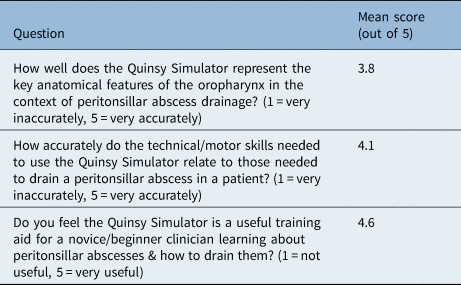
Thirteen ‘expert’ ENT practitioners evaluated the simulator: Four consultants, and six middle-grade and three advanced nurse practitioners (Table 1). Most of the expert cohort felt the simulator accurately represented the key anatomical features of the oropharynx in the context of peritonsillar abscess drainage, with a mean rating of 3.8 out of 5. The expert group agreed that the technical skills needed to use the simulator related well to those required to drain a real peritonsillar abscess, with a mean rating of 4.1 out of 5.
Questionnaire results for ‘expert’ group*

*The ‘expert’ group comprised ENT consultants, and middle-grade and advanced nurse practitioners (n = 13).
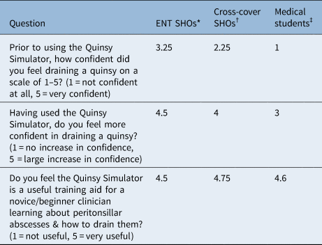
Eleven novice practitioners evaluated the model (four ENT SHOs, four cross-cover SHOs and three medical students). The ENT SHOs and cross-cover SHOs had minimal experience of peritonsillar abscess drainage, with all but one respondent having attempted drainage between one and five times previously. No medical students had attempted drainage. The ENT SHOs were somewhat confident in draining peritonsillar abscesses prior to simulator exposure, with mean rating of 3.25 out of 5, and reported a strong increase in confidence after using the simulator, with a mean rating of 4.5 out of 5 (Table 2). The cross-cover SHOs and students reported low confidence prior to simulator exposure and reported a strong increase in confidence following simulator use, with mean increases in confidence of 4 and 3 out of 5, respectively.
Questionnaire results for ‘novice’ group

Data represent mean scores (out of 5). *n = 4; †n = 4; ‡n = 3. SHO = senior house officer
When asked about the simulator's usefulness as a training aid for the novice clinician, the majority of respondents in both the expert and the novice groups rated the simulator as ‘very useful’, with the remaining respondents describing it as ‘useful’. The mean rating for this question was 4.6 out of 5.
The free-text qualitative responses of both groups were mostly positive, with common themes identified. Many evaluators highlighted: the simulator's usefulness to new clinicians, the simplicity of its construction and ingenuity of design, as well as the relative realism achieved. Evaluators noted that the Newport Quinsy Simulator filled a gap needed for teaching this procedure to new otolaryngologists. In areas identified for improvement, a few individuals in the expert group and one novice felt the mouth opening was too large without the wooden tongue depressor, and that the peritonsillar abscess was too accessible in this configuration. Others indicated this was ameliorated when the wooden tongue depressor was used to simulate trismus, presenting a suitable challenge for beginners.
Discussion
Simulator use is becoming a common part of otolaryngology training.Reference Thone, Winter, García-Matte and González4 Simulators allow junior clinicians to learn skills, acquire knowledge, and prepare for and perform new procedures. In addition, they provide an opportunity for repetition, to consolidate learning. Simulator use builds confidence. This is important when draining a peritonsillar abscess, as patients are often distressed and anxious, and may present out of hours. The benefits of simulation are amplified by the coronavirus pandemic, as emergency ENT presentations have decreased,Reference Gelardi, Iannuzzi, Trecca, Kim, Quaranta and Cassano9 negatively impacting the opportunity for trainees to practise abscess drainage, or to safely observe an expert clinician performing it.
For a simulator to be widely adopted, it must strike a balance between ease of preparation and use, cost-effectiveness, and educational validity.Reference Van Nortwick, Lendvay, Jensen, Wright, Horvath and Kim10 From an educational perspective, evaluation of the Newport Quinsy Simulator indicates a reasonable level of face validity, with good representation of key anatomical features of a peritonsillar abscess. The simulator shows high levels of content validity, as evaluators thought it a very useful training aid for the novice. It offers the opportunity to assemble and use all of the requisite equipment, as well as to co-ordinate the bimanual skills needed to drain a peritonsillar abscess. Further work may assess whether simulator use has an effect on clinician performance, perhaps by considering success rates in clinical practice.
The simulator's ability to differentiate between skilled and novice performers (construct validity) was not directly evaluated. This was because peritonsillar abscess drainage is performed by novice clinicians in practice and has a modest learning curve. Qualitative analysis did suggest that experts found the simulator too easy to use, but the authors note that some novices found it challenging, suggesting that the model possesses some construct validity; this could be explored in further studies.
Limitations include the non-robustness of the simulator, which is a direct consequence of it being made out of disposable hospital goods; this necessitates the construction of a fresh simulator after a number of uses. The simulator does not include potential additional features such as a carotid artery, which was omitted to keep construction simple. We also note that the evaluators were small in number, and mostly otolaryngologists who are intimately familiar with the anatomy and pathology, which may predispose the evaluation to bias.
In designing the simulator, the authors prioritised simplicity and convenience, alongside educational value. We feel it is reasonable to presume that the equipment needed for the simulator would be available in almost every hospital, and the party balloons can be purchased in any supermarket, at minimal cost. Construction is straightforward, can be performed solo, and three or four of the simulators can be made in half an hour. The minimal outlay of time and money make this useful simulator accessible to any otolaryngology department in the developed or developing world.
Conclusion
The Newport Quinsy Simulator is affordable, requires little effort to construct, and is portable and easy to use. Initial evaluation suggests it is a useful training aid with high validity, providing an enjoyable educational experience.
We encourage our colleagues who wish to construct the Newport Quinsy Simulator to contact the authors to request an assembly guide.
Acknowledgements
We thank Maureen Dobbins of the Royal Gwent Hospital, Newport, South Wales, UK, for her invaluable advice when conceptualising the simulator. Thanks to the otolaryngologists of the Royal Gwent Hospital for their support, and to Myles Sheehan for artistic logistics.
Competing interests
None declared