Introduction
The cytoskeleton (CSK) is a highly cross-linked scaffold that binds to organelles and molecules, such as polysomes, mRNAs and other factors including translation initiation and elongation factors. It has been postulated that the CSK ensures that a critical mass of translation factors and RNAs comes in contact with each other to facilitate efficient translation (Heuijerjans et al., Reference Heuijerjans, Pieper, Ramaekers, Timmermans, Kuijpers, Bloemendal and van Venrooij1989; Hesketh et al., Reference Hesketh, Campbell, Piechaczyk and Blanchard1994; Hovland et al., Reference Hovland, Hesketh and Pryme1995; Jansen, Reference Jansen1999). However, presently, many questions remain to be answered regarding the role of cytoskeletal components in supporting the proteins related to discrete steps of the translation process.
In this study, we examined the interaction between the CSK and components of the maturation/translation process during early oogenesis (stage I) of Xenopus laevis. In stage I oocytes, in which the yolk has not yet been deposited, the CSK forms a supple network that gradually spreads throughout the cell and is primarily composed of actin microfilaments, microtubules, vimentin and cytokeratin (Godsave et al., Reference Godsave, Wylie, Lane and Anderton1984a, Reference Godsave, Anderton, Heasman and Wylieb; Gard et al., Reference Gard, Cha and King1997). Actin-binding spectrin is associated with the cell membrane, the nuclear envelope and the mitochondrial cloud (MC) (Carotenuto et al., Reference Carotenuto, Maturi, Infante, Capriglione, Petrucci and Campanella1997, Reference Carotenuto, Vaccaro, Capriglione, Petrucci and Campanella2000), similar to associations of intermediate filaments (Gard et al., Reference Gard, Cha and King1997; Klymkowsky, Reference Klymkowsky1995). Tubulin gradually becomes organized into bundles that form large corridors between the nuclear envelope and the cortex (Gard et al., Reference Gard, Affleck and Error1995).
Ribosomal subunits form in the nucleolus and are then exported to the cytoplasm, where they associate with a vast number of essential factors, a process particularly relevant in X. laevis, and which contain more than 1000 nucleoli. Our study was predominantly focused on two proteins involved in 60S maturation and translation, rpl10 and the eukaryotic initiation factor 6 (Eif6 or Tif6 in yeast; Basu et al., Reference Basu, Si, Warner and Maitra2001).
Further data are produced regarding thesaurins, homologs of the eEF1α elongation factor in X. laevis oocytes (Viel et al., Reference Viel, Le Mair, Morales and Denis1991), and P0, the ribosomal stalk protein that binds to the GTPase centre and eEF1α during translation (Lo et al., Reference Lo, Li, Bussiere, Bresson, Marcotte and Johnson2010).
rpl10 is a highly conserved phosphoprotein encoded by a member of the Qm gene family and is a homologue of bacterial rpl16 (Ban et al., Reference Ban, Nissen, Hansen, Moore and Steitz2000). rpl10 localizes to the periphery of the 60 subunit in a slit among its central protuberance and the P0, P1 and P2 ribosome stalk. A protrusion of rpl10 contacts the peptidyl transferase centre to appropriately position it to interact with elongation factors (Uchiumi et al., Reference Uchiumi, Honma, Endo and Hachimori2002). rpl10 is necessary for the formation of an active ribosome, as it participates in the maturation of 60S by organizing the architecture of the aminoacyl-tRNA-binding site on the ribosome (Dick & Trumpower, Reference Dick and Trumpower1998). Moreover, rpl10 is involved in a variety of additional cell functions (see Dowdy et al., Reference Dowdy, Lai, Weissman, Matsui, Hogan and Stanbridge1991). The transport of 60S from the nucleus to the cytoplasm has been described in animal cells including Xenopus oocytes (Bataille et al., Reference Bataille, Helsen and Fried1990). In preparation for 60S nuclear export, proteins such as Eif6, Arx1, Nmd3 associate with pre-60S (Gadal et al., Reference Gadal, Strauß, Kessl, Trumpower and Tollervey2001; Nissan et al., Reference Nissan, Bassler, Petfalski, Tollervey and Hurt2002; Lo et al., Reference Lo, Li, Bussiere, Bresson, Marcotte and Johnson2010). The import of the 60S subunit into the cytoplasm is then assisted by CRM1 exportin, which is associated with Nmd3 (Fornerod et al., Reference Fornerod, Ohno, Yoshida and Mattaj1997), where it binds to rpl10 in support of the chaperone Sqt1 (West et al., Reference West, Hedges, Chen and Johnson2005). In particular, Lsg1 GTPase (Kallstrom et al., Reference Kallstrom, Hedges and Johnson2003), when GTP-linked, also binds to 60S subunit. GTP hydrolysis to GDP causes a conformational change in 60S that facilitates the insertion of rpl10 and the release of Nmd3–Sqt1 (West et al., Reference West, Hedges, Chen and Johnson2005; Lo et al., Reference Lo, Li, Bussiere, Bresson, Marcotte and Johnson2010).
Eif6 binds to the 60S ribosomal subunits in the nucleolus (Sanvito et al., Reference Sanvito, Piatti, Villa, Bossi, Lucchini, Marchisio and Biffo1999). In Saccharomyces, once bound to the 60S subunit, Tif6 is active in keeping 60S separate from the 40S ribosomal subunit in both the nucleus and the cytoplasm (Senger et al., Reference Senger, Lafontaine, Graindorge, Gadal, Camasses, Sanni, Garnier, Breitenbach, Hurt and Fasiolo2001; Lebreton et al., Reference Lebreton, Saveanu, Decourty, Rain, Jacquier and Fromont-Racine2006). The larger pool of Eif6 is located in the cytoplasm. Eif6 binds to the 60S subunit near the ribosomal proteins rpL23 and rpL24 and the sarcine ricine loop at the GTPase Centre (Gartman et al., Reference Gartman, Blau, Armache, Mielke, Topf and Beckman2010). Interestingly, under in vitro conditions, the release of 60S-bound Eif6 in the cytoplasm at maturation specifically requires SBD1 (Shwahman-Bodian-Diamond Syndrome 1) and the GTPase Efl1 (Gartman et al., Reference Gartman, Blau, Armache, Mielke, Topf and Beckman2010; Finch et al., Reference Finch, Hilcenko, Basse, Drynan, Goyenechea and Menne2011) and precedes the release of Nmd3 (Senger et al., Reference Senger, Lafontaine, Graindorge, Gadal, Camasses, Sanni, Garnier, Breitenbach, Hurt and Fasiolo2001). Therefore, this process appears to be strictly linked to the modulation of ribosome conformational change and rpl10 insertion. At present, it is not understood whether Efl1 and SBD1 also participate in the process of translation, i.e. whether the regulation of maturation and translation is linked. Conversely, the role of Eif6 in regulating translation was determined. Indeed, Eif6 also binds to mature 60S, thus regulating 60S availability (Brina et al., Reference Brina, Grosso, Miluzio and Biffo2011); if protein kinase C (PKC) phosphorylates Eif6 at serine 235, the affinity of Eif6 for 60S is reduced, and the availability of 60S for translation and the formation of the active 80S ribosome is increased (Ceci et al., Reference Ceci, Gaviraghi, Gorrini, Sala, Offenhauser and Marchisio2003). Accordingly, in vivo, Eif6 acts as a rate-limiting initiation factor, and its expression level influences the translation rate (Basu et al., Reference Basu, Si, Warner and Maitra2001, Ceci et al., Reference Ceci, Gaviraghi, Gorrini, Sala, Offenhauser and Marchisio2003; Gandin et al., Reference Gandin, Miluzio, Barbieri, Magri, Kiyokawa and Marchisio2008).
Does the CSK interact with components involved in translation in oocytes? Vaccaro et al. (Reference Vaccaro, Gigliotti, Graziani, Carotenuto, De Angelis, Tussellino and Campanella2010, Reference Vaccaro, Wilding, Dale, Campanella and Carotenuto2012) demonstrated that actin-bound spectrin interacts with XNOA 36 mRNA in Xenopus stage I oocytes. Actin microfilaments co-localize with poly(A) RNA and are necessary for efficient protein synthesis (Taneja et al., Reference Taneja, Lifshitz, Fay and Singer1992). mRNA-CSK complexes contain components of the translation-regulating machinery (Jansen, Reference Jansen1999). In stages II and III Xenopus oocytes, microtubules are also involved in mRNA transport via protein linkers (for reviews: see Kloc et al., Reference Kloc, Zearfoss and Etkin2002; King et al., Reference King, Messit and Mowry2005). Our study showed that both eif6 and rpl10 associate with spectrin, cytokeratin and tubulin, whereas thesaurins and P0 interact only with spectrin and cytokeratin. Hypotheses are discussed regarding the involvement of the investigated proteins in ribosomal maturation and/or translation.
Materials and methods
Animals
Adult X. laevis females were obtained from ‘Rettili’, Varese, Italy. They were maintained and utilised at the Department of Biology of the University of Naples, Federico II, according to the guidelines and policies dictated by the University Animal Welfare Office and in agreement with international laws.
Previtellogenic oocytes at stage I of oogenesis (stage I oocytes) with a diameter of approximately 150–200 μm were excised from the ovaries of females anaesthetised using MS222 (Sigma Chemical Co, St. Louis, MO, USA). Somatic tissue (outer ovarian epithelium, theca and follicle cells) was removed from the oocytes via exposure to 0.04% collagenase type XI (Sigma), 20 UI/ml hyaluronidase and 10 μg/ml proteinase K (Sigma) in suitable Ringer solution (111 mM NaCl, 1.3 mM CaCl2, 2 mM KCl, 0.8 mM MgSO4, 25 mM HEPES, pH 7.8) for 90 min with gentle rocking at room temperature. Following several rinses, the oocytes were processed for electrophoresis.
Antibodies
eif6 immunostaining was performed using an anti-Eif6 rabbit polyclonal antiserum as previously described (Carotenuto et al., Reference Carotenuto, Vaccaro, De Marco, Wilding, Biffo, Marchisio, Capriglione and Campanella2005). Monoclonal antibodies against the ribosomal protein rpl10 were purchased from Sigma-Aldrich (WH0006134M1) and utilised at a 1:100 (v/v) dilution for both western blot and immunofluorescence.
The anti-P0 polyclonal antibody was a kind gift from Dr Jorge Bolivar (Cadix University, Spain), and the polyclonal anti-thesaurin A/B antibody was a gift from BIOGEM (Ariano Irpino, Italy). The proteins identified by both antibodies were tested via MALDI mass spectrometry (analysis kindly performed by Angela Flagiello at Ceinge, Naples, Italy).
The following anti-cytoskeletal protein antibodies were obtained from Sigma: anti-pan-cytokeratin monoclonal antibody (C2931), cross-reacting with type I cytokeratin (K10,13,18) and type II cytokeratins (K4,5,6,8) of Xenopus oocytes, utilised at a 1:200 (v/v) dilution (see Carotenuto et al., Reference Carotenuto, Petrucci, Correas, Vaccaro, Marco, Dale and Wilding2009); monoclonal antibody against anti-acetylated-α-tubulin (T6793), a broadly distributed form of tubulin in stage I oocytes (Gard et al., Reference Gard, Affleck and Error1995), was utilised at a 1:100 dilution. The anti-βIIΣI human β-spectrin polyclonal antibody was a gift from J. Morrow (Department of Pathology of Yale University School of Medicine, New Haven, CT, USA) and was utilised at a dilution of 1:100 for immunostaining and 1:500 for western blot.
Immunofluorescence microscopy
Clusters of Xenopus oocytes in early stage I of oogenesis were fixed in a solution containing 2% paraformaldehyde (PFA), 250 mM NaCl and 5% acetic acid for 50 min at 4°C. Following rinses in phosphate-buffered saline (PBS), the oocytes were embedded in Killik (BioOptica, Italy), frozen and cryostat-sectioned at a thickness of 7 μm. The sections were placed on Superfrost Plus slides (Carlo Erba, Italy). Non-specific background was blocked by incubating the sections for 20 min in PBS containing 3% normal goat serum, 0.5% bovine serum albumin (BSA), 0.1% Triton-X100 (PBT), prior to exposure overnight at 4°C to the primary antibodies (anti-β-spectrin; anti-pan-cytokeratin; anti-tubulin, anti-rpl10; anti-Eif6; anti-P0; or anti-thesaurins) diluted in PBT. Control sections incubated in rabbit IgG (Sigma, I5006) or in the absence of a primary antibody were included in each experiment. BODIPY FL goat anti-rabbit IgG (H+L) (Molecular Probes, B2766) or Texas Red goat anti-mouse IgG (H+L) (Molecular Probes, T-862) diluted 1:200 in PBT were used as the secondary antibodies. 4’,6-Diamidino-2-phenylindole (DAPI) was used for nuclear staining. For double-staining, the sections were incubated in succession with the anti-rpl10 monoclonal antibody and the anti-spectrin polyclonal antibody followed by the anti-mouse and anti-rabbit antibodies.
Ultimately, the sections were mounted in 9:1 glycerol:PBS (v:v). Images were captured using a Leica DM6000B photomicroscope equipped with a digital camera.
Sample preparation for SDS-PAGE and western blot
Stage I oocytes were homogenised in HEPES buffer, pH 7.5, containing 900 mM glycerol, 0.02 mM NaN3, 1 mM ATP, 1 mM dithiothreitol (DTT), 5 mM ethylene glycol tetraacetic acid (EGTA) (buffer I) and the following protease inhibitors (Sigma): 2 mM tosylarginine methyl ester (TAME), 5 mg/ml soybean trypsin inhibitor (SBTI), 5 μg/ml aprotinin and 10 μM E64. After centrifugation at 15,000 g for 40 min at 4°C, the protein concentration of the supernatants was determined using the bicinchoninic acid (BCA) protein assay reagent (Pierce, Rockford, IL, USA). After boiling in sample buffer with DTT, 30–60 μg aliquots of the proteins were analysed via sodium dodecyl sulphate polyacrylamide gel electrophoresis (SDS-PAGE) (7, 12 or 15% polyacrylamide) along with molecular mass standards (200, 116, 97, 45, 31, 21 and 14 kDa) (Bio-Rad, Hercules, CA, USA). The proteins were detected using coomassie blue stain. Western blot on Whatman Protan nitrocellulose transfer membranes (Whatman, GmbH, Dassel, Germany) was performed as previously described (Maturi et al., Reference Maturi, Infante, Carotenuto, Focarelli, Caputo and Campanella1998) using secondary anti-mouse or anti-rabbit IgGs coupled with alkaline phosphatase and revealed using 5-bromo-4-chloro-3-indolyl phosphate/p-nitroblue tetrazolium chloride (NBT/BCIP). Alternatively, the proteins were detected using secondary antibodies coupled to horseradish peroxidase (Pierce) and revealed using the enhanced chemiluminescence kit (Pierce).
Immunoprecipitation
The samples for immunoprecipitation were homogenised in buffer I or, in the case of co-IP, in buffer II, i.e. 1% Igepal (Sigma 17771) in PBS supplemented with 100 mM NaF and a protease inhibitor cocktail (Sigma P8340), prior to centrifugation at 15,000 g for 45 min. Undiluted anti-rpl10, anti-Eif6, anti-P0, anti-thesaurins, anti-spectrin, anti-cytokeratin (anti-CK), anti-tubulin or pre-immune serum were exposed to supernatants (1:75 v/v) for 1 h under constant agitation. Protein A Sepharose Cl4B (Pharmacia Biotech) was added to the sample (1:1 or 1:3 v/v) and incubated under agitation for 30 min. Following several rinses in buffer, the recovered pellet was boiled in sample buffer under reducing conditions.
Protein nomenclature
We use official names (www.ncbi.nlm.nih.gov/sites/gquery) for proteins according to the mammalian or the amphibian species to which they refer.
Results
Interaction of rpl10 and eif6 with cytoskeletal proteins
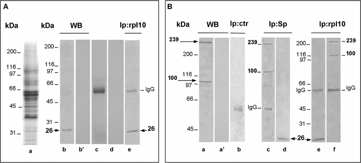
Western blot analyses of stage I oocyte protein lysates (Fig. 1A, lane a) incubated in anti-rpl10 revealed a specific band of the expected molecular weight (Mr; 26 kDa) (Fig. 1A, lanes b and b′). The 26 kDa band was also present in the western blot of a sample immunoprecipitated using anti-rpl10 (Fig. 1A, lane e). In Fig. 1A, standard controls of the immunoprecipitation are also shown, i.e. omission of the oocyte sample and omission of IgGs in Fig. 1A, lanes c and d, respectively. To investigate the possible interaction between rpl10 and the primary cytoskeletal constituents of the early oocyte, co-IPs using anti-rpl10 were performed using anti-spectrin, anti-CK or anti-tubulin.
Co-immunoprecipitation (Co-IP) of rpl10 with cytoskeletal proteins and related controls. (A) Lane a: Oocyte lysates stained with coomassie blue. Lane b: Western blot of the oocyte lysate, revealing the expected band of 26 kDa upon monoclonal anti-rpl10 incubation. Lane b′: Western blot in the absence of the primary antibody: no protein bands are observed, indicating that the mouse IgGs do not cross-react with any proteins present in the lysate. Lane c: Protein A sepharose incubated in anti-rpl10 in the absence of the oocyte sample: only the IgG heavy chain band is present. Lane d: The oocyte sample incubated in Sepharose beads in the absence of IgGs: no immunoprecipitated proteins are detected. Lane e: Upon immunoprecipitation using the anti-rpl10 antibody and incubation in the same antibody, the oocyte sample displays the 26 kDa band. (B) Co-IP using monoclonal anti-rpl10 antibodies and polyclonal anti-β-spectrin antibodies: half of the sample was used for anti-rpl10 IP, and half of the sample was used for anti-β-spectrin IP, which was the case for all subsequent co-IPs. Lane a: Western blot of the oocyte lysate revealing the two major bands of spectrin (239 kDa and 100 kDa). Lane a′: Western blot incubated in pre-immune serum: no band was detected. Lane b: Immunoprecipitation using the pre-immune serum: the IgG band was detected. Lane c: Oocyte sample immunoprecipitated using the anti-β-spectrin antibody and revealed using the same antibody: the 239 kDa and 100 kDa proteins are detected. Lane d: Oocyte sample immunoprecipitated using the anti-β-spectrin antibody and revealed using anti-rpl10: the 26 kDa band of rpl10 was detected. Lane e: Oocyte sample was immunoprecipitated using the rpl10 antibody and revealed using anti-rpl10: the 26 kDa band was detected. Lane f: Oocyte sample immunoprecipitated using the anti-rpl10 antibody and revealed using anti-β-spectrin: the two spectrin bands were detected.

In Fig. 1B, the anti-spectrin western blot of the oocyte lysate revealed two specific bands of 239 and 100 kDa (see Carotenuto et al., Reference Carotenuto, Petrucci, Correas, Vaccaro, Marco, Dale and Wilding2009) (Fig. 1B, lanes a and a′). In the co-IPs, one half of the anti-spectrin immunoprecipitate (IP) was revealed using anti-β-spectrin and the other half using anti-rpl10. Upon western blot using anti-spectrin, the 239 and 100 kDa bands were detected, and these bands were absent in control IP, in which immunoprecipitation using pre-immune serum was performed (Fig. 1B, lanes b and c). After IP using anti-spectrin followed by western blot using anti-rpl10, the 26 kDa band was present (Fig. 1B, lane d). The reciprocal co-IP performed using anti-rpl10 indicated the presence of the rpl10 band (Fig. 1B, lane e) and the two X. laevis β-spectrin bands of 100 and 239 kDa (Fig. 1B, lane f).
For rpl10 and spectrin, we could perform co-localization experiments on frozen sections of stage I oocytes because the corresponding antibodies are monoclonal and polyclonal, respectively (Fig. 2). Co-localization of the two proteins was detected, in particular, in the perinuclear region and in the MC, which contains RNA, mitochondria and cytoskeletal proteins (Heasman et al., Reference Heasman, Quarmby and Wylie1984; King et al., Reference King, Messit and Mowry2005) (Fig. 2c, e). In control sections, this immunostaining was absent (Fig. 2f, g).
(a) Immunofluorescence in stage I oocytes using the monoclonal anti-rpl10 (revealed using anti-mouse IgG conjugated with Texas Red). The heavy fluorescent rim around the oocyte labels the somatic tissue that typically surrounds oocytes. The immunofluorescence was localized to the mitochondrial cloud (MC), around the nuclear envelope (arrow) and mildly at the oocyte periphery (small arrow), similar to the immunolocalization of spectrin using polyclonal anti-spectrin and revealed using anti-rabbit IgG conjugated with BODIPY®FL (b), in green. (a, b) Bars = 21 μm. (c, e) Co-localization of the two proteins in the MC and the nuclear envelope (arrow). Some immunostaining was also detected at the oocyte periphery (small arrow) and in the cytoplasm (arrowhead). (c) Bar = 21 μm; (e) bar = 4 μm. In controls, in which the primary antibody was omitted (f) or substituted with pre-immune serum (g), the immunostaining was absent. (f) Bar = 25 μm; (g) bar = 46 μm. The sections were counterstained with DAPI.
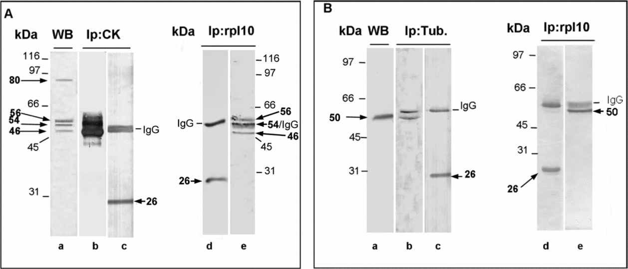
Then, we used anti-CK antibodies that can immunolocalize several CK isoforms in Xenopus oocytes (Mr of 42, 44, 54, 56 and 80 kDa, see Carotenuto et al., Reference Carotenuto, Petrucci, Correas, Vaccaro, Marco, Dale and Wilding2009; for detailed localization of CK, see Gard and Klymkowsky, 1998) (Fig. 3A, lane a) and anti-tubulin antibodies that can immunolocalize acetylated α-tubulin, the most widespread form of tubulin during early X. laevis oogenesis (Gard et al., Reference Gard, Affleck and Error1995; Vaccaro et al., Reference Vaccaro, Gigliotti, Graziani, Carotenuto, De Angelis, Tussellino and Campanella2010) (Fig. 3B, lane a). Co-immunoprecipitations utilising anti-rpl10 and anti-CK or anti-rpl10 and anti-tubulin (Fig. 3A, B) indicated that rpl10 physically interacts with some cytokeratin isoforms and with tubulin.
(A) Co-immunoprecipitation (Co-IP) using monoclonal anti-cytokeratin (anti-CK) or anti-rpl10. Lane a: Crude oocyte lysate incubated in anti-CK, revealing the presence of several CK isoforms of 46, 54, 56 and 80 kDa. The oocyte lysate immunoprecipitated using anti-CK and revealed using anti-CK showed several CK isoforms partially overlapping with IgG band (lane b) or the rpl10 band when incubated in anti-rpl10 (lane c). The oocyte sample was immunoprecipitated using anti-rpl10 and revealed using anti-CK, displaying the CK isoforms (lane e) or the rpl10 band when revealed using anti-rpl10 (lane d). (B) Co-IP using monoclonal anti-tubulin or anti-rp110. Lane a: Western blot of the oocyte lysate using the anti-tubulin antibody: a band of approximately 50 kDa was present. Lane b: Oocyte sample immunoprecipitated using the anti-tubulin antibody and revealed using the same antibody: a band of approximately 50 kDa was detected. Lane c: Oocyte sample immunoprecipitated using the anti-tubulin antibody and revealed using the anti-rpl10 antibody: the 26 kDa was present. Lane d: Oocyte sample immunoprecipitated using the anti-rpl10 antibody and revealed using the same antibody: the 26 kDa band was detected. Lane e: Oocyte sample immunoprecipitated using the anti-rpl10 antibody and revealed using the anti-tubulin antibody: the 50 kDa band was present.
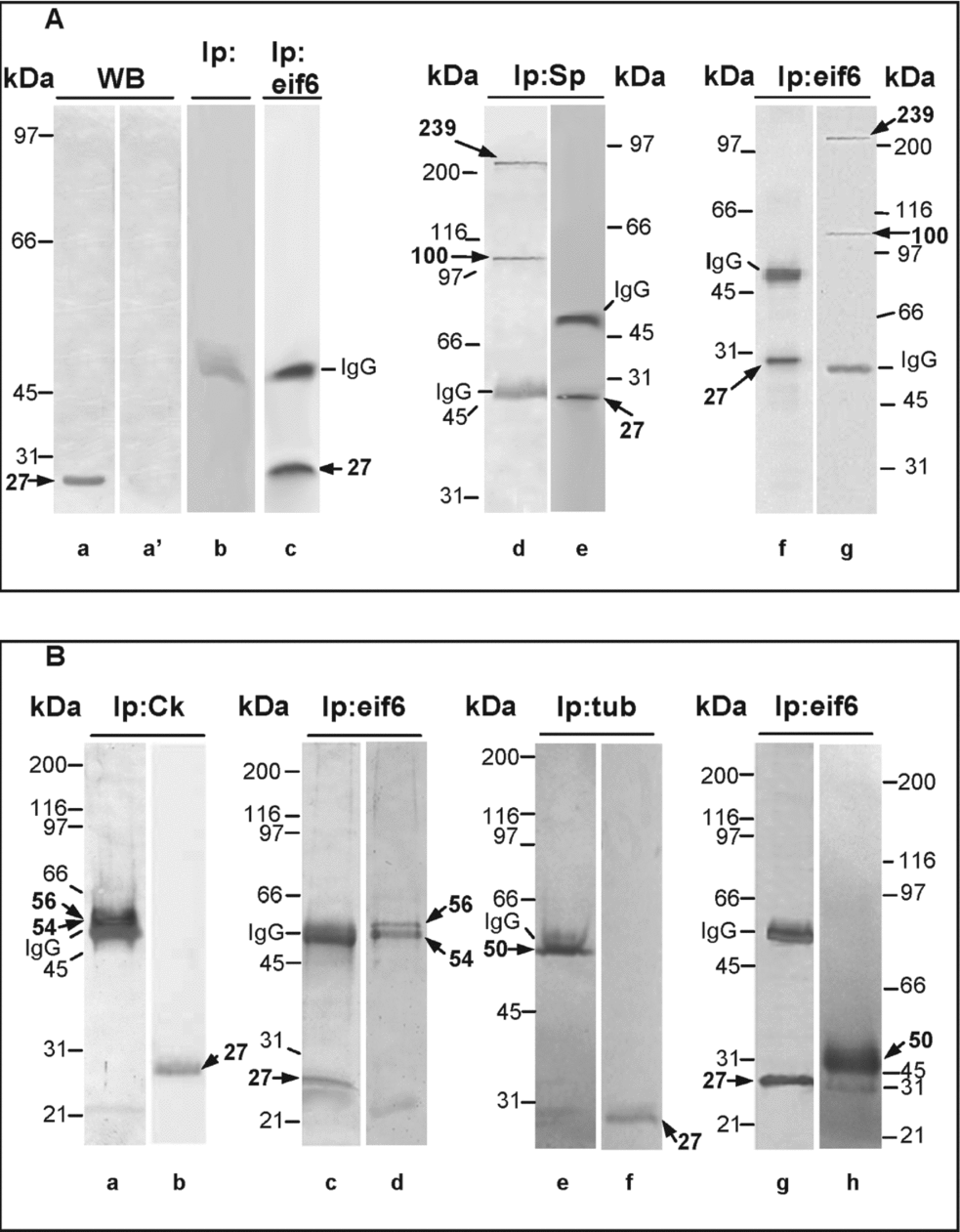
Based on immunofluorescence using anti-Eif6, immunolabelling was present in the MC and in the perinuclear region of the early oocytes (Fig. 4). Western blot of stage I oocytes lysates incubated in the anti-Eif6 antibody revealed a single band of 27 kDa (Fig. 5A, lanes a and a′), which was in agreement with previous data indicating that during X. laevis oogenesis, the 29 kDa form of eif6, phosphorylated at serine 235, appears only following germinal vesicle breakdown (Carotenuto et al., Reference Carotenuto, Vaccaro, De Marco, Wilding, Biffo, Marchisio, Capriglione and Campanella2005). Similarly, the 27 kDa band was present in the IPs using the same antibody, and the immunoreactivity was specific (Fig. 5A, lanes b and c). Co-IPs using anti-spectrin, anti-cytokeratin, or anti-tubulin indicated that eif6 physically interacts with the antibodies used and, in the case of CKs, with the 54 and 56 kDa isoforms (Fig. 5A, B).
Immunostaining of stage I oocytes using polyclonal anti-Eif6. (a, b) The mitochondrial cloud (MC) is immunostained. Mild staining was present in particular around the nuclei (arrows). (a) Bar = 45 μm; (b) bar = 25 μm. The sections were counterstained with DAPI. (c) In controls, in which the primary antibody was omitted, the immunostaining was absent. (c) Bar = 30 μm.
Co-immunoprecipitation (co-IP) of the oocyte samples using Eif6 and cytoskeletal protein antibodies. (A) Lane a: Western blot of the oocyte lysate revealed the expected band of 27 kDa upon anti-Eif6 incubation. Lane a′: Western blot incubated in pre-immune serum: no band was detected. Lane b: Immunoprecipitation using pre-immune serum: the IgG band was detected. Lane c: Oocyte sample immunoprecipitated using the anti-Eif6 antibody and revealed using the same antibody: the 27 kDa protein was detected. Lane d: Oocyte sample immunoprecipitated using the anti-β-spectrin antibody and revealed using the same antibody: the two spectrin bands were detected. Lane e: Oocyte sample immunoprecipitated using the anti-β-spectrin antibody and revealed using anti-Eif6: the 27 kDa eif6 band was detected. Lane f: Oocyte sample immunoprecipitated using the anti-Eif6 antibody and revealed using the same antibody: the 27 kDa band was detected. Lane g: Oocyte sample immunoprecipitated using the anti-Eif6 antibody and revealed using the anti-spectrin antibody: the 239 and 100 kDa bands were detected. (B) Co-immunoprecipitation using anti-cytokeratin (anti-CK) or anti-tubulin. Controls for monoclonal antibody specificity and IP specificity provided the same results as those of Fig. 1(A) lanes b′, c and d and are not shown here. The oocyte sample immunoprecipitated using anti-CK and revealed using anti-CK displayed some CK isoforms (lane a) or the eif6 band when incubated in anti-Eif6 (lane b). The oocyte sample immunoprecipitated using anti-Eif6 and revealed using anti-Eif6 displayed the 27 kDa band (lane c) or the CK bands when revealed using anti-CK (lane d). Lane e: The oocyte sample immunoprecipitated using anti-tubulin and revealed using the same antibody displayed a tubulin band of approximately 50 kDa or the 27 kDa band when incubated in anti-Eif6 (lane f). Lane g: Western blot of the oocyte sample was immunoprecipitated using the anti-Eif6 antibody and revealed using the same antibody; the 27 kDa band was detected. Lane h: Western blot of the oocyte sample was immunoprecipitated using the anti-Eif6 antibody and revealed using anti-tubulin; the 50 kDa band was detected.
Interactions between cytoskeletal proteins and P0 and thesaurins
To further investigate the interaction between the CSK and the translation machinery, we evaluated P0 and thesaurins A and B.
P0 is a phosphoprotein located on the 60S subunit in the ribosomal stalk together with P1 and P2 and is contiguous with the GTPase Centre. Therefore, P0 is involved in the interaction with elongation factors and in the movements of tRNA on the ribosome, conferring specificity to the ribosome for interaction and translocation (Uchiumi et al., Reference Uchiumi, Honma, Endo and Hachimori2002). Thesaurins A and B are elongation factors that are active during protein synthesis and are stored in the early oocyte in thesaurisomes, which can also store 5S RNA and aminoacyl-tRNAs for extended periods. Thesaurin A is homologous to eukaryotic EF-1α differing only in that it binds to uncharged tRNA more tightly than charged RNA and to GTP more tightly than GDP (Viel et al., Reference Viel, Le Mair, Morales and Denis1991).
Immunofluorescence using anti-P0 indicated that in stage I oocytes, this protein is present in the perinuclear region, at the oocyte periphery, in the MC and in thin cytoplasmic strands (Fig. 6a, b), similar to the localization of cytokeratins and spectrin (see Carotenuto et al., Reference Carotenuto, Vaccaro, Capriglione, Petrucci and Campanella2000; Vaccaro et al., Reference Vaccaro, Wilding, Dale, Campanella and Carotenuto2012). In control sections, this immunostaining was absent (Fig. 6c).
Immunostaining of stage I oocytes using anti-P0. (a) Some labelling was found at the oocyte periphery (small arrow), in cytoplasmic aggregates (arrowhead) and at the nuclear envelope (arrow). Bar = 40 μm. (b) Immunolabelling was also present in the mitochondrial cloud (MC). Bar = 36 μm. (c) In control sections, in which the primary antibody was substituted with pre-immune serum, the immunolabelling was absent. Bar = 36 μm.
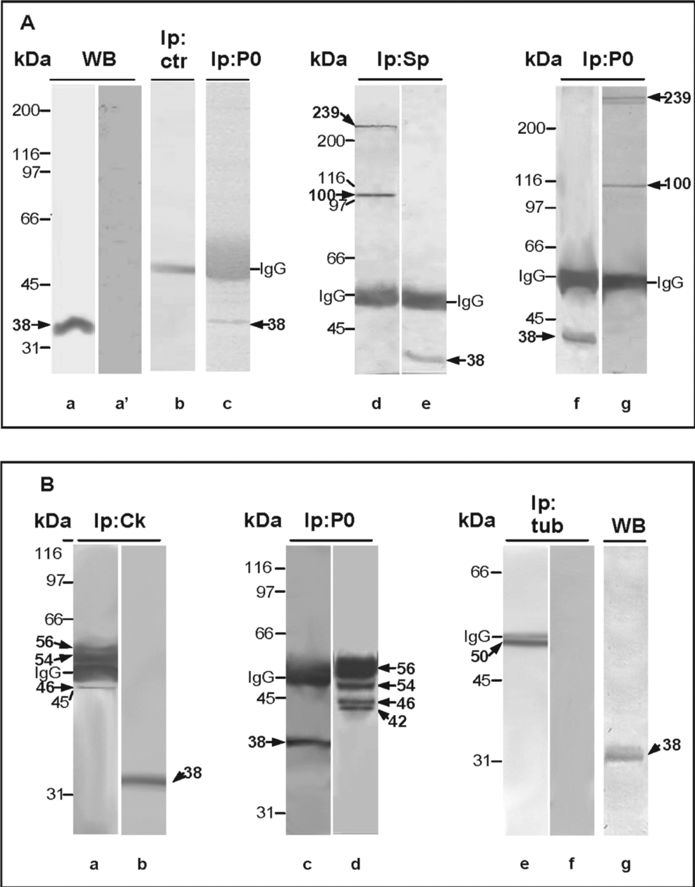
Anti-P0 specifically identifies a band of 38 kDa in western blots of both oocyte lysates and IPs using anti-P0. Co-immunoprecipitation using anti-P0, anti-spectrin, anti-CK or anti-tubulin indicated that P0 does interact with spectrins or keratins of 56, 54, 46 and 42 kDa (Fig. 7A, B). In contrast, this phosphoprotein did not appear to interact with tubulin (Fig. 7B, lanes e–g), although this protein was present in the same oocyte lysates as those used for co-IP (Fig. 7Bg).
In the oocyte lysates, P0 co-immunoprecipitated (co-IP) with cytoskeletal proteins. (A) Separation and immunoprecipitation of P0. Lane a: Western blot of the oocyte lysate revealing the 38 kDa P0 band. (a′) Western blot incubated in pre-immune serum. Lane b: Control IP incubated in pre-immune serum. Lane c: Oocyte sample immunoprecipitated using the anti-P0 antibody and revealed using anti-P0, displaying the 38 kDa band. Lane d: Oocyte sample immunoprecipitated using the anti-spectrin antibody and revealed using anti-spectrin, displaying the 100 and 239 bands. Lane e: Oocyte sample immunoprecipitated using the anti-β-spectrin antibody and revealed using anti-P0: the 38 kDa band was detected. Lane f: Oocyte sample immunoprecipitated using the anti-P0 antibody and revealed using the anti-P0 antibody: the 38 kDa band was present. Lane g: Oocyte sample immunoprecipitated using the anti-P0 antibody and revealed using the anti-β-spectrin antibody: the 239 kDa and 100 kDa bands were detected. (B) Co-IP using the anti-cytokeratin (anti-CK) or anti-tubulin antibody. Oocyte lysate immunoprecipitated using anti-CK and revealed using anti-CK, displaying CK isoforms (lane a) or the P0 band when incubated in anti-P0 (lane b). The oocyte lysate immunoprecipitated using anti-P0 and revealed using anti-P0 displayed the 38 kDa band (lane c) or CK isoform bands when revealed using anti-CK (lane d). Lane e: The oocyte lysate immunoprecipitated using anti-tubulin and revealed using anti-tubulin displayed the 50 kDa tubulin band (lane f), but the P0 band was not detected when revealed using anti-P0. Lane g: Western blot of the same oocyte lysate used for the co-IPs; the 38 kDa band was present.
Immunofluorescence utilising anti-thesaurin A and B revealed immunostaining in the perinuclear region, in the MC, and in the cytoplasm, in particular in the oocyte periphery, where the protein forms conspicuous aggregates or strands (Fig. 8a–d). Accordingly, Viel et al. (1990) found that thesaurin A (of approximately 50 kDa) is concentrated in the MC, whereas thesaurin B (of approximately 40 kDa) is scattered throughout the cytoplasm. In control sections, this immunostaining was absent (Fig 8e).
Immunostaining of stage I oocytes using anti-thesaurins. (a–c) Immunolabelling was detected in the nuclear envelope (arrow), in cytoplasmic aggregates at the periphery of larger oocytes (small arrow), and in the mitochondrial cloud (MC). (a) Bar = 42 μm; (b) bar = 52 μm; (c) bar = 85 μm. (d) Immunolabelling was detected as characteristic strands radiating from the cortex (double arrow). Bar = 27 μm. (e) In control sections, in which the primary antibody was substituted with pre-immune serum, the immunolabelling was absent. Bar = 75 μm.
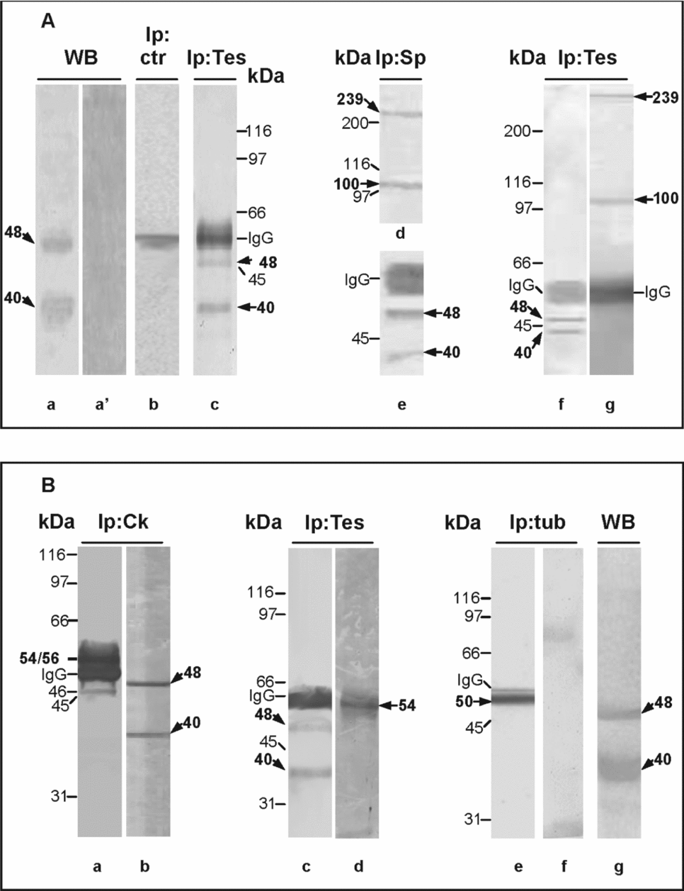
Co-immunoprecipitation using anti-thesaurin A and B and the antibodies against the cytoskeletal proteins revealed that thesaurins physically interact with spectrin and with cytokeratin (Fig. 9A, B), but they do not appear to interact with tubulin (Fig. 9B, lanes e and f), although they were present in the same oocyte lysates as those used for co-IP (Fig. 9B, lane g).
Thesaurins A and B in the oocyte lysate and co-immunoprecipitation (co-IP) of thesaurin A and B with cytoskeletal protein antibodies. (A) Separation and co-IP of thesaurins with spectrin. Lane a: Western blot of the oocyte lysate incubated in anti-thesaurins displaying thesaurin A and B bands of approximately 48 and 40 kDa. Lane a′: Western blot incubated in pre-immune serum: no band was detected. Lane b: Control IP incubated in pre-immune serum. Lane c: Oocyte sample immunoprecipitated using the anti-thesaurins antibody and revealed using the same antibody, displaying the 40 and 48 kDa bands. Lanes d and e: Oocyte sample immunoprecipitated using the anti-spectrin antibody. The nitrocellulose membrane was cut into two portions: the upper portion (lane d) was revealed using the anti-spectrin antibody, displaying the 100 and 239 kDa bands, while (lane e) the lower portion was revealed using anti-thesaurins: the 48 and40 kDa bands were detected. Lane f: Oocyte sample immunoprecipitated using the anti-thesaurins antibody and revealed using the same antibody, displaying the 48 and 40 kDa bands. Lane g: Oocyte sample was immunoprecipitated using the anti-thesaurins antibody and revealed using anti-spectrin: the 100 and 239 kDa bands were detected. (B) Co-IP using the anti-CK or anti-α-tubulin antibody. The oocyte lysate immunoprecipitated using anti-CK and revealed using anti-CK displayed CK isoforms partially masked by the IgG band (lane a) or the thesaurins bands when incubated in anti-thesaurins (lane b). The oocyte sample immunoprecipitated using anti-thesaurins and revealed using the same antibody displayed the 48 and 40 kDa bands (lane c) or the CK band when revealed using anti-CK (lane d). Lane e: The oocyte sample immunoprecipitated using anti-tubulin and revealed using anti-tubulin displayed the tubulin band, but the thesaurins bands were not detected when revealed using anti-thesaurins (lane f), although these proteins were present in western blots (lane g) of the same oocyte lysate used for the co-IPs.
Discussion
RNA, elongation factors and polysomes are found in clusters at microfilament intersections termed ‘vertices’ (Bassell et al., Reference Bassell, Power, Taneja and Singer1994) or between loosely bundled microtubules (for X. laevis oocytes, see Oberman & Yisraeli, Reference Oberman and Yisraeli1995). Moreover, elongation factors, such as EF-2, associate with microfilaments or intermediate filaments depending on the functional stage of the cell (Shestakova et al., Reference Shestakova, Motuz and Gavrilova1993).
Microfilaments play the predominant role in the RNA–cytoskeletal association in most cell types, playing active roles in regulating cell functions. For example, in growth cone guidance of Xenopus embryos, beta-actin synthesis contributes to the directional motility of growth cones (Leung et al., Reference Leung, van Horck, Lin, Allison, Standart and Holt2006; Yao et al., Reference Yao, Sasaki, Wen, Bassell and Zheng2006). Actin also facilitates myosin-dependent RNA transport, and microtubule networks play roles in RNA binding in oocytes, as the transport of oskar mRNA in Drosophila (Ephrussi et al., Reference Ephrussi, Dickinsons and Lehman1991) and of Vg1 and VegT mRNAs in X. laevis (see King et al., Reference King, Messit and Mowry2005). Intermediate filaments play a role in the storage of ribonucleoproteins (RNPs) (Traub et al., Reference Traub, Bauer, Harting, Grub and Stahl1998), and their co-alignment with microtubules and microfilaments, as found in X. laevis vitellogenic oocytes (Gard et al., Reference Gard, Affleck and Error1995, Carotenuto et al., Reference Carotenuto, Vaccaro, De Marco, Wilding, Biffo, Marchisio, Capriglione and Campanella2005), might facilitate the directional transport of RNPs.
Actin cables, intermediate filaments and microtubules are present in oocytes beginning in the early stages of oogenesis (Gard Reference Gard, Affleck and Error1995; Gard et al., Reference Gard, Affleck and Error1995). Spectrin is broadly distributed during Xenopus oogenesis (Ryabova et al., Reference Ryabova, Virtanen, Wartiovaara and Vassetzky1994; Carotenuto et al., Reference Carotenuto, Maturi, Infante, Capriglione, Petrucci and Campanella1997, Reference Carotenuto, Vaccaro, Capriglione, Petrucci and Campanella2000, Reference Carotenuto, Petrucci, Correas, Vaccaro, Marco, Dale and Wilding2009; Kloc & Etkin, Reference Kloc and Etkin1998), and its presence correlates to the distribution of actin microfilaments (Bennett & Gilligan, Reference Bennett and Gilligan1993; Vaccaro et al., Reference Vaccaro, Gigliotti, Graziani, Carotenuto, De Angelis, Tussellino and Campanella2010). The distribution of CK also correlates to that of actin microfilaments (Gard, Reference Gard, Affleck and Error1995).
Our data indicated that both eif6 and rpl10 associate with spectrin, cytokeratin and tubulin, whereas thesaurins and P0 interact only with spectrin and cytokeratin. However, it should be remembered that microtubules may interact with other molecules via connectors (King et al., Reference King, Messit and Mowry2005).
Intermediate filaments associate with Eif6 in mouse fibroblasts (Biffo et al., Reference Biffo, Sanvito, Costa, Preve, Pignatelli and Spinardi1997). Confocal microscopy using anti-CK antibodies revealed that in Xenopus oocytes, the association between eif6 and the CSK correlates to the phosphorylation of eif6 at serine 235 following maturation and fertilization. During earlier stages of X. laevis oogenesis, the CSK appears to sparsely associate with eif6, suggesting a scant or dynamic association that is modulated according to the functional stages, but this pattern could not be deduced based on static confocal images (Carotenuto et al., Reference Carotenuto, Vaccaro, De Marco, Wilding, Biffo, Marchisio, Capriglione and Campanella2005). Our data indicate that, at least under in vitro conditions, this interaction may occur and may extend to spectrin and tubulin. eif6 is predominantly expressed around the nucleus and in the MC, where cytoskeletal components (i.e. tubulin, CK and spectrin) are also present (Gard et al., Reference Gard, Affleck and Error1995; Carotenuto et al., Reference Carotenuto, Petrucci, Correas, Vaccaro, Marco, Dale and Wilding2009; Vaccaro et al., Reference Vaccaro, Gigliotti, Graziani, Carotenuto, De Angelis, Tussellino and Campanella2010). In these regions, co-IP using anti-rp110 and anti-spectrin revealed superimposed immunolabelling. Given the functional relationship between Eif6 and rpl10, i.e. at ribosome maturation, the insertion of rpl10 into the 60S subunit is correlated to the release of Eif6, one can hypothesise that the CSK provides the scaffold necessary for the insertion/release of the two molecules and subsequently for eif6 transport and binding to the mature 60S subunit (see Brina et al., Reference Brina, Grosso, Miluzio and Biffo2011). Interestingly, according to Hovland et al. (Reference Hovland, Hesketh and Pryme1995), the association between ribosomal subunits and initiation factors and the CSK might be transiently required to increase the local concentration of translation factors. Moreover, assembly of the ribosome stalk, where P0 is located, to the 60S subunit is essential for the release of Tif6/Eif6 (Lo et al., Reference Lo, Li, Bussiere, Bresson, Marcotte and Johnson2010), and the CSK, particularly rich in CK isoforms due to the presence of P0 (this paper), could facilitate the approach of the stalk to the maturing 60S subunit.
Interestingly, actin appears to be essential for the organization of translational complexes (Jansen, Reference Jansen1999), as efficient translation is lost upon F-actin disruption (Stapulionis et al., Reference Stapulionis, Kolli and Deutscher1997). In fibroblasts, EF1α cross-links actin filaments in a specific manner (Negrutskii et al., Reference Negrutskii, Stapulionis and Deutscher1994), thereby creating microcompartments that are important for mRNA anchoring and/or cytoskeletal facilitation of protein synthesis. EF1α from X. laevis oocytes (i.e. thesaurins) can sever microtubules via unknown mechanisms (Shiina et al., Reference Shiina, Gotoh, Kubomura, Iwamatsu and Nishida1994 and for a detailed discussion see Jansen, 1999) and may play a similar role. This finding is in agreement with our data indicating that thesaurins bind to spectrin and cytokeratins and but not to tubulin. The CSK may provide support to thesaurins for their interaction with sites of the translating ribosome and in particular with rpl10 (see Uchiumi et al., Reference Uchiumi, Honma, Endo and Hachimori2002). The broad distribution of thesaurins that we detected in the cortical cytoplasm and in the perinuclear region may indicate that thesaurins render these regions as favourable sites for active protein synthesis.
At present, our interpretation of the role of the association between these maturation and translational components and the CSK is only circumstantial. However, our data demonstrate that the actin-based CSK and, in some cases, the microtubules associate with factors implicated in the transport of 60S or in the export of the 60S subunit during different stages, strongly suggesting that in early X. laevis oocytes, the CSK stabilises maturation/translation processes, providing new topics for future investigation.
Acknowledgements
This study was funded by a grant from the ‘Compagnia di San Paolo’ (‘Integration of spatial and biochemical clues during brain development’ Project) to CC. We wish to thank the Elsevier Editorial Service.