Implications
In bringing together the results of recent research on the cryopreservation of ram semen and the relaxation of the ewe cervix, this review provides a basis for identifying opportunities for improving the efficiency of cervical insemination and transcervical embryo recovery and transfer in sheep genetic improvement programmes.
Introduction
In the 1970s, the introduction of cattle transcervical embryo recovery and transfer procedures removed the need for major surgery in the form of mid-ventral laparotomy and flank incision for multiple ovulation and embryo transfer (MOET) programmes and led to widespread use of MOET for genetic improvement (Gordon, Reference Gordon1994). It also acted as a stimulus for the pursuit of similar procedures in small ruminants, notably sheep. The ewe cervix, however, has proved to be substantially more difficult than the cow cervix to penetrate and traverse. The inability to manipulate the ewe cervix per rectum, in the same way as in the cow, is a further difficulty. Thus, the application of MOET programmes, and indeed the use of frozen–thawed semen, which requires deposition within the uterus for successful fertilisation, still relies on laparoscopic penetration of the abdomen in ewes (Cognie, Reference Cognie1999; Cognie et al., Reference Cognie, Baril, Poulin and Mermillod2003; Menchaca et al., Reference Menchaca, Vilarino, Crispo, de Castro and Rubianies2009; Fukui et al., Reference Fukui, Kohno, Okabe, Katsuki, Yoshizawa, Togari and Watanabe2010).
In current commercially operated embryo transfer programmes, the degree of laparoscopic involvement is still generally less than that shown to be successful under experimental conditions. Thus, despite the successful commercial application by McKelvey at Edinburgh Genetics of an intra-abdominal laparoscopic procedure for the transfer of embryos (McKelvey et al., Reference McKelvey, Robinson and Aitken1985), most operators use a modification of the technique, which involves exteriorising the tip of the uterine horn (Haresign et al., Reference Haresign, Merrell and Richards1994; McMillan and Hall, Reference McMillan and Hall1994; Wolf and McDougall, Reference Wolf and McDougall1994; Wolf and Mylne, Reference Wolf and Mylne1994; Li et al., Reference Li, Guan, Hou, An and Chen2008). The same is true for embryo recovery from superovulated donor ewes in which the complete intra-abdominal flushing of each uterine horn by laparoscopy as described by McKelvey et al. (Reference McKelvey, Robinson, Aitken and Robertson1986) and Nellenschulte and Niemann (Reference Nellenschulte and Niemann1992) appears to have only been used, on a commercial scale, by Lamb XL in New Zealand (P. Lynch, personal communication, 1990) and by Rowett Research Services in the United Kingdom (Scudamore et al., Reference Scudamore, Robinson, Aitken, Kennedy, Ireland and Robertson1991). Despite the success of a modified version of the intra-abdominal flushing technique involving the exteriorisation of the tip of the uterine horn for ease of insertion of the flushing medium (Bari et al., Reference Bari, Khalid, Haresign, Merrell, Murray and Richards1999), the majority of recoveries of sheep embryos in the United Kingdom still involve mid-line laparotomy.
Research into laparoscopic techniques, and the introduction of the laparoscope to commercial sheep breeding for genetic improvement, has been accompanied by numerous other research studies aimed at capturing the benefits of artificial insemination and MOET in sheep breeding. These studies have been wide ranging and have involved cervical dilation by both physical and pharmacological means in order to achieve transcervical artificial insemination with frozen–thawed semen and transcervical embryo recovery and transfer. They have also involved investigations into methods for improved cryopreservation of semen and semen sexing by flow cytometry. The experimental approaches vary from applied to fundamental. This review brings together the results of studies aimed at increasing the post-thaw viability of ram semen and enhancing understanding of cervical dilation in the ewe as means of improving the success of cervical insemination and transcervical embryo recovery and transfer.
Recent studies in semen freezing
Improving the conception rates of ewes following cervical insemination with frozen–thawed semen has proved difficult. The finding that the consistently high conception rates achieved in Norway are not due to Norwegian semen or its freezing protocol (Hanrahan, Reference Hanrahan2003), eliminated the possibility of an instant improvement in lambing rates elsewhere by the adoption of the Norwegian cryopreservation procedure. There is also the problem that changes to the freezing protocol that enhance post-thaw motility do not necessarily translate into improved lambing rates (Sanchez-Partida et al., Reference Sanchez-Partida, Windsor, Eppleston, Setchell and Maxwell1999). Variation in fertility between rams and between ejaculates within rams (Windsor, Reference Windsor1997) adds to the complexity. Even an evaluation of semen quality based on in vitro fertilisation proved ineffective in predicting pregnancy rates following cervical insemination with frozen–thawed semen (Papadopoulos et al., Reference Papadopoulos, Hanrahan, Donovan, Duffy, Boland and Lonergan2005), although a reduction in sperm concentration from 2 × 10⁶ to 0.0625 × 10⁶ per ml (O'Meara et al., Reference O'Meara, Hanrahan, Donovan, Fair, Rizos, Wade, Boland, Evans and Lonergan2005) resulted in a significant correlation (P = 0.013) between fertility in vivo and fertility in vitro. However, defining a sensitive critical threshold for sperm numbers that is applicable to all breeds and conditions presents its own problems.
The need for more sophisticated studies to identify how the effects of freezing and thawing compromise pregnancy rates has long been recognised. Windsor and White (Reference Windsor and White1995) reported severe mitochondrial damage of ram spermatozoa following freezing and thawing and reasoned that the associated attenuation of respiratory metabolism may compromise their cervical passage. Recently, Nur et al. (Reference Nur, Zik, Ustumer, Sagirkaya and Ozguden2010) reported the effects of a range of cryoprotective agents (glycerol, propanediol, sucrose and trehalose) on DNA integrity. All of them induced apoptosis and DNA damage. Although glycerol gave the best post-thaw motility, this was not accompanied by better DNA integrity; indeed the opposite, albeit not significant statistically, tended to be the case. Thus, the authors suggest that there may be merit in studying novel combinations of glycerol with sucrose or trehalose in order to maximise DNA integrity.
In a recent attempt to improve sperm membrane integrity during cryopreservation, by the incorporation of cholesterol into the membrane, Mocé et al. (Reference Mocé, Purdy and Graham2010) tested the effects of six cholesterol-loaded cyclodextrins on the cryosurvival of ram spermatozoa. The pre-freezing addition of cholesterol-loaded 2-hyroxypropyl-β-cyclodextrin increased the post-thaw percentage of motile sperm from 43% to 62%. A similar improvement occurred with methyl-β-cyclodextrin but not with the other 4 cyclodextrins (α-cylcodextrin, α-cylcodextrin hydrate, β-cyclodextrin, β-cyclodextrin hydrate). The enhanced post-thaw sperm motility was sustained through a much wider range of osmotic solutions (150 to 425 mOsm) compared with control sperm (270 to 370 mOsm). The number of sperm binding to zonae pellucida also was increased by previous inclusion of cholesterol-loaded 2-hydroxypropyl-β-cyclodextrins.
The report by Maxwell et al. (Reference Maxwell, Evans, Mortimer, Gillon, Gellatly and McPhie1999) that normal fertility was achieved following cervical insemination with frozen–thawed semen that had been supplemented, post thawing, with seminal plasma brought new hope that technically, sheep breeding using frozen–thawed semen would soon be at par with cattle breeding. The studies indicated that the seminal plasma met the two essential requirements for ensuring high pregnancy rates following cervical insemination, namely, stabilisation of the sperm membranes and improved sperm transport through cervical mucus. As it is the vesicle-free seminal plasma that confers the improvement in membrane integrity (El-Hajj Ghaoui et al., Reference El-Hajj Ghaoui, Gillan, Thomson, Evans and Maxwell2007a), seminal plasma from vasectomised rams, which is naturally vesicle-free, is as effective as that from entire rams. Not all protein fractions in seminal plasma, however, have a beneficial effect; indeed, some can be detrimental. In a recent study using SDS-polyacrylamide gel electrophoresis, Yue et al. (Reference Yue, Shi, Bai, Ren and Zhao2009) found eight protein bands that were different between rams of high natural fertility as opposed to low natural fertility. Of these, a 116 kDa protein band was either absent or poorly expressed in low-fertility rams implying that, by its presence in significant quantities in all high-fertility rams, it conferred a beneficial effect. There was also a high content of a 72.45 kDa band in the seminal plasma of high-fertility rams and a high positive correlation between its content and sperm viability, suggesting a protective role in sperm membrane integrity. Low-fertility rams had a high content of a 15.9 kDa band, corresponding in molecular weight to TIMP-2, the tissue inhibitor of metalloproteinase-2, shown by Cardozo et al. (Reference Cardozo, Fernández-Juan, Forcada, Abecia, Muiño-Blanco and Cebrián-Pérez2006) to be more highly expressed during the non-breeding season and to be associated with poor sperm viability.
The variable mix of beneficial and detrimental proteins in seminal plasma implies that there may be a range of outcomes following its post-thaw addition to semen and indeed this is proving to be the case. In contrast to the beneficial effect observed by Maxwell et al. (Reference Maxwell, Evans, Mortimer, Gillon, Gellatly and McPhie1999), O'Meara et al. (Reference O'Meara, Donovan, Hanrahan, Duff, Fair, Evans and Lonergan2007) found no improvement in pregnancy rate when ewes were cervically inseminated with frozen–thawed semen resuspended in seminal plasma from high-fertility rams as compared with low-fertility rams, indicating that, in this instance, the historical record of ram fertility was an inadequate selection criterion. In a study by El-Hajj Ghaoui et al. (Reference El-Hajj Ghaoui, Thomson, Leahy, Evans and Maxwell2007b), autologous seminal plasma improved sperm motility and membrane integrity but, following cervical insemination, failed to improve on the abnormally low (∼12%) pregnancy rate among ewes inseminated without additional autologous seminal plasma. On the other hand, Leahy et al. (Reference Leahy, Evans, Maxwell and Marti2010a) found that, for cervically inseminated ewes, the addition of seminal plasma post-thawing was as effective as laparoscopic intrauterine insemination in raising pregnancy rates on the first day of a two-day insemination programme, but not on the second day. This observation typifies the elusive and transitory nature of the beneficial effect of post-thaw supplementation.
From an on-farm practical viewpoint, pre-freezing supplementation with seminal plasma is a lot more attractive than post-thaw supplementation. It could also have the added benefit of preventing, rather than of reversing, the damaging effects of freezing and thawing. Evidence for this comes from the recent study of Leahy et al. (Reference Leahy, Marti, Evans and Maxwell2010b) in which pre-freezing addition of crude seminal plasma or its >10 kDa protein component improved sperm motility and the proportion of acrosome-intact sperm. The beneficial effects were greater when the supplementation comprised seminal plasma collected during the breeding season rather than in the non-breeding season, fractionated using >19 kDa filters rather than as the crude plasma and added directly to the spermatozoa rather than via the diluent. Pre-freezing supplementation of flow-cytometrically sex-sorted sperm with crude seminal plasma, or its low-molecular-weight (<10 kDa) or high-molecular-weight (>10 kDa) fraction, improved its functional integrity, whereas post-thaw supplementation had no beneficial effect (Leahy et al., Reference Leahy, Marti, Evans and Maxwell2009). Extensive field testing is required to determine whether the improved functional integrity arising from pre-freezing seminal plasma supplementation is translated into improved pregnancy rates. Meanwhile, the relatively high sperm concentration used when cryopreserving, in the belief that it avoids negative effects of high dilution, has been questioned by Leahy et al. (Reference Leahy, Marti, Mendoza, Pérez-Pé, Muiño-Blanco, Cabrián-Pérez, Evans and Maxwell2010c); after all, sex sorting by flow cytometry, of necessity, involves a much higher dilution rate than that currently used for cryopreservation. The first record of a lamb born from sexed semen involved intracytoplasmic sperm injection (Catt et al., Reference Catt, Catt, Gomez, Maxwell and Evans1996) and was soon followed by intrauterine insemination using fresh semen (Cran et al., Reference Cran, McKelvey, King, Dolman, McEvoy, Broadbent and Robinson1997) at a dilution rate that was approximately 800 times that used in cryopreservation. In their study of the effect of pre-freezing dilution to 20 million sperm per ml, that is, the current freezing concentration for sex-sorted semen, and some 40 to 50 times the current dilution rate for freezing non-sorted semen, Leahy et al. (Reference Leahy, Marti, Mendoza, Pérez-Pé, Muiño-Blanco, Cabrián-Pérez, Evans and Maxwell2010c) observed improved post-thaw sperm viability and acrosome integrity. As frozen–thawed semen destined for intrauterine insemination currently involves low total sperm numbers and high sperm concentrations inseminate volumes are inevitably very small. Therefore, in terms of sperm quality and on-farm accuracy in measuring inseminate volumes, higher pre-freezing dilutions than are currently used may be warranted. Indeed, the remarkably low dose of motile sperm (3.5 million in each of two 0.2 ml inseminations) used in the first report of pregnancies in ewes cervically inseminated with frozen–thawed sex-sorted semen (Leahy et al., Reference Leahy, Evans, Maxwell and Marti2010a) adds to the practical relevance of investigations aimed at improving understanding of the effects of dilution and sex sorting by flow cytometry on sperm viability and membrane integrity. On the basis of their data demonstrating the success of very-low-dose (1 × 10⁶ motile sperm) intrauterine insemination of frozen sexed semen, de Graaf et al. (Reference De Graaf, Evans, Maxwell, Downing and O'Brien2007b) and Beilby et al. (Reference Beilby, Grupen, Thomson, Maxwell and Evans2009) put forward the intriguing hypothesis that the sex-sorting procedure may result in a highly resilient and fertile sub-population of sperm. If this is the case, an alternative practical method for identifying and isolating such a population, with a view to enhancing cervical transport, could be a major advance in improving fertility following low-dose cervical insemination.
Transcervical insemination
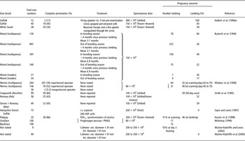
In their comprehensive review of the literature on artificial insemination, Salamon and Maxwell (Reference Salamon and Maxwell1995) concluded that, given further research and improved understanding of what was needed to achieve cervical penetration, transcervical insemination using frozen and thawed semen had the ‘potential to replace the laparoscopic method in some situations’. Much of the optimism in this view, at that time, was based on the improvement in complete cervical penetration provided by the Guelph equipment and insemination technique (see Table 1; Halbert et al., Reference Halbert, Dobson, Walton, Sharpe and Buckrell1990a; Buckrell et al., Reference Buckrell, Buschbeck, Gartley, Kroetsch, McCutcheon, Martin J Penner and Walton1994) and by the knowledge that complete penetration of the cervix at insemination increased the numbers and the quality of spermatozoa reaching the oviducts (Fukui and Roberts, Reference Fukui and Roberts1977). In the Halbert et al. (Reference Halbert, Dobson, Walton, Sharpe and Buckrell1990a) study, complete cervical penetration was achieved in 49 of 90 ewes (54%) and 40 of the 49 ewes (82%) lambed. In the much larger Buckrell et al. (Reference Buckrell, Buschbeck, Gartley, Kroetsch, McCutcheon, Martin J Penner and Walton1994) study, the corresponding percentages for 2060 ewes were 88% and 33%, with significant effects of the time of year (breeding v. non-breeding season) and interval from previous lambing, Table 1, on lambing outcome. Complete penetration rates increased from 76% for the first 500 ewes inseminated in the study to 98% for the last 500 and the corresponding mean times to achieve complete penetration decreased from 8.6 to 3.6 min, reflecting the importance of operator experience.
Summary of pregnancy outcomes following complete transcervical penetration and intrauterine deposition of chilled or frozen–thawed semen in oestrous-synchronised ewes

Reports of attempts (Table 1) to extend the use of the Guelph inseminating equipment and technique (Windsor et al., Reference Windsor, Szell, Buschbeck, Edward, Milton and Buckrell1994 in Australia; Smith et al., Reference Smith, Parr, Beaumont, Oliver and Upreti1995 in New Zealand; McKelvey, Reference McKelvey1999 in the United Kingdom) or a modified form of the inseminating equipment (Campbell et al., Reference Campbell, Harvey, McDonald and Sparksman1996 in New Zealand) have thwarted rather than promoted its application. In the Windsor et al. (Reference Windsor, Szell, Buschbeck, Edward, Milton and Buckrell1994) study, the combined lower success rate in traversing the cervix and in establishing pregnancy, compared with the Buckrell et al. (Reference Buckrell, Buschbeck, Gartley, Kroetsch, McCutcheon, Martin J Penner and Walton1994) study, resulted in overall lambing rates of less than 1 in 4. The technique was therefore unable to compete with the success of the already well-established laparoscopic intrauterine technique in Australia. The same was true for the United Kingdom (McKelvey Reference McKelvey1999; Table 1), but here there was the added complication of invasion of the peritoneal cavity from accidental puncturing of the reproductive tract with the inseminating pipette. Similar inadvertent damage occurred in the New Zealand studies of Campbell et al. (Reference Campbell, Harvey, McDonald and Sparksman1996).
Although the incidence of penetration of the cervical wall was reduced from 31% to 10% by a ball tip addition to the Guelph insemination pipette, histological observations showed that there was still damage to the epithelial lining over the entire length of the cervix in ewes for which penetration was complete. In the separate New Zealand study of Smith et al. (Reference Smith, Parr, Beaumont, Oliver and Upreti1995; Table 1), complete penetration of the cervix, using the Guelph inseminating pipette, was also much lower than that achieved by Buckrell et al. (Reference Buckrell, Buschbeck, Gartley, Kroetsch, McCutcheon, Martin J Penner and Walton1994); however, conception rates for those ewes that were completely penetrated were similar to those achieved by Buckrell et al. (Reference Buckrell, Buschbeck, Gartley, Kroetsch, McCutcheon, Martin J Penner and Walton1994) using laparoscopy. The same is true for the observations of Windsor et al. (Reference Windsor, Szell, Buschbeck, Edward, Milton and Buckrell1994). Thus, not surprisingly, Smith et al. (Reference Smith, Parr, Beaumont, Oliver and Upreti1995) concluded that research should concentrate on ways of increasing the incidence and ease of cervical penetration.
Evidence for an adverse effect of transcervical procedures on fertilisation and lambing rates is equivocal. The observations of Halbert et al. (Reference Halbert, Dobson, Walton, Sharpe and Buckrell1990a), Buckrell et al. (Reference Buckrell, Buschbeck, Gartley, Kroetsch, McCutcheon, Martin J Penner and Walton1994), Windsor et al. (Reference Windsor, Szell, Buschbeck, Edward, Milton and Buckrell1994) and Husein et al. (Reference Husein, Romano, Bailey, Ababneh, Crabo, Godfrey, Head and Wheaton1998) imply that there is no adverse effect, and with no accompanying information on fertility it is not known whether the damage to the cervical epithelial lining observed by Campbell et al. (Reference Campbell, Harvey, McDonald and Sparksman1996) has a detrimental effect on lambing rate. The lower lambing rates observed for transcervical than laparoscopic insemination by McKelvey (Reference McKelvey1999) and included in Table 1, and the associated trauma to the cervix, imply an adverse effect. Sayre and Lewis (Reference Sayre and Lewis1997) used oxytocin to dilate the cervix. Although they found no detrimental effect of oxytocin on fertilisation rate, the transcervical technique per se appeared to reduce fertilisation rate. In contrast, Stellflug et al. (Reference Stellflug, Wulster-Radcliffe, Hensley, Cowardin, Seals and Lewis2001) found no adverse effect of experimental cervical manipulation on fertility in laparoscopically inseminated ewes, but lambing rates were decreased by oxytocin-induced cervical dilation. Using a brass bulb tip to decrease cervical trauma and to guide the inseminating pipette, Wulster-Radcliffe and Lewis (Reference Wulster-Radcliffe and Lewis2002) found no adverse effect of transcervical insemination on fertility. In a subsequent study, however (Wulster-Radcliffe et al., Reference Wulster-Radcliffe, Wang and Lewis2004; see Table 1), only 4% of ewes lambed following transcervical insemination. Yet when sham transcervical insemination was immediately followed by ram mating, lambing rate was 63%, implying that the relatively small numbers of spermatozoa used in transcervical insemination may have been too low to ensure normal rates of fertilisation and/or embryo survival.
A recent experiment by Gündüz et al. (Reference Gündüz, Turna, Circt, Uçmak, Çağatay, Sabuncu and Bacinoğlu2010) aimed to combat the adverse effects of stress and its associated adrenalin-induced stimulation of beta 2 adrenoreceptors in the myometrium that suppresses the natural oxytocin-induced myometrial contractions, which promote the transport of spermatozoa. Treatment involved an intramuscular injection of 0.5 mg of the beta adrenergic blocking agent, carazolol (1-[carbazol 4-yloxy]-3-isopropylamino-2-propanol) half-an-hour before insemination. Carazolol did not permit complete penetration, and despite deep cervical penetration being increased in carazolol-treated ewes (48% v. 33%), there was no overall statistically significant improvement in lambing rate or litter size.
In addition to the skill and experience of the inseminator, ewe maturity (maiden v. multiparous), time of year (breeding v. non-breeding season) and the interval from previous lambing (Table 1), other factors influence cervical penetration rates. Kershaw et al. (Reference Kershaw, Khalid, McGowan, Ingram, Leethongdee, Wax and Scaramuzzi2005) achieved deeper penetration in ewes during the follicular than luteal phase of the oestrous cycle. Furthermore, ewes in the non-aligned, incomplete, interdigitating cervical ring category, were the most difficult to penetrate, whereas those in the aligned, complete, non-interdigitating cervical ring category, were the easiest. These links between cervical anatomy and the ease, or otherwise, of penetration no doubt account for its high within-ewe repeatability. For example, using the Guelph technique on Merino ewes, Windsor et al. (Reference Windsor, Szell, Buschbeck, Edward, Milton and Buckrell1994) found that over 75% of ewes gave the same penetration outcome at each of three attempts, implying merit in selecting ewes for ease of penetration and therefore suitability for transcervical insemination. However, in the absence of any evidence (Halbert et al., Reference Halbert, Dobson, Walton and Buckrell1990b; Naqvi et al., Reference Naqvi, Pandey, Gautam, Joshi, Geethalakshmi and Mittal2005) for an association between an easily observed feature, such as the morphology of the os cervix (duckbill, rosette, flap, spiral) and those characteristics of the cervical canal (length, width at narrowest point, number and alignment/completeness of rings or folds) that influence the ease of penetration, a preliminary penetration test seems to be the only reliable way to select those animals best suited for transcervical insemination.
Breed effects
Although the study of Halbert et al. (Reference Halbert, Dobson, Walton and Buckrell1990b) involved seven breeds/crosses (Suffolk, Cheviot, Suffolk cross, Dorset, Leicester, Clun Forest and Hampshire), the total number, 79, and the numbers for each breed were too small to identify breed differences in cervical morphology and ease of penetration. In a post-mortem study involving 365 cervices from four breeds (Churra, Assaf, Merino and Castellana), Kaabi et al. (Reference Kaabi, Alvarez, Anel, Chamorro, Boixo, de Paz and Anel2006) reported both age and breed effects on the morphology of the cervix, which were linked to the degree of cervical penetration. The Churra, a milk breed, had shorter narrower cervices with more folds or rings than the other breeds making its cervix most difficult to penetrate. In all breeds, there were increases in length and width and reductions in the number of folds with age, all of which made penetration easier. The impenetrable nature of the cervix of the Churra is associated with low conception rates following the deposition of semen in the os cervix. Thus, intrauterine insemination by laparoscopy involving frozen–thawed semen is now the preferred option for genetic improvement in this breed (Anel et al., Reference Anel, Kaabi, Abroug, Alvarez, Anel, Boixo, de la Fuente and de Paz2005).
In the absence of any attempt to traverse the cervix at insemination, breed differences in terms of pregnancy rates (8% for Suffolk, 28% for Texel, 44% for Belclare and 77% for Finnish Landrace) were observed by Donovan et al. (Reference Donovan, Hanrahan, Duffy, Byrne and Boland1999) using frozen–thawed semen. These breed effects did not appear to be linked to breed differences in the gross anatomy of the cervix (Donovan et al., Reference Donovan, Hanrahan, Duffy and Boland2000) or peri-ovulatory hormone (LH, oestradiol and progesterone) concentrations (Fair et al., Reference Fair, Hanrahan, Donovan, Duffy, O'Meara, Lonergan and Evans2007). In a separate study involving cervical insemination of Suffolk and Belclare ewes with frozen–thawed semen, Fair et al. (Reference Fair, Hanrahan, O'Meara, Duffy, Rizos, Wade, Donovan, Boland, Lonergan and Evans2005) found that a greater proportion of spermatozoa reached the fallopian tubes leading to a higher incidence of fertilisation in the Belclare than in the Suffolk breed.
Cervical mucus
With regard to the possibility that the breed effects on fertility (Fair et al., Reference Fair, Hanrahan, O'Meara, Duffy, Rizos, Wade, Donovan, Boland, Lonergan and Evans2005) may be due to hormonal influences on the quantity and composition of cervical mucus, data on this subject are equivocal. Ahmadi et al. (Reference Ahmadi, Nazifi, Sajedianfard and Moathari2007), albeit in cows, found no effect of a progesterone-releasing controlled intravaginal drug release (CIDR) device for oestrous synchronisation when given alone or in combination with injected oestradiol benzoate, on the cellular (neutrophil) content of mucus. They concluded that the marked CIDR-induced increase in neutrophils was mechanical in origin and probably masked any endocrine effects. In ewes, Rexroad and Barb (Reference Rexroad and Barb1977) found that the use of oestrous-synchronising pessaries impregnated with either 6-α-methyl-17α-acetoxyprogesterone or 9-flouro-11β-17-dihydroxyprogesterone (FGA) increased mucus secretion at oestrus, whereas Smith and Allison (Reference Smith and Allison1971) using FGA found the opposite to be the case. In the Rexroad and Barb study, subcutaneous oestradiol-17β enhanced the protein concentration in mucus. Within treatments, spinnbarkeit (the ability of mucus to be drawn into a thread) was negatively correlated with the protein concentration in mucus. Although Smith (Reference Smith1971) noted a negative effect of large amounts of mucus on ewe fertility, Fair et al. (Reference Fair, Hanrahan, Donovan, Duffy, O'Meara, Lonergan and Evans2007) found that the higher fertility of the Finnish Landrace than Suffolk or Texel breeds following cervical insemination with frozen–thawed semen occurred in the presence of the Finnish Landrace's higher oestradiol and progesterone concentrations. Although Fair et al. (Reference Fair, Hanrahan, Donovan, Duffy, O'Meara, Lonergan and Evans2007) provide no data for mucus production, it seems reasonable to assume that it too would have been increased by the Finnish Landrace's high steroid concentrations. On the other hand, there was no link between steroid concentrations and fertility following cervical insemination with frozen–thawed semen when the low-fertility Suffolk was compared with the Belclare, a breed intermediate in fertility between the Suffolk and Finnish Landrace (Donovan et al., Reference Donovan, Hanrahan, Duffy, Byrne and Boland1999). However, at 48 h after pessary withdrawal, Richardson et al. (Reference Richardson, Hanrahan, Donovan, Fair, O'Hara, O'Sullivan, Lonergan and Evans2009) observed that more spermatozoa penetrated the cervical mucus of Belclare than Suffolk ewes. There was no accompanying breed effect on the viscoelastic properties of mucus.
Cervical activity
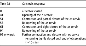
Although cervical or cervical-associated features such as muscle contractions and mucus characteristics that may contribute to between-ewe differences in sperm transport have not been identified, hysteroscopic observations have revealed that the deposition of semen with an angle-tipped insemination pipette in the os cervix initiates changes in cervical activity (King et al., Reference King, McKelvey, Ranilla and Robinson1998). These changes involve contractions of the anterior vaginal wall and the opening and closing of the os cervix, which appear to draw retrograde-flow semen from the anterior vagina into the cervical canal (Table 2).
An example of temporal changes in the activity of the os cervix following cervical artificial insemination (Data source: King et al., Reference King, McKelvey, Ranilla and Robinson1998)

Although oestrous synchronisation with a progestagen pessary does not appear to interfere with the natural pattern of cervical myoelectrical activity that occurs during spontaneous oestrus (Cavaco-Goncalves et al., Reference Cavaco-Goncalves, Marques, Horta and Figueroa2006), further hysteroscope-observed contractions of the os cervix occurred following exposure to the ram, natural mating and systemic oxytocin administration (King et al., Reference King, McKelvey, Ranilla and Robinson1998). In all cases, there is considerable between-ewe variation in the occurrence and strength of the cervical response, which perhaps may be a clue to why conception, following cervical deposition of frozen–thawed semen, seems to occur with apparent ease in some ewes but not others. The stimulatory effect of oxytocin is particularly interesting in that Houdeau et al. (Reference Houdeau, Raynal, Marnet, Germain, Mormede, Rossano, Monnerie and Prud'homme2002) found that a significant increase in oxytocin release only occurred following poor, as opposed to rapid, access to the cervix at insemination with the effect on uterine activity lasting for a mean ± s.e.m., of 15 ± 4 min following a difficult insemination compared with 2 ± 1 for an easy one. Although the stress response, as measured by the elevation in plasma cortisol was similar for both categories, it may be that the oxytocin-induced extension of uterine activity had an adverse effect on cervical sperm transport. Such a conclusion is in keeping with the adverse effect on conception noted by King et al. (Reference King, McKelvey, Dingwall, Matthews, Gebbie, Mylne, Stewart and Robinson2004) following systemic oxytocin administration before cervical insemination using frozen–thawed semen and the absence of a deleterious effect of the oxytocin on conception when the cervix was by-passed by laparoscopic intrauterine insemination. It is also in keeping with the data of Stellflug et al. (Reference Stellflug, Wulster-Radcliffe, Hensley, Cowardin, Seals and Lewis2001), which imply that a combination of exogenous oxytocin and cervical manipulation accentuates the oxytocin-mediated impairment of fertility following cervical insemination. Thus, King et al. (Reference King, McKelvey, Dingwall, Matthews, Gebbie, Mylne, Stewart and Robinson2004) suggested that, when using oxytocin for enhancing cervical dilation and os cervix activity, it may be advantageous to replace a single relatively large bolus dose of oxytocin, which results in an extended period of elevated oxytocin concentrations, with a series of smaller doses that more closely mimic, in number, amplitude and duration, the pulses in plasma concentrations that occur during natural oestrus (Sheldrick and Flint, Reference Sheldrick and Flint1981; Wathes et al., Reference Wathes, Guldenaar, Swann, Webb, Porter and Pickering1986; Gilbert et al., Reference Gilbert, Jenkins and Wathes1991).
Transcervical embryo recovery
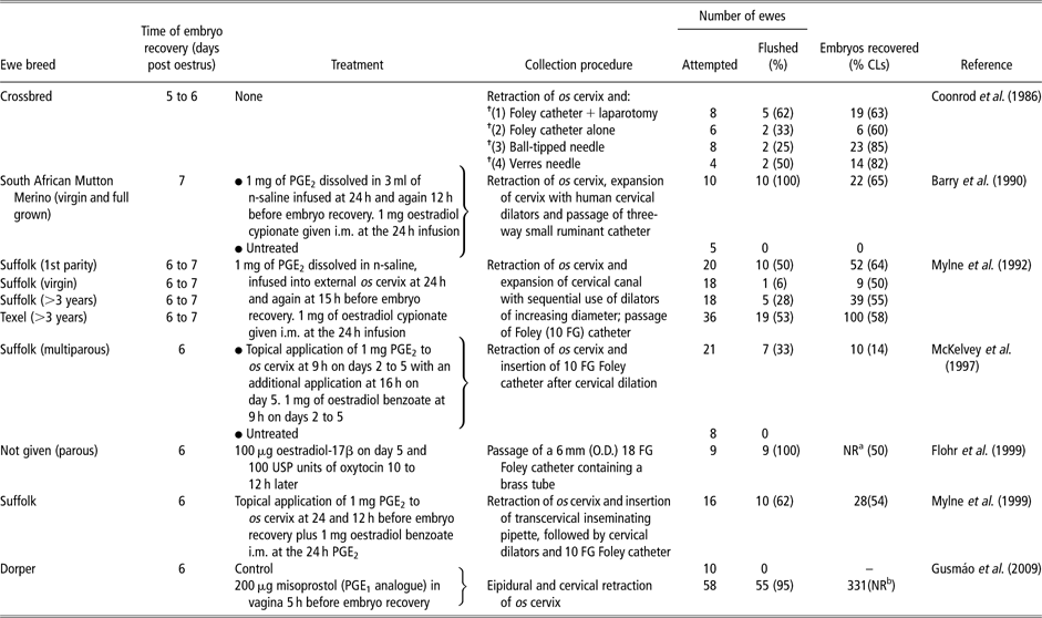
Coincident with the report by McKelvey et al. (Reference McKelvey, Robinson, Aitken and Robertson1986) of successful embryo recoveries from superovulated donor ewes by laparoscopy, Coonrod et al. (Reference Coonrod, Coren, McBride, Bowen and Kraemer1986) reported data on transcervical embryo recovery rates using either a Foley catheter or needle. No attempt was made to stimulate the relaxation of the cervix before embryo recovery. Subsequent studies by other researchers have involved a range of cervical relaxation treatments before attempted embryo recovery. Information on the treatments used and the results obtained in these studies, together with the data of Coonrod et al. (Reference Coonrod, Coren, McBride, Bowen and Kraemer1986), is set out in Table 3. Pre-treatment with systemic oestradiol combined with the topical application of prostaglandin (PGE2) to the os cervix resulted in a marked increase in the proportion of ewes for which the cervix was successfully traversed, and it did not seem to have an adverse effect on the percentage of embryos recovered (data of Coonrod et al., Reference Coonrod, Coren, McBride, Bowen and Kraemer1986v. data of others in Table 3). In ewes treated with oestradiol, systemic oxytocin appeared to be an effective and a much cheaper alternative to topical PGE2 (Flohr et al., Reference Flohr, Wulster-Radcliffe and Lewis1999v. others in Table 3). Data for an age effect on transcervical penetration were equivocal; Mylne et al. (Reference Mylne, McKelvey, Fernie and Matthews1992) recorded a much lower success rate in virgin as opposed to parous Suffolks, whereas Barry et al. (Reference Barry, van Niekerk, Rust and van der Walt1990) found no effect of age in South African Mutton Merinos. Although Flohr et al. (Reference Flohr, Wulster-Radcliffe and Lewis1999) achieved 100% success in traversing the cervix of parous ewes, without resorting to general anaesthesia, cervical retraction and physical dilation, and a 50% embryo recovery rate (range 0% to 100%), they indicated that their procedure was not suitable for nulliparous ewes. Their results for parous ewes however demonstrate the value of embryo collection transcervically from conscious animals. A skilled operator required only about 15 min from speculum insertion to ewe release. The technique also avoided the risk associated with the laparoscopic procedure, that of post-operative protrusion of the uterine endometrium (Nellenschulte and Niemann, Reference Nellenschulte and Niemann1992) at the catheter insertion point.
A summary of the treatments involved and the data obtained in studies investigating transcervical recovery of sheep embryos
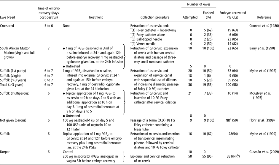
CL = corpora lutea.
aNumber not reported.
bCL not counted.
†15 to 20 flushes each of 5 to 10 ml.
In the report by Gusmáo et al. (Reference Gusmáo, Silva, Bittencourt, Martins, Fordiano and Barbosa2009) on the transcervical flushing of Dorper ewes, Table 3, an epidural was used and the mean ± s.e.m. time taken for the procedure was 30.1 ± 9.00 min or twice as long as that required by Flohr et al. (Reference Flohr, Wulster-Radcliffe and Lewis1999). However, the procedure was highly effective with 95% of ewes successfully flushed. Although corpora lutea were not counted, the mean ± s.e.m. of 6.02 ± 3.61 embyos recovered per ewe is impressive for a technique in which its repeated use is not hampered by the adhesions that accompany flushing via mid-ventral laparotomy.
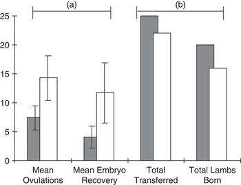
An important aspect of transcervical recovery is the quality and viability of the embryos. McEvoy et al. (Reference McEvoy, McKelvey, Robinson, Dingwall, Lindsay, King and FitzSimons1996) found no evidence of altered metabolic activity in terms of glucose uptake in morula-stage and blastocyst-stage embryos recovered transcervically from ewes treated with systemic oestradiol and topical PGE2 to dilate the cervix. For expanded blastocyst-stage embryos, glucose uptake and cell numbers were enhanced. Wulster-Radcliffe et al. (Reference Wulster-Radcliffe, Costine and Lewis1999) noted no adverse effect of systemic oestradiol (100 μg) and oxytocin (400 USP units) treatments on embryo survival at days 12 to 14. In fact, the treatment enhanced embryo development. Further evidence that the cervical dilating agents and the transcervical recovery procedure had no adverse effects is seen in the data of Mylne et al. (Reference Mylne, Dingwall, King, McKelvey and Robinson1999) in Figure 1. From 25 embryos recovered transcervically from seven ewes and transferred by laparoscopy, 20 lambs were born. In the case of embryos recovered from three ewes by mid-line laparotomy, 16 lambs were born from 22 transferred embryos.
(a) Mean (±s.e.m.) number of ovulations and embryos recovered for ewes flushed cervically (▪) or surgically (□) and (b) the effect of recovery method on total number of lambs born following laparoscopic embryo transfer (Data source: Mylne et al., Reference Mylne, Dingwall, King, McKelvey and Robinson1999).

Transcervical embryo transfer
Probably because of the relative ease with which embryos can be transferred by laparoscopy using neuroleptic anaesthesia with supplementary local anaesthesia and the high success rate for the procedure, little emphasis has been placed on their transcervical transfer. On days 12 to 14 of pregnancy, Wulster-Radcliffe et al. (Reference Wulster-Radcliffe, Costine and Lewis1999) recovered 54% of 28 embryos transferred via the cervix on days 5 to 6. The transfers had been achieved using 100 μg of oestradiol-17β and 400 USP units of oxytocin to dilate the cervix. A 17.5 cm long, 17-gauge semi-flexible steel tube with a 2 mm brass bulb tip was used to transfer the embryos. In contrast, in the absence of any pharmacological cervical dilating agents, we recorded a pregnancy rate of only 20% following the transcervical transfer of two embryos to each of 15 ewes, 5 to 6 days after oestrus (Robinson et al., Reference Robinson, King, Williams, Mitchell, Mylne and McKelvey1999) and the success rate was even lower when we used oestradiol and oxytocin to dilate the cervix. There was no evidence in Wulster-Radcliffe et al.'s (Reference Wulster-Radcliffe, Costine and Lewis1999) observations of an adverse effect of the oestradiol and oxytocin treatment on luteal progesterone concentrations or on the percentage of laparoscopically transferred embryos recovered on days 12 to 14 (12 of 16, 75% v. 22 of 32, 69% for treated v. control ewes). These higher percentages for the laparoscopic as opposed to the transcervical (54%) procedure, however, are important, particularly when transferring high-value embryos.
Cervical dilating agents
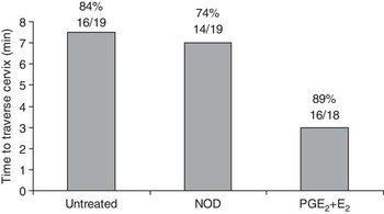
Many of the studies referred to in the preceding sections of this review involved the use of cervical dilating agents, notably systemic oestradiol and/or oxytocin, and topical application of prostaglandin E2 to the os cervix in the form of fixed-time infusions as described in the Mylne et al. (Reference Mylne, McKelvey, Fernie and Matthews1992) study referred to in Table 3. More recently, Candappa et al. (Reference Candappa, Bainbridge, Price, Hourigan and Bartlewski2009) have used a constant-release PGE2 (0.3 mg per hour) vaginal insert (Cervidil®) developed for use in women. Other dilating agents have also been studied. Robinson et al. (Reference Robinson, King, Williams, Mitchell, Mylne and McKelvey1999) tested the efficacy of nitric oxide for embryo recovery from Scottish Blackface ewes on days 5 and 6 post oestrus. The test compared the insertion of a 60 mg slow-release capsule of the nitric oxide donor, isosorbide-5-mononitrate, in the os cervix, 6 to 10 h before attempted cervical penetration with topical application of prostaglandin E2 (PGE2) to the os cervix at 24 and 12 h before attempted penetration plus 1 mg of systemic oestradiol (E2) given at the time of the first PGE2 treatment. Despite its ability to (i) stimulate blood flow, (ii) enhance prostaglandin F2α production (Ledingham et al., Reference Ledingham, Denison, Kelly, Young and Norman1999), (iii) induce an inflammatory reaction and (iv) accentuate the natural increase in nitric oxide synthase expression during human cervical ripening (Tschugguel et al., Reference Tschugguel, Schneeberger, Lass, Stonek, Zaghlula, Czerwenka, Schatten, Kaider, Husslein and Huber1999), the nitric oxide generated from the isosorbide-5-mononitrate did not improve penetration rates and did little to reduce penetration time. In contrast, the prostaglandin plus oestradiol treatment produced a dramatic reduction in penetration time (Figure 2) with full penetration into the uterine lumen being achieved within 1 min in 20% of the ewes.
The effect of topical treatment of the os cervix of Scottish Blackface ewes on days 5 and 6 post oestrus with nitric oxide donor isosorbide-5-mononitrate, or prostaglandin E2 (PGE2) in combination with oestradiol (E2), on the number and the percentage of ewes for which the cervix was successfully traversed and on the mean duration (in minutes) of successful attempts to traverse the cervix (Data source: Robinson et al., Reference Robinson, King, Williams, Mitchell, Mylne and McKelvey1999).

In a study of the effects of inserting, into the anterior vagina at progestagen pessary withdrawal, a wax suppository that released, over a 48-h period, 0.6 μg of human interleukin 8, Croy et al. (Reference Croy, Prudencio, Minhas, Ashkar, Galligan, Foster, Buckrell and Coomber1999) found enhanced neutrophil recruitment leading to cervical relaxation and increased penetration. In contrast, there was no effect on neutrophil recruitment or cervical dilation in dioestrous ewes. A notable feature of the cervical dilation following progestagen withdrawal (i.e. at oestrus) was its variation between ewes. Thus, complete cervical penetration was only achieved in two out of five oestrous ewes, prompting the authors to conclude that interleukin (IL; IL-8) alone is not a ‘useful clinical product’ for transcervical intrauterine insemination.
Mitchell et al. (Reference Mitchell, Robinson, King, McKelvey and Williams2002) investigated IL-8 gene expression in the luminal epithelium and fibroblastic cells of the cervix during both natural and artificially induced oestrous cycles. Expression was highest at oestrus and at day 5 of the oestrous cycle. Expression was also higher in ewes with a naturally occurring oestrus than in those artificially induced, either by progesterone pessaries and gonadotrophin injection or by prostaglandin F2α injection. The deposition of 0.2 ml of fresh semen in the os cervix, 36 h after progestagen withdrawal further enhanced expression. All of these observations demonstrate the reliance of IL-8 on naturally occurring events for eliciting its maximum inflammatory response and expression, which in turn limits its usefulness as a sole cervical dilating agent in a pre-programmed fixed-time artificial insemination programme.
In an attempt to identify pro-inflammatory agents to ameliorate the anti-inflammatory effects of oestrous-synchronising agents, Mitchell et al. (Reference Mitchell, Robinson, King and Williams2005) investigated the proteinase-activated receptors (PAR-1 and PAR-2), which have an important role in the early stages of inflammation (Macfarlane et al., Reference Macfarlane, Seatter, Kanke, Hunter and Plevin2001). Both receptors were expressed in the luminal epithelium of the cervix throughout the oestrous cycle and the level of expression was highest at oestrus. Unlike their suppression of IL gene expression, the oestrous-synchronising protocols (progesterone plus gonadotrophin and prostaglandin F2α) did not inhibit the expression of the PAR receptors at oestrus. Indeed, PAR-2 expression at oestrus was enhanced by progesterone synchronisation, and both synchronising protocols (progesterone and prostaglandin) enhanced both PAR-1 and PAR-2 receptor expression during the luteal phase, implying that there may be a role for PAR agonists in the initiation of the inflammatory response that leads to dilation.
The natural partial relaxation of the cervix that occurs at oestrus is generally regarded to be the result of changes in reproductive hormones, notably progesterone, oestradiol and oxytocin, at this time. Receptors for all three hormones are abundantly expressed in the cervix at oestrus (Zhao et al., Reference Zhao, Williams, Hannah, Ross, McKelvey and Robinson1999 for progesterone and oestradiol; Matthews and Ayad, Reference Matthews and Ayad1994 and Ayad et al., Reference Ayad, Leung, Parkinson and Wathes2004 for oxytocin). There is now evidence that gonadotrophins are also involved in cervical relaxation at oestrus, as intracervically administered follicle stimulating hormone (FSH) either alone or in combination with prostaglandin E1 (Misoprostol) resulted in increased penetrability (Leethongdee et al., Reference Leethongdee, Khalid, Bhatti, Ponglowhapan, Kershaw and Scaramuzzi2007). Furthermore, judicious timing of their administration (24 and 48 h after progestagen pessary withdrawal for the FSH and prostaglandin E1, respectively) advanced the time of maximum cervical dilation from 72 h after pessary withdrawal in untreated ewes to coincide more closely with the preferred optimum time for insemination of 54 h. The amounts of FSH and Misoprostol administered per ewe intracervically were 2 and 1 mg, respectively.
Mechanisms and pathways involved in cervical relaxation
Advances in understanding the mechanisms involved in cervical relaxation come from studies on a wide range of species, including laboratory animal and human as well as the farmed species (see reviews by Leppert, Reference Leppert1995 and Timmons et al., Reference Timmons, Akins and Mahendroo2010). They also range from the anatomical, histological and biochemical (Moré, Reference Moré1984; Dobson, Reference Dobson1988) to the molecular (Read et al., Reference Read, Word, Ruscheinsky, Timmons and Mahendoo2007) studies. Species differences in terms of the hormonal mechanisms involved in the relaxation process add complexity. For example, while relaxin softens the human cervix in late pregnancy, its relaxation properties as measured by the improved ease in passing a catheter through the cervix of oestradiol-treated ovariectomised ewes (Nemec et al., Reference Nemec, Loskutoff, Lesley, Fields, Bowen and Kraemer1988) or ewes that had lambed more than 30 days previously (Akinbami et al., Reference Akinbami, Meredith, Warren, Anthony and Day1990) appear to have been too minor to warrant further studies of its use for transcervical insemination and embryo transfer in ewes. Nonetheless, improved understanding of the molecular processes involved in remodelling of the extracellular matrix of the cervix during its relaxation is revealing that they are highly conserved in species as anatomically different as humans and mice (Timmons et al., Reference Timmons, Akins and Mahendroo2010). They may therefore yet provide useful information for improving transcervical insemination in ewes.
The major component of the cervix is the extracellular matrix consisting of collagen (Types l, lll and lV) fibres and elastin, which confer tensile strength and elasticity, respectively. These are bound together by a matrix whose main constituents are proteoglycans, glycosaminoglycans and water (Dobson, Reference Dobson1988). The cellular component, which is home to the hormone changes that drive the remodelling of the extracellular matrix needed for cervical relaxation, comprises epithelial and smooth muscle cells and fibroblasts. Much of the early research on cervical relaxation concentrated on the nature of the anatomical changes that occur in the remodelling process and the importance of quantitative changes in the rates of synthesis of glycosaminoglycans in conferring plasticity. For example, hyaluronic acid, the main glycosaminoglycan in the ewe cervix, was shown to be present in greater abundance in late-pregnant than early-pregnant ewes. Its high affinity for water and enhanced accumulation between the collagen fibrils accompanies late-pregnancy cervical softening and ripening (Dobson, Reference Dobson1988). In a recent study, Perry et al. (Reference Perry, Haresign, Wathes and Khalid2010a) found that intracervical application of 25 mg of the low-molecular-weight form of hyaluronan at either 50 or 52 h after progestagen pessary withdrawal and PMSG injection in Welsh Mountain ewes, increased the depth of cervical penetration from 1.2 to 3.7 cm within 2 to 4 h.
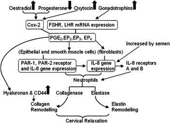
All the evidence suggests that the hyaluronan content of the cervix is enhanced by the high-oestrogen, low-progesterone status of the ewe during the pre-LH surge phase. An accompanying feature at this time is the abundant expression of the hyaluronan receptor, CD44, in the epithelial layer (Perry et al., Reference Perry, Haresign, Wathes and Khalid2010b). In view of the central role that peri-oestrous endocrine changes play in the collagen and elastin remodelling processes that promote cervical relaxation, the research emphasis in ewes in recent years has been on improving understanding of their mechanistic pathways (Mitchell et al., Reference Mitchell, Robinson, King, McKelvey and Williams2002 and Reference Mitchell, Robinson, King and Williams2005; Kershaw et al., Reference Kershaw, Scaramuzzi, McGowan, Wheeler-Jones and Khalid2007; Leethongdee et al., Reference Leethongdee, Khalid, Bhatti, Ponglowhapan, Kershaw and Scaramuzzi2007 and Reference Leethongdee, Kershaw-Young, Scaramuzzi and Khalid2010; Kershaw-Young et al., Reference Kershaw-Young, Khalid, McGowan, Pitsillides and Scaramuzzi2009 and Reference Kershaw-Young, Scaramuzzi, McGowan, Pitsillides, Wheeler-Jones and Khalid2010). An overview of the findings from these studies is illustrated in Figure 3.
Overview of recent findings, in the ewe, on endocrine stimulation of cervical relaxation at oestrus. Cox-2 = cyclooxygenase-2; FSHR = follicle-stimulating hormone receptor; LHR = luteinizing hormone receptor; PGE2 = prostaglandin; EP = prostaglandin E receptors; PAR = proteinase-activated receptor; IL = interleukin; CD44 = hyaluronan receptor.

The overview incorporates the recent observation that gonadotrophins play a permissive role in the steroid-activated cervical relaxation process and that judicious timing of intracervical FSH administration may be a useful practical method for ensuring that maximum cervical relaxation coincides with the optimum time for transcervical insemination (Leethongdee et al., Reference Leethongdee, Khalid, Bhatti, Ponglowhapan, Kershaw and Scaramuzzi2007 and Reference Leethongdee, Kershaw-Young, Scaramuzzi and Khalid2010). The overview also includes the role of IL and the inflammatory response in cervical softening and relaxation. Mitchell et al. (Reference Mitchell, Robinson, King, McKelvey and Williams2002) observed that IL-8 gene expression in the luminal epithelium of the cervix was highest at oestrus and at day 5 of the oestrous cycle, this latter time being coincident with increased oestradiol concentrations coincident with the first post-ovulatory wave of ovarian follicular activity. Conveniently, it is also coincident with the preferred time for embryo recovery and transfer. It is also close to the time when Audicana et al. (Reference Audicana, Aughey and O'Shaughnessy1998) observed enhanced sensitivity of the cervix, in terms of collagen composition, to PGE2. In so far as PGE2 is a potent EP3 receptor agonist that, in turn, is involved in the activation of immune cells, these authors speculated that by acting on the EP3 receptor, PGE2 may stimulate neutrophil-induced degradation of the extracellular matrix of the cervix.
The studies on the PARs (Figure 3) were prompted by the finding that IL-8 gene expression is significantly decreased by the synchronisation of oestrus via either progestagen pessaries or prostaglandin (PGF2α) injection (Mitchell et al., Reference Mitchell, Robinson, King, McKelvey and Williams2002); yet oestrous synchronisation is a practical necessity for fixed-time artificial insemination. Moreover the cost of IL for cervical relaxation would prohibit its widespread use in practice. In common with IL-8, the PARs, notably PAR-1 and PAR-2 are also important in the initiation of the inflammatory response. Unlike IL-8, oestrous synchronisation did not inhibit PAR-1 or PAR-2 expression at oestrus; indeed in the case of progesterone pessary-mediated synchronisation, their expression was increased. Moreover both methods of oestrous synchronisation increased the expression of PAR-1 and PAR-2 during the subsequent luteal phase, coincident with the preferred time for embryo recovery and transfer (Mitchell et al., Reference Mitchell, Robinson, King and Williams2005). Unlike the normal ligand–receptor interaction that occurs in other G-protein-coupled receptors, the PARs are irreversibly activated by extracellular proteases, which cleave the N-terminus of the receptor, which acts as a tethered ligand binding to and activating the cleaved receptor (Vu et al., Reference Vu, Hung, Wheaton and Coughlin1991). Readily available agonists make the PARs attractive agents for studies of their effect on cervical penetration. At the same time, the rapidly expanding database for the alterations in cervical tissue gene expression, albeit mostly in relation to ripening and relaxation during pregnancy and at parturition in laboratory species (Read et al., Reference Read, Word, Ruscheinsky, Timmons and Mahendoo2007), may provide new avenues of investigation that are relevant to the ewe at oestrus and during the early luteal phase.
Conclusions and perspectives
The benefits of artificial insemination and embryo transfer to genetic improvement in the cattle industry, made possible by the relative ease in traversing the bovine cervix, have kept alive the desire to, similarly, make the cervix a practical route for intrauterine insemination and embryo transfer in sheep. Advances have been made in the science of ovine cervical relaxation and its pharmacological stimulation with associated improvements in physical penetration that have facilitated transcervical embryo recovery and that have provided optimism for success in transcervical artificial insemination. At the same time, progress is being made in improving semen processing and in reducing the damaging effects of freezing and thawing on sperm viability and membrane integrity. For semen transport from collection stations to user farms, short-term liquid storage at 4°C to 5°C, rather than the generally recommended 15°C can give acceptable pregnancy rates up to 24 h after semen collection (O'Hara et al., Reference O'Hara, Hanrahan, Richardson, Donovan, Fair, Evans and Lonergan2010). However, the recent study by Gundogan et al. (Reference Gundogan, Yeni, Avdatek and Fidan2010) on DNA integrity and oxidative stress indicates that deterioration in sperm quality during low-temperature liquid storage is greater at a concentration suitable for intrauterine insemination (25 × 106 spermatozoa per ml) than cervical insemination (100 × 106 per ml). Of course, 24 h is still only a tiny fraction of the time that semen can be stored in female reptiles, amphibians, fish and birds, prompting Holt and Lloyd (Reference Holt and Lloyd2010) to express surprise that the mechanisms used by such species have not been investigated by those researching the long-term storage of fresh semen. There has also been success in producing pre-sexed offspring (86 of 93) from superovulated ewes, albeit following laparoscopic intrauterine insemination rather than deposition in the os cervix (De Graaf et al., Reference de Graaf, Beilby, O'Brien, Osborn, Downing, Maxwell and Evans2007a). The goal now is to achieve this through transcervical insemination by creative science, which builds on the advances in stimulating cervical relaxation and in improving semen quality that have accrued from applied through to molecular research. The challenge for the combined research disciplines is to bring reliability and robustness to what are currently promising but, in a number of respects, still fragile techniques.
Acknowledgements
Research carried out at the Scottish Agricultural College and at the Rowett Institute of Nutrition and Health, University of Aberdeen, and referred to in this review, was supported by the Scottish Government Rural and Environment Research and Analysis Directorate (RERAD) and by the UK Meat and Livestock Commission.