We present the case of girl with a history of unrestrictive perimembranous ventricular septal defect, severe valvular and subvalvular aortic stenosis, and type B interrupted aortic arch.
The patient underwent Damus-Stensel-Kaye surgery and aortic arch repair as a neonate. The status of the patient after surgery was uneventful. At 30-month-old, the child represented with arterial hypertension. There were no signs of re-coarctation.
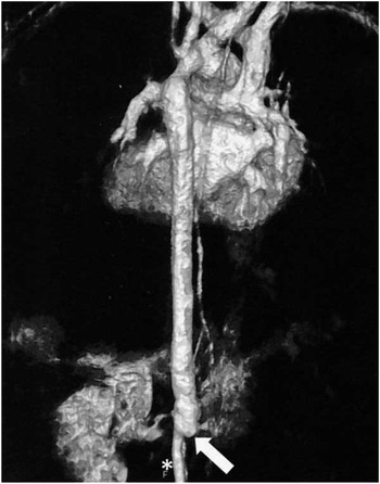
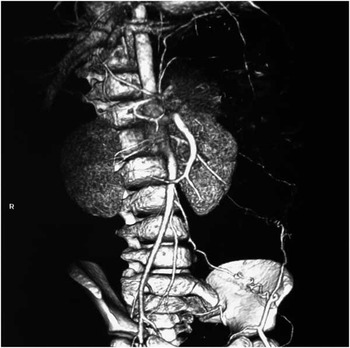
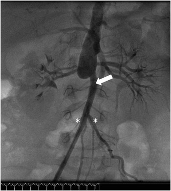
A magnetic resonance angiography of the aorta showed a right renal artery stenosis and a dilated sac-like termination below the renal arteries. The abdominal aorta continued through a smaller calibre vessel emerging from its anterior surface (Fig 1), giving origin to both iliac arteries. A CT angiography confirmed this diagnosis (Fig 2) and an angiography was performed, which confirmed the aortic malformation and right renal artery duplication, with diffuse hypoplastic arteries (Fig 3). An isotopic renal scan revealed a decrease in right kidney function (40%). Screening for systemic diseases was negative. Arterial hypertension was controlled pharmacologically and the child remains asymptomatic, and thus surgical bypass has not yet been considered.
Magnetic resonance angiography, posterior view, showing the abdominal aorta terminating in a larger sac-like structure (arrow) and its continuation through a smaller calibre vessel (*). Perfusion of the right kidney is decreased.

CT angio-scan, confirming the diagnosis of abdominal aorta malformation.

Aortography, anterior view, confirmed the aortic malformation, after the origin of the renal arteries, and a collateral vessel (arrow) arising from the anterior surface of the aorta, and from which both common iliac arteries emerged (*). There is duplication and hypoplasia of right renal vessels.

This case is a very uncommon association, and its origin is not clear: congenital or acquired. A dissection/thrombosis of the distal part of the aorta could have been the trigger, as the patient had a femoral arterial line inserted at the time of cardiac surgery. Mid-aortic syndrome, characterised by thoracic or abdominal aortic narrowing or occlusion, and often associated with stenosis of efferent vessels,Reference Levart and Klokocovnik 1 including the renal arteries, could also be a possibility, although this is not a typical presentation.
Acknowledgements
The authors acknowledge the support of the Hospital de Santa Cruz (Centro Hospitalar Lisboa Ocidental, Lisboa) for providing supporting images.
Financial Support
This research received no specific grant from any funding agency, commercial, or not-for-profit sectors.
Conflicts of Interest
None.