Introduction
Currently, many investigations indicate that silk fibroin (SF), a fibrous protein present in silkworm cocoons (Bombyx mori), is a promising source of biotechnological and biomedical materials because of its properties, including good biocompatibility, permeability of oxygen and water vapor, biodegradability, and minimal inflammatory reaction [Reference Arai, Freddi, Innocenti and Tsukada1]. SF has been used to produce a variety of biomaterials, usually mixed with other polymers (blends), in injectable form: hydrogels and microspheres; transdermal: microneedles; implants: sponges, films, porous structures, and nanofibers, among others [Reference Yucel, Lovett and Kaplan2]. The processing of silkworm cocoons in the laboratory starts with the process of degumming or removing sericin, a protein present on silkworm cocoons to which are assigned the problems of silk biocompatibility [Reference Altman, Diaz, Jakuba, Calabro, Horan, Chen, Lu, Richmond and Kaplan3]. To solubilize the SF yarn, a dispersion is prepared, usually with the use of concentrated salt solutions in the presence of organic solvents [Reference Ki, Um and Park4]. Thus, both degumming and dispersion are critical steps in the development of fibroin-based materials, which could influence the final product characteristics. Finally, the SF dispersion is dialyzed with the purpose of removing the ions present in the dispersion and obtain an aqueous dispersion with which the fibroin-based materials are prepared [Reference Kundu, Kurland, Bano, Patra, Engel, Yadavalli and Kundu5].
Our research group observed experimental differences in the processing of SF films with cocoons from the same batch but at different periods of time (years): During the process of preparation of SF films by casting using old cocoons, it was observed a decrease of solubility, an excessive formation of precipitates and agglomerates within the dialysis tube, and a longer time to complete film formation (solvent evaporation). These experimental observations motivated the present work, because any difference in the physicochemical properties of fibroin dispersion could affect the final properties of fibroin-based materials (films, hydrogels, particles, and sponges, among others) for various proposes, from biotechnology to biomedical applications. Studies by Ahmed and Darwish [Reference Ahmed and Darwish6] mention that SF deteriorates as a result of exposure to environmental conditions such as light, temperature, and humidity. Koperska et al. [Reference Koperska, Pawcenis, Bagniuk, Zaitz, Missori, Łojewski and Łojewska7] experimentally investigated the influence of these factors on degummed SF fibers through artificial aging procedures. However, no studies have yet reported the importance of assessing the age of the cocoons, emphasizing the novelty of this work, because it could cause changes in the properties of fibroin during processing and influence on the characteristics of the final product.
It is known that some natural polymers show significant changes over time because of physical and chemical deterioration or aging [Reference Wang, Rudym, Walsh, Abrahamsen, Kim, Kim, Kirker-Head and Kaplan8]. SF, in addition to deterioration by aging, may suffer as a whole protein, a process of denaturation either by adding solvents, variations in pH, or by mechanical means. Furthermore, the SF amino acids such as glycine, alanine, serine, and tyrosine are photo-oxidized in the presence of ultraviolet light [Reference Gong and Yang9]. When degummed, SF is exposed to degradation by light and oxygen, its yarns easily become yellow with time [Reference Koperska, Lojewski and Lojewska10]. The presence of the yellow color is due to the tyrosine reaction: There is a decrease in the percentage of tyrosine from 6.16 to 2.72% depending on the time of exposure to light of SF [Reference Ahmed and Darwish6]. Mamedov et al. [Reference Mamedov, Akta, Cantürk, Aksakal, Alekperov, Bülbül, Yilgin and Aslanov11] identified the free radicals from SF yarns by the ESR technique (electron spin resonance). The radicals are formed by the breaking of C–C bonds, when induced by UV radiation, whereas the variations in the amount of free radicals depend on the humidity, mechanical stress, and temperature to which the wires are exposed.
Considering the above, our main objective was to study the influence of age of cocoons produced by the silkworm Bombyx mori on their properties during all stages of processing SF films. In this work, we used two batches of cocoons of different ages, named C0 (aged zero, fresh cocoons) and C6 (six-year-old cocoons), stored in hermetic manner, away from sources of heat and light. The evaluations were done simultaneously in the batches in two stages: (i) evaluation of the dialyzed dispersions of SF and (ii) evaluation of the films generated from the dialyzed dispersions. It is noteworthy that, in this study, the influence of age is the sum of aging and batch production factors, and inferred that the aging factor has a greater influence on the evaluation, although it is likely also to influence the production batch.
Results and discussion
SF dispersion
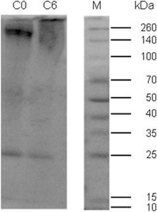
The molecular weight of the dialyzed dispersion of SF prepared from C0 and C6 cocoons is shown in Fig. 1. There were two bands in the sample C0, at ∼26 and 260 kDa, which could be associated to the light and heavy chains of SF, typically present at 25 and 350 kDa, respectively [Reference Jin and Kaplan12].
SDS-PAGE analysis of SF dialyzed dispersion prepared from C6 and C0 cocoons.

It is also possible to observe a diffuse band (which may be caused by the breaking of peptide bonds), above 260 kDa for C6 sample. These results indicated that SF heavy chain was probably degraded into polypeptides of lower size during dissolution process. Yamada et al. [Reference Yamada, Nakao, Takasu and Tsubouchi13] and Cheng et al. [Reference Cheng, Wang, Tao, Xia and Xu14], in their studies about the differences in degummed and solvent systems of SF using SDS-PAGE, show that CaCl2:CH3CH2OH:H2O causes degradation of the SF molecular chains.
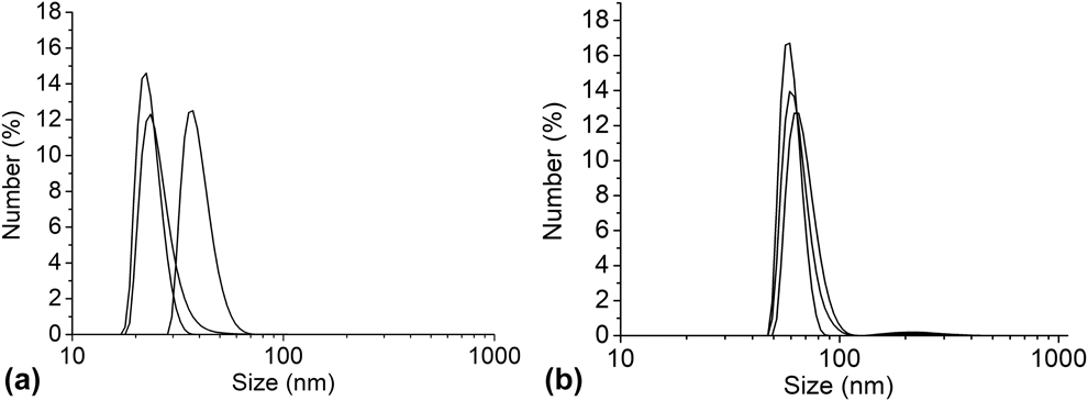
Previous study by Lu et al. [Reference Lu, Zhu, Zhang, Zhang, Zhang and Kaplan15] proposed that the SF molecules in aqueous dispersion form colloidal aggregates of spherical shape due to hydrophobic interactions of its chains. Under this argument, the particles analyzed by dynamic light scattering (DLS) were called colloidal aggregates. The particle size distribution by number to the sample C6 [Fig. 2(a)] indicates that, on average, 13% of the total colloidal aggregates measured 27.42 ± 6 nm. The value of the polydispersity index was 0.427. For the dispersions prepared from cocoons C0 [Fig. 2(b)], the particle size curves by number present two intervals. The first interval between 47.7 and 109.6 nm indicates that, on average, 14.6% of the total aggregates measured 60 ± 2 nm.
Particle size distribution by number of the SF dialyzed dispersion prepared from cocoons C6 (a) and C0 (b).

The second range, from 140.0 to 373.0 nm, indicates that, on average, 0.15% of total aggregates measured 233.7 ± 14.4 nm. The PDI was 0.412 confirming that the system is polydispersed.
There is a phenomenon called Ostwald aging [Reference Squier16], which is the main mechanism for the destabilization of nanoemulsions; it shows polydispersity of emulsions and solubility difference between the particles of large and small sizes. In this phenomenon, there is an increase in the size of the larger particles by agglomeration of smaller particles through electrostatic forces.
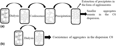
This phenomenon could be applied in the dialyzed dispersions of SF, to try to understand what happens in the system. Figure 3 shows an illustration of how the SF colloidal aggregates within the dispersions would be formed and distributed from the results obtained by DLS analysis. Our hypothesis is that, for the dispersions prepared from cocoons C6, SF molecules are solvated by ions and, during dialysis, ions are removed and the molecules start to form the colloidal aggregates [Fig. 3(a)]. These colloidal aggregates form big agglomerates with time, which are removed during sample dilution for DLS, remaining only the small agglomerates. The agglomerate is characterized by the irregular shape of the surface of the (non-spherical) particles. Therefore, this could explain the irregularity of the intensity in the dispersions prepared from cocoons C6. For the case of the dispersions prepared from cocoons C0 [Fig. 3(b)], there was coexistence of large and small colloidal aggregates. Thus, apparently, the age of the cocoon influences the size distribution and formation of colloidal aggregates in the SF-dialyzed dispersions.
Illustrative scheme of the possible formation of aggregates in the dispersions prepared from cocoons C6 (a) and C0 (b).

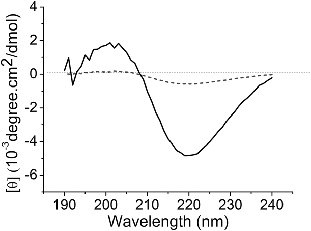
Figure 4 shows the circular dichroism (CD) spectra of dispersions prepared from C6 and C0 cocoons. The spectrum of dispersion from C6 showed a band located at 219 nm, starting the negative value at 208 nm. This spectrum is typical of β-sheet secondary structures. For the dialyzed dispersion, C0 spectrum refers to a conformation of random structures.
CD spectra of the dialyzed dispersions of SF prepared from cocoons C6 (solid line) and C0 (dashed line).

Molar ellipticity values were located at −4.83 × 10⁻³ degree cm2/dmol to C6 dispersion. These values indicate the presence of higher amounts of β-sheets in the C6 samples that may be related to the concentration of the samples, because in the C6 dialyzed dispersions, agglomerates were observed macroscopically and were removed by filtration, decreasing the final concentration of the dispersion.
Furthermore, we hypothesize that the agglomerates formed could present β-sheet structures. Therefore, the results for the C6 dispersion samples could be attributed to an early formation of β-sheet conformation [Reference Li, Zhou, Shao, Xie, Chen, Wang, Chunyu and Yu17].
SF films
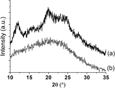
In this section, we evaluated SF films. X-ray diffraction (XRD) is shown in Fig. 5, where it is possible to see that the film prepared from cocoons C0, Fig. 5(b), showed amorphous structure, with absence of crystallinity peaks. Nonetheless, the film prepared from cocoons C6, Fig. 5(a), showed diffraction intensity halos at 2θ of 12.0 and 19.9°, which could indicate the organization of molecular structure, with interplanar distance of 7.3 and 4.4 Å, respectively. The same film C6 showed weaken halos observed at 16.1 and 24.7°, whose interplanar distances are 5.4 and 3.6 Å, respectively.
X-ray diffractograms of SF films prepared from cocoons C6 (a) and C0 (b).

It is assumed from these results that the film prepared from cocoons C6 showed higher crystallinity, probably because of the presence of agglomerates that formed again and rapidly during the preparation of the film, and these agglomerates are likely to have β-sheet structures, which are more organized structures, thus causing a higher crystallinity of the film. The degree of crystallinity for SF films was calculated from the intensity of the halos and showed values of 19.3% and 0.0% for films prepared from cocoons C6 and C0, respectively.
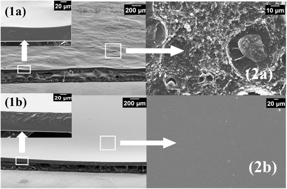
It can be observed an irregular and rough surface of the film prepared from cocoons C6, with formation of microstructures, as shown in Figs. 6(2a). The fracture surface [Figs. 6(1a)] was dense and homogenous with a thickness of 46.7 ± 1.8 µm. For the SF films prepared from C0 cocoons, the surface [Figs. 6(2b)] and fracture surface [Figs. 6(1b)] presented smooth and homogeneous morphology, with a thickness of 81.2 ± 1.2 µm.
SEM micrographs of fracture surface [Reference Arai, Freddi, Innocenti and Tsukada1] and surface [Reference Yucel, Lovett and Kaplan2] of the films prepared from C6 (a) and C0 (b) cocoons.

The film prepared from cocoons C6 had a smaller thickness compared to the film prepared from cocoons C0, probably because of the lower concentration of C6 sample. The preparation of C6 and C0 films started simultaneously and with the same concentrations of salt dispersions. There was a decrease in the final concentration of C6 film (concentration of 24.1 g/L and thickness of approximately 45 µm) caused by the poor solubility and formation of precipitates in the dialysis tube, when compared to the C0 film (concentration of 26.3 g/L and thickness of approximately 81 µm). In the latter case, the dispersion was complete and there was no formation of precipitates, so the raw material (silk fibroin) was not discarded and the concentration of the dispersion was higher, thus resulting in a higher thickness.
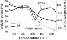
Figure 7 shows the thermogravimetric curves for SF films prepared from C6 and C0 cocoons.
Thermogravimetric curves of SF films prepared from C6 and C0 cocoons and their derivative.

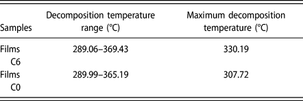
The maximum temperature of degradation from Fig. 7 can be evident in Table I. The C6 film showed a higher degradation temperature of 330.19 °C compared to the C0 film (307.72 °C). These values are associated with the breakdown of the side chain of residual amino acids [Reference Um, Kweon, Park and Hudson18]. This variation may be related to the higher crystallinity of the C6 films, as observed on XRD analysis.
Data from thermal analysis of SF films prepared from cocoons C6 and C0, obtained from thermogravimetric and its derivative curves.

Conclusions
The results obtained in this study confirm that cocoons age is a key factor to be considered when preparing SF-based materials. Differences on molecular weight, particle size, and secondary structure conformation were observed for the dialyzed dispersions of SF. Furthermore, SF films prepared from 6-year-old cocoons showed higher crystallinity and higher thermal resistance than SF films prepared from fresh cocoons. Finally, it can be concluded that all studies addressing SF-based materials should consider and state the age of the cocoons, to obtain well-defined and reproducible materials for several applications, from biotechnology to biomaterials.
Materials and methods
Cocoons degumming
Cocoons of Bombyx mori silkworm were supplied by Bratac SA (Bastos, SP, Brazil). Fresh and old cocoons were stored into polystyrene bags at 23 °C, relative humidity (RH) of approximately 43%, and away from light exposure. In this work, six-year-old cocoons were called C6, whereas fresh cocoons were called C0. All the cocoons, C0 or C6, received the same treatment, briefly: cocoons were cut using scissor and washed with water, boiled in a 1 g/L Na2CO3 solution at 85 °C (to three degumming cycles, 30-min changes), and then rinsed thoroughly with water to extract sericin proteins. After degumming, the SF fibers were air-dried at room temperature (23 °C, relative humidity = 43%). Each degumming treatment was conducted in triplicate for C0 and C6 cocoons.
Preparation of SF dispersion
The SF fibers degummed were stretched manually along with tweezers, milled, and then dissolved in a ternary solvent system of CaCl2:CH3CH2OH:H2O (1:2:8 molar ratio) at 85 °C for 90 min to obtain SF concentration of ∼50 g/L. The dispersion was prefiltered and about 6 mL of the filtrate was added into dialysis tube (3500 MWCO). The SF dispersion was dialyzed against ultrapure water, with water changes every 24 over 72 h. Once dialyzed, the SF dispersion concentration was about 25 g/L. Each preparation was conducted in triplicate for C0 sample and C6 sample.
Film formation
Films were prepared by casting using 20 mL of SF dispersion, poured into polystyrene dishes (d = 8.5 cm) and dried at room temperature (25 °C), until no mass variation was observed. Each film formation from C0 and C6 sample was conducted in triplicate.
Characterization of SF dispersion
The molecular weight of SF dispersion prepared from C0 or C6 cocoons was determined with sodium dodecyl sulfate (SDS)–polyacrylamide gel electrophoresis (PAGE) and 12% gradient gel for electrophoresis. Supernatant was mixed with an equivalent amount of buffer:sample 1:1. Then, 20 µL of supernatant was used as sample. The gel was stained with Coomassie Brilliant Blue R-250.
Particle size of SF dispersion was analyzed by dynamic light scattering (DLS). Samples of SF dispersion were diluted with ultrapure water to 1 g/mL concentration and analyzed by Zetasizer Nano ZS of Malvern Instruments (Malvern, UK).
Circular dichroism (CD) experiments were conducted using spectropolarimeter model Jasco J-720 with N2 gas. The spectra were recorded from 190 to 240 nm, accumulation of eight scans. The scan speed was 20 nm/s with a quartz cuvette of 0.05 cm path length. The mean residue molar ellipticity values [θ] were expressed in degree cm2/dmol and were calculated using Eq. (1);
 $$\theta = {{{\theta _{{\rm{obs}}}} \times 100 \times {M_{\rm{w}}}} \over {l \times c \times n}}\quad ,$$
where θobs is the observed ellipticity value in degrees, “l” is the optical path in cm, “c” is the concentration in g/mL, “M w” is the molecular mass, and “n” is the number of residues in the protein.
Each characterization of SF dispersion was conducted in triplicate.
Characterization of SF films
Films were structural characterized by X-ray diffraction (XRD), with Analytical X-Ray (X’Pert-MPD, Philips) operating at 40 kV and 40 mA with a Cu Kα target. The samples were run at diffraction angles (2θs) from 10 to 60° with a step size of 0.02° (2θ) and a scanning rate of 0.0166°/s.
Scanning electron microscopy (SEM) was conducted on the surface and fracture surface. Morphology of SF films were imaged with a Leo 440i (LEO Electron Microscopy, Cambridge, UK) with an acceleration voltage of 10 kV. SF films thickness were measured from SEM images using ImageJ software (National Institute of Health, Bethesda, MD). The final concentration of SF films was determined by pouring 5 mL of the dispersion into polystyrene Petri dishes of 3.5 cm in diameter. After complete evaporation of the solvent under room conditions within the hood (23 °C and 33% RH) for a period of 48–120 h, the Petri dishes were weighed again and the remaining dry mass value was obtained.
Thermogravimetric analysis (TGA) was performed with SHIMADZU model TGA-50M (Shimadzu, Kyoto, Japan), in a temperature range of 25–550 °C with a ramp rate of 10 °C/min and O2 atmosphere flow of 50 mL/min.
Acknowledgments
The authors thank Coordenação de Aperfeiçoamento de Pessoal de Nível Superior—Brasil (CAPES)—Finance Code 001, for financial support.