1. Introduction
1.a. Rhachiberothidae
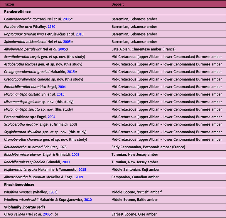
Rhachiberothidae, also known as thorny lacewings, is a small group of Neuroptera consisting of 13 extant species in three genera, with 17 extinct taxa, within two subfamilies (Table 1) (Aspöck & Mansell, Reference Aspöck and Mansell1994; Aspöck & Aspöck, Reference Aspöck and Aspöck1997; Makarkin & Kupryjanowicz, Reference Makarkin and Kupryjanowicz2010; Makarkin, Reference Makarkin2015a; Nakamine & Yamamoto, Reference Nakamine and Yamamoto2018; Oswald, Reference Oswald2019). Of note, the distribution of modern rhachiberothids is restricted to sub-Saharan Africa, with records from Ethiopia, Zimbabwe, Angola, Namibia and South Africa (Aspöck & Aspöck, Reference Aspöck and Aspöck1997); however, they are relatively abundant in various Cretaceous amber deposits in the Northern Hemisphere, with only a few fossil records from some Eocene European ambers (Table 1; see following section). Adult rhachiberothids are small in size, but they are active predators, having specialized raptorial forelegs. However, little other ecological information is available, except that they are generally collected at high altitudes (400–1800 m, mostly above 1000 m; Aspöck & Mansell, Reference Aspöck and Mansell1994; Aspöck & Aspöck, Reference Aspöck and Aspöck1997).
List of the fossil Rhachiberothidae of the world

a This amber is contemporaneous with Baltic amber (Jarzembowski, Reference Jarzembowski1999).
This family is an enigmatic lineage of insects whose phylogenetic relationships and systematic status have long been debated (Aspöck & Mansell, Reference Aspöck and Mansell1994). Rhachiberothidae was originally established as a subfamily of Berothidae because of similar venational characters on the fore- and hindwings (Tjeder, Reference Tjeder, Hanström, Brinck and Rudebeck1959). Since then, several authors followed this taxonomic treatment and placed it within Berothidae (e.g. Winterton et al. Reference Winterton, Hardy and Wiegmann2010; Makarkin, Reference Makarkin2015a). Nevertheless, two recent extensive studies refuted this hypothesis, and assigned family-rank status for this taxon (Engel et al. Reference Engel, Winterton and Breitkreuz2018; Winterton et al. Reference Winterton, Lemmon, Gillung, Garzon, Badano, Bakkes, Breitkreuz, Engel, Lemmon, Liu, Machado, Skevington and Oswald2018). In the present study, we tentatively follow and maintain the familial status of Rhachiberothidae.
The phylogenetic relationships among Rhachiberothidae and related groups are controversial. Rhachiberothids can be recognized as a sister taxon to either Berothidae (Aspöck & Mansell, Reference Aspöck and Mansell1994; Aspöck et al. Reference Aspöck, Plant and Nemeschkal2001, Reference Aspöck, Haring and Aspöck2012) or Mantispidae (Liu et al. Reference Liu, Winterton, Wu, Piper and Ohl2015; Engel et al. Reference Engel, Winterton and Breitkreuz2018). In a more recent molecular phylogenetic study, the relationship of (Mantispidae + [Rhachiberothidae + Berothidae]) was supported (Song et al. Reference Song, Li, Zhai, Bozdoğan and Yin2019). These families, and the extinct family Mesoberothidae, constitute the superfamily Mantispoidea (Engel et al. Reference Engel, Winterton and Breitkreuz2018; Winterton et al. Reference Winterton, Lemmon, Gillung, Garzon, Badano, Bakkes, Breitkreuz, Engel, Lemmon, Liu, Machado, Skevington and Oswald2018). Mesoberothidae was established by Riek (Reference Riek1955) based on two forewing fossils from the Upper Triassic Mount Crosby Formation in Australia. Furthermore, there is another extinct Mantispoidea family, Mesithonidae (Panfilov, Reference Panfilov, Dolin, Panfilov, Ponomarenko and Pritykina1980), but it has been suggested as either a synonym of Mesoberothidae (Engel et al. Reference Engel, Winterton and Breitkreuz2018) or a subfamily of Berothidae (Jepson, Reference Jepson2015). The entirely extinct family Mesoberothidae is considered to be a stem group of Berothidae or a sister group to the rest of Mantispoidea (Engel et al. Reference Engel, Winterton and Breitkreuz2018).
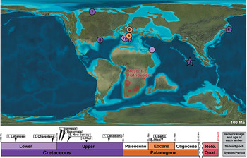
Rhachiberothidae comprises two subfamilies: Rhachiberothinae and Paraberothinae. Rhachiberothinae includes 13 extant species in three genera and 2 extinct species in one genus from middle Eocene Baltic amber (Whalley, Reference Whalley1983; Engel Reference Engel2004; Makarkin & Kupryjanowicz, Reference Makarkin and Kupryjanowicz2010). In contrast, the subfamily Paraberothinae is uniformly extinct, known to occur only during the Cretaceous Period. To date, Paraberothinae contains 14 described species in 13 extinct genera, and a single undescribed species of uncertain generic placement (Table 1). This group is found in a series of different Cretaceous amber deposits, namely Burmese, Canadian, French, Kuji, Lebanese and New Jersey ambers (Schlüter, 1978; Whalley, Reference Whalley1980; Grimaldi, Reference Grimaldi and Grimaldi2000; Engel, Reference Engel2004; Nel et al. Reference Nel, Perrichot, Azar and Néraudeau2005a; Engel & Grimaldi, Reference Engel and Grimaldi2008; McKellar & Engel, Reference Mc Kellar and Engel2009; Petrulevičius et al. Reference Petrulevičius, Azar and Nel2010; Shi et al. Reference Shi, Ohl, Wunderrich and Ren2015; Makarkin, Reference Makarkin2015a; Nakamine & Yamamoto, Reference Nakamine and Yamamoto2018; Table 1). The taxonomic position of the monotypic species Oisea celinea (Nel et al. Reference Nel, Perrichot, Azar and Néraudeau2005a, b), from the earliest Eocene Oise amber of France, remains uncertain within Rhachiberothidae (Makarkin & Kupryjanowicz, Reference Makarkin and Kupryjanowicz2010).
In the present study, a remarkable assemblage of Cretaceous paraberothines is documented in the mid-Cretaceous amber of northern Myanmar (i.e. Burmese amber) (earliest Cenomanian age, c. 99 Ma; Shi et al. Reference Shi, Grimaldi, Harlow, Wang, Wang, Yang, Lei, Li and Li2012), with descriptions of four new genera and seven new species. Our study sheds further light on the unexpected diversity and abundance of thorny lacewings in Burmese amber. Additionally, this study contributes to the elucidation of the morphological diversity of paraberothine insects in the Cretaceous System based on a series of well-preserved amber fossils, allowing us to observe fine details of the raptorial forelegs.
1.b. The fossil record of Paraberothinae
To date, the fossil records of rhachiberothids are limited to amber fossils, possibly because of their small and fragile bodies (Petrulevičius et al. Reference Petrulevičius, Azar and Nel2010). As mentioned in the previous section, the subfamily Paraberothinae is an entirely extinct group that occurred only during the Cretaceous Period. The earliest fossil record of paraberothines is from Lower Cretaceous (Barremian) Lebanese amber, consisting of four species in four genera (Whalley, Reference Whalley1980; Nel et al. Reference Nel, Perrichot, Azar and Néraudeau2005a; Petrulevičius et al. Reference Petrulevičius, Azar and Nel2010): Chimerhachiberotha acrasarii Nel et al. Reference Nel, Perrichot, Azar and Néraudeau2005a; Paraberotha acra Whalley, Reference Whalley1980; Raptorapax terribilissima Petrulevičius et al. Reference Petrulevičius, Azar and Nel2010; and Spinoberotha mickaelacrai Nel et al. Reference Nel, Perrichot, Azar and Néraudeau2005a. The fossil records of Paraberothinae are more extensive in the mid-Cretaceous deposits, ranging from the upper Albian to the lowest Cenomanian stages. For example, Alboberotha petrulevicii Nel et al. Reference Nel, Perrichot, Azar and Néraudeau2005a is the only species recorded from the upper Albian Charentese amber of France. In contrast, a great diversity of paraberothines is known from the lowest Cenomanian Burmese amber of northern Myanmar, with four known species in four genera (Engel, Reference Engel2004; Engel & Grimaldi, Reference Engel and Grimaldi2008; Makarkin, Reference Makarkin2015a; Shi et al. Reference Shi, Ohl, Wunderrich and Ren2015): Creagroparaberotha groehni Makarkin, Reference Makarkin2015a; Eorhachiberotha burmitica Engel, Reference Engel2004; Micromantispa cristata Shi et al. Reference Shi, Ohl, Wunderrich and Ren2015; and Scoloberotha necatrix Engel & Grimaldi, Reference Engel and Grimaldi2008. In addition, another undetermined, poorly preserved species, with only the head, anterior part of the prothorax and right foreleg being present, was recorded from the same amber deposit (Engel, Reference Engel2004). Retinoberotha stuermeri Schlüter, 1978 is the only species known from the lower Cenomanian Bezonnais amber of France (Schlüter, 1978). Although this specimen is poorly preserved and incomplete, its main hindwing characters are relatively well preserved (Schlüter, 1978). The same trend may also have continued during Late Cretaceous time, but with less diversity compared with during mid-Cretaceous time. From the Turonian New Jersey amber of the USA, there are two described species (Grimaldi, Reference Grimaldi and Grimaldi2000; Engel & Grimaldi, Reference Engel and Grimaldi2008): Rhachibermissa splendida Grimaldi, Reference Grimaldi and Grimaldi2000 and R. phenax Engel & Grimaldi, Reference Engel and Grimaldi2008. The recent finding of Kujiberotha teruyukii Nakamine & Yamamoto, Reference Nakamine and Yamamoto2018 from the middle Santonian Kuji amber of Japan was unexpected and noteworthy (Nakamine & Yamamoto, Reference Nakamine and Yamamoto2018). Although the holotype of K. teruyukii is poorly preserved, this taxon represents the first and oldest fossil record of Paraberothinae from East Asia, suggesting a potentially wider distribution of paraberothines in the eastern part of Laurasia during the Late Cretaceous Period (Nakamine & Yamamoto, Reference Nakamine and Yamamoto2018). Another Late Cretaceous paraberothine, Albertoberotha leuckorum McKellar & Engel, Reference Mc Kellar and Engel2009, was described from the Campanian Canadian amber (McKellar & Engel, Reference Mc Kellar and Engel2009). There is no fossil record of Paraberothinae from the Cenozoic Erathem. However, there is a single species of Rhachiberothidae, Oisea celinea (Nel et al. Reference Nel, Perrichot, Azar and Néraudeau2005a, b) from the lowest Eocene Oise amber of France, whose taxonomic position remains uncertain within the family (Makarkin & Kupryjanowicz, Reference Makarkin and Kupryjanowicz2010). These fossil records indicate a much wider range and higher diversity of Rhachiberothidae during the Cretaceous Period, with a subsequent decline or extinction before early Cenozoic time.
2. Materials and methods
All fossil specimens used in this study are deposited in the American Museum of Natural History (AMNH, New York, NY, USA; D. Grimaldi). They were mined from amber deposits in the Hukawng Valley of Kachin State, northern Myanmar (26° 21' 33.41'' N, 96° 43' 11.88'' E; Cruickshank & Ko, Reference Cruickshank and Ko2003; Grimaldi & Ross, Reference Grimaldi, Ross, Fraser and Sues2017). The so-called Burmese amber, also known as Kachin amber, is yielded by nearshore deposits characterized by coalified plant fragments and coal laminations in the fine clastic facies (Cruickshank & Ko, Reference Cruickshank and Ko2003). Mao et al. (Reference Mao, Liang, Su, Li, Rao, Zhang, Xia, Fu, Cai and Huang2018) also suggested that the amber-secreting trees were coastally situated because of the shapes of the amber, and the presence of pholadid bivalve borings and sessile marine organism on the amber surface. The earliest Cenomanian (mid-Cretaceous age, 98.79 ± 0.62 Ma) northern Myanmar amber was identified by uranium–lead (U–Pb) dating of zircon crystals from the amber matrix (Shi et al. Reference Shi, Grimaldi, Harlow, Wang, Wang, Yang, Lei, Li and Li2012). However, other published works suggest slightly older ages, ranging from late Albian to near the Albian–Cenomanian boundary (e.g. Cruickshank & Ko, Reference Cruickshank and Ko2003; Mao et al. Reference Mao, Liang, Su, Li, Rao, Zhang, Xia, Fu, Cai and Huang2018). Very recently, a Puzosia ammonite was found to occur within the inclusion of Burmese amber, reinforcing this hypothesis and further suggesting a mid-Cretaceous age for the amber (Yu et al. Reference Yu, Kelly, Mu, Ross, Kennedy, Broly, Xia, Zhang, Wang and Dilcher2019). Burmese amber exceedingly preserves rich terrestrial biota from Cretaceous tropical forests (Grimaldi et al. Reference Grimaldi, Engel and Nascimbene2002). In fact, according to the most recent checklist (Ross, Reference Ross2019), 881 genera and 1192 species belonging 535 families mostly occupied by arthropods have been described from this amber deposit so far.
The amber fossils were cut and polished by the second and third authors with waterproof sandpapers of different grain sizes, and finally polished with plastic buffing cloths. Observations were conducted under an MZ16 stereomicroscope (Leica Microsystems GmbH, Wetzlar, Germany) and SMZ800 stereomicroscope (Nikon Corporation, Tokyo, Japan). Photographs were produced using an 80D digital camera (Canon Inc., Tokyo, Japan), and later processed with the auto-montage software CombineZM (Alan Hadley, Sheffield, UK). During photography, the amber specimens were completely submerged in clove oil (Wako Pure Chemical Industries, Osaka, Japan; refractive index, 1.52–1.55) to avoid reflection of light by the amber surface and increase the visibility of the amber inclusions (Crighton & Carrio, Reference Crighton and Carrio2007; Penney & Green, Reference Penney, Green and Penney2010). All images were prepared using Adobe Photoshop and Illustrator CC 2019 (Adobe Inc., San Jose, CA, USA).
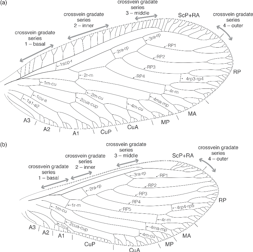
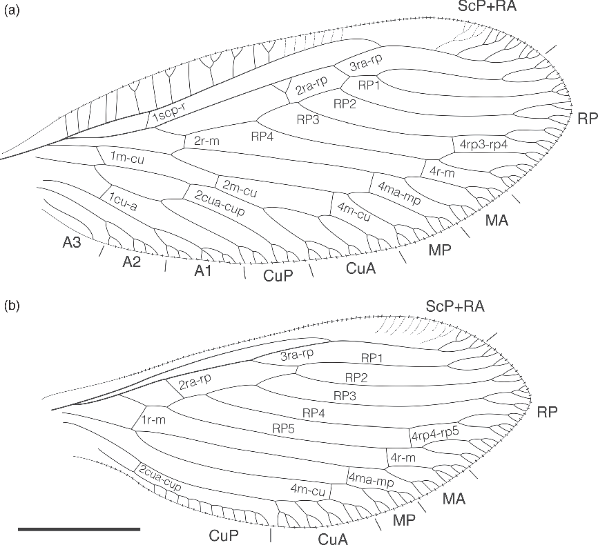
The morphological terminology follows Aspöck & Mansell (Reference Aspöck and Mansell1994) and Aspöck & Aspöck (Reference Aspöck and Aspöck1997, Reference Aspöck and Aspöck2008). The terminology of wing venations generally follows Breitkreuz et al. (Reference Breitkreuz, Winterton and Engel2017) and Oswald (Reference Oswald1993) (summarized in Fig. 1).
Generalized framework of the wing venation used in this study, modified from Oswald (Reference Oswald1993), Makarkin (Reference Makarkin2015a) and Wichard (Reference Wichard2016): (a) forewing; (b) hindwing. A1–A3 (a1–a3) – first to third anal vein; CuA (cua) – cubitus anterior; CuP (cup) – cubitus posterior; MA (ma) – media anterior; MP (mp) – media posterior; RA (ra) – radius anterior; RP (rp) – radius posterior; RP1–RP5 (rp1–rp5) – anterior to posterior branch of RP; ScP (scp) – subcosta posterior.

This published work and the nomenclatural acts it contains have been registered in ZooBank, the proposed online registration system for the International Code of Zoological Nomenclature (ICZN). The ZooBank LSIDs (Life Science Identifiers) for this publication is: urn:lsid:zoobank.org:pub:8CA3CB3C-DDB9-4808-A963-D279BD40F565.
3. Systematic palaeontology
Order NEUROPTERA Linnaeus, Reference Linnaeus1758
Superfamily MANTISPOIDEA Leach, Reference Leach and Brewster1815
Family RHACHIBEROTHIDAE Tjeder, Reference Tjeder, Hanström, Brinck and Rudebeck1959
Subfamily PARABEROTHINAE Nel et al. Reference Nel, Perrichot, Azar and Néraudeau2005a
Remarks. Rhachiberothids can be distinguished from Berothidae by the presence of developed raptorial forelegs. Further, this family can be discriminated from Mantispidae based on elongated pronotum anterior to the forelegs (they are elongated posteriorly to forelegs in Mantispidae) (Aspöck & Mansell, Reference Aspöck and Mansell1994). Paraberothinae can be separated from Rhachiberothinae by the presence of at least two (and usually numerous) spines on the inner edge of the protibia, and the absence of the intermediate subcostal crossvein in the distal part (2scp-r) of the forewing.
According to Makarkin (Reference Makarkin2015a), the Cretaceous paraberothines are characterized by a combination of the following (slightly revised) 11 morphological states: (1) small body size (forewing 2.9–5.4 mm long); (2) postocular lobe reduced (or at least not inflated); (3) antennal scapus long to very long (c. 2–10 times longer than the width); (4) forelegs raptorial; (5) protarsus of both female and male 5-segmented; (6) at least two spines present on the inner edge of the protibia (synapomorphy); (7) subcosta posterior (ScP) and radius anterior (RA) fused distally in both fore- and hindwings; (8) loss of the intermediate subcostal crossvein in the distal part (2scp-r) of the forewing (Fig. 1a); (9) humeral veinlet relatively short, none or few branched in the forewing; (10) basal crossvein between radius posterior (RP) and media (M) (1r-m) straight (if present) in the hindwing (Fig. 1b); and (11) cubitus posterior (CuP) present in the hindwing (Fig. 1b).
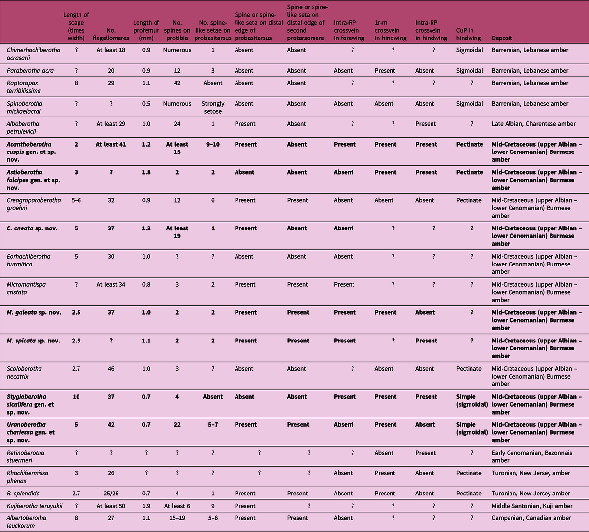
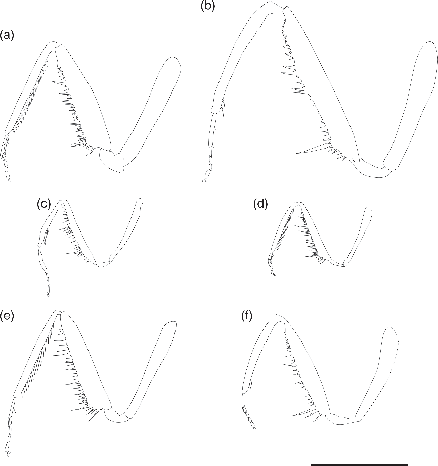
The foreleg structures of Paraberothinae are very diverse and noteworthy. The protarsus of all paraberothine species is 5-segmented in both sexes, and most paraberothines bear spine-like setae on the ventral edge of the probasitarsus (Table 2). Furthermore, Paraberothinae possess at least two spine-like setae on the inner (ventral) edge of the protibia. All fossil and extant species of Rhachiberothinae, Oisea, Berothidae and Mantispidae bear no spine-like setae on the inner edge of the protibia (Makarkin, Reference Makarkin2015a). However, a series of numerous strong setae (prostrate setae) on the inner edge of the protibia are present in the following three subfamilies in Mantispidae: Symphrasinae, Drepanicinae and Calomantispinae (Symphrasinae and Drepancinae include fossil and modern species) (Tjeder, Reference Tjeder, Hanström, Brinck and Rudebeck1959; Lambkin, Reference Lambkin1986; Makarkin, Reference Makarkin2015a; Pérez-de la Fuente & Peñalver, Reference Pérez-de la Fuente and Peñalver2019). In the extinct subfamily Mesomantispinae and the Burmese amber mantispid Dratomantispa burmanica Poinar & Buckley, 2011, they also bear peg-like protrusions on the inner edges of the protibiae (Poinar & Buckley, 2011; Jepson et al. Reference Jepson, Heads, Makarkin and Ren2013, Reference Jepson, Khramov and Ohl2018).
Evolutionary trends of key characters in Paraberothinae. 1r-m, basal crossvein between radius posterior (RP) and media (M); CuP– cubitus posterior; RP– radius posterior. Bold formatting indicates species described in this paper

The wing venation of the paraberothines is also diverse. The presence of a CuP in the hindwing is one of the diagnostic features of Paraberothinae. Makarkin (Reference Makarkin2015a) mentioned that this character can be considered as a plesiomorphic condition. CuP in the hindwing are partly reduced in all Rhachiberothinae and strongly reduced in most taxa of Mantispidae (except Sympharasinae) and Berothidae (Makarkin, Reference Makarkin2015b). Nearly all paraberothines have a developed CuP in the hindwing, and Acanthoberotha gen. nov. and Astioberotha gen. nov. also have a pectinately branched CuP. However, Stygioberotha gen. nov. and Uranoberotha gen. nov. have a simple CuP in the hindwing.
Genus Acanthoberotha gen. nov.
Zoobank LSID. urn:lsid:zoobank.org:act:0274B052-82AE-44F4-927F-AB41464E45DC
Type species. Acanthoberotha cuspis sp. nov., here designated.
Derivation of name. Combination of the Greek ‘akantha’ [ἄκανθα], meaning ‘thorn’, and the generic name Berotha. Gender feminine.
Diagnosis. Antennae with scapus relatively short, about twice as long as maximum width. Protibia bearing with at least 15 slightly curved thin spines toward apex on ventral edge. Protarsus: probasitarsus with 9–10 spine-like setae on external edge and one slightly curved claw-like spine at apically, lacking spine or spine-like setae on second to fifth tarsomeres. Forewing hyaline, elongate oval; RP divided into four branches, intra-RP crossvein between RP3 and RP4 present. Hindwing hyaline, ovoid; RP divided into four branches, intra-RP crossvein between RP3 and RP4 present; basal crossvein between RP and M present; anal vein absent.
Differential diagnosis. Acanthoberotha gen. nov. can be distinguished from six paraberothine genera (Albertoberotha, Creagroparaberotha, Eorhachiberotha, Raptorapax, Stygioberotha gen. nov. and Uranoberotha gen. nov.) by length of scape; in Acanthoberotha, it is twice as long as maximum width, while 5–6 times width in Creagroparaberotha, Eorhachiberotha and Uranoberotha or 8–10 times width in Albertoberotha, Raptorapax and Stygioberotha. Acanthoberotha can be discriminated from Kujiberotha by number of spines on protibia (in Acanthoberotha, at least 15 spines, while at least 6 in Kujiberotha). Acanthoberotha can be separated from six paraberothine genera (Alboberotha, Astioberotha gen. nov., Chimerhachiberotha, Micromantispa, Paraberotha and Rhachibermissa) by spine(s) or spine-like setae on probasitarsus (Acanthoberotha has 9–10 spine-like setae on probasitarsus, while 1–3 in Alboberotha, Astioberotha, Chimerhachiberotha, Micromantispa, Paraberotha and Rhachibermissa). Furthermore, Acanthoberotha can be distinguished from three paraberothine genera (Retinoberotha, Scoloberotha and Spinoberotha) by the hindwing venation (Acanthoberotha has basal 1r-m crossvein present, while absent in these genera) (see details in Table 2).
Acanthoberotha cuspis sp. nov.
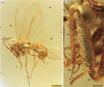
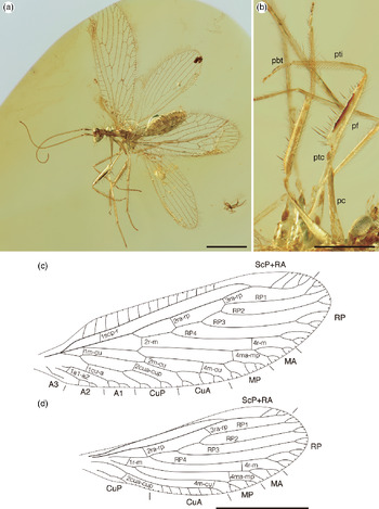
Habitus and foreleg of Acanthoberotha cuspis gen. et sp. nov. (holotype, AMNH Bu-SY16) in mid-Cretaceous amber from northern Myanmar: (a) left lateral view (scale bar 1.0 mm); and (b) left foreleg, arrow showing claw-like spine (scale bar 0.2 mm). pbt – probasitarsus; pf – profemur; ptc – protrochoanter; pti – protibia.

Wing venation of Acanthoberotha cuspis gen. et sp. nov. (holotype, AMNH Bu-SY16) in mid-Cretaceous amber from northern Myanmar: (a) left forewing; and (b) right hindwing. Scale bar represent 0.5 mm. Abbreviations as for Figure 1.

Zoobank LSID. urn:lsid:zoobank.org:act:057ADBEB-22A7-4664-8936-8984F13B907A
Derivation of name. From the Latin ‘cuspis’ meaning ‘sharp point’.
Holotype. A nearly complete female adult (Fig. 2a), partly invisible due to bubbles and cracks, preserved in a c. 9.1 mm × 13.1 mm × 3.5 mm flattened, clear yellow, transparent amber piece; specimen accession number AMNH Bu-SY16, deposited in AMNH.
Diagnosis. As for the genus (vide supra).
Description. Body. Length c. 3.7 mm as preserved (measured from vertex to apex of abdomen).
Head. Vertex with dense fine setae. Eyes large, protruding laterally; postocular lobe reduced. Antennae partly lost; scapus clavate, slightly elongate, about twice as long as width; pedicellus c. 0.3 times length of scape; both covered with fine dense setae; flagellum moniliform, right antenna with 37 and left with 41 flagellomeres, with scattered fine setae on each segment.
Thorax. Pronotum length c. 0.6 mm, elongate, slightly narrower than maximum width of head, covered with fine setae. Mesothorax and metathorax not clearly visible due to cracks, bubbles and position of wings.
Legs. Foreleg. (Fig. 2b) Procoxa elongate, c. 1.2 mm, nearly as long as profemur, slightly narrowed distally. Protrochanter elongate. Profemur long, c. 1.2 mm, slightly broadened, covered with dense fine setae on surface, with spines of variable lengths forming two rows on ventral edge, only slightly curved distally. Protibia long, c. 1.0 mm, slender, covered with fine setae on dorsal and lateral edges, with single row of proximally inclined blade-shape of at least 15 spines regularly spaced on external ventral edge. Protarsus: all protarsomeres covered with fine setae; probasitarsus longest, relative length of tarsomeres 5–1.5–1.5–1–1.5, with 9–10 spine-like setae on external ventral edge and one long curved claw-like spine distally; claws curved.
Midleg. Mesocoxa stout and conical. Mesotrochanter elongate, slightly curved. Mesofemur long, c. 1.0 mm, slender, covered with dense long setae. Mesotibia long, c. 1.2 mm, slender, covered with dense long setae, several long stronger setae. Mesotarsus: all mesotarsomeres covered with fine setae; first tarsomere longest, relative length of tarsomeres 6.9–2.5–1.3–1–1.3; claws small, curved.
Hindleg. Metacoxa similar to mesocoxa. Metatrochanter elongate, slightly curved. Metafemur long, c. 1.1 mm, slender, covered with dense long setae. Metatibia long, c. 1.8 mm, slender, covered with dense long setae, several long stronger setae. Metatarsus: all metatarsomeres covered with fine setae; first tarsomere longest, relative length of tarsomeres 7.5–2.3–1.3–1–1.3; claws small, curved.
Wings (Fig. 3). Forewing hyaline, elongate-ovoid, c. 4.5 mm length, c. 1.8 mm width. Trichosors present along the whole wing margin; marginal portions of veins and trichosors with tufts of thin setae; thin setae sparsely present on dorsal and ventral surfaces of longitudinal veins, but absent on crossveins. Costal space slightly broad; about half of subcostal veinlets once forked. ScP fused with RA at apical third. Subcostal space broad; proximal crossvein (1scp-r) located proximal forth. RP divided into four branches; RP1 deeply forked; intra-crossvein (4rp3-rp4) present. Two ra-rp crossveins present; 2ra-rp located between origin of RP3 and RP4, while once forked in left forewing; 3ra-rp located between origin of RP1 and RP2, whereas once branched in right forewing. M basally fused with ScP, divided into media anterior (MA) and media posterior (MP); one crossvein (4ma-mp) present. Two r-m crossveins present; 2r-m located slightly distal to RP and MA origin; 4r-m located between RP4 and MA. Cu divided into CuA and CuP; one crossvein (2cua-cup) present. Three m-cu crossveins present; 1m-cu located slightly distal to origin of M and CuA; 2m-cu between distal to stem of MP and CuA; 4m-cu between MP and CuA. Three anal veins present: A1, A2 and A3; A3 simple. One cu-a crossvein (1cu-a) present between stem of CuP and A1.
Hindwing hyaline, elongate-ovoid, c. 3.8 mm length, c. 1.6 mm width. Trichosors present on wing margin; marginal portions of veins and trichosors with tufts of thin setae; thin setae sparsely present on dorsal and ventral surfaces of longitudinal veins, but absent from crossveins. Costal space narrowed; subcostal veinlets poorly preserved. ScP fused with RA at middle slightly distal. Subcostal space no crossvein. RP divided into five branches; intra-RP crossvein (4rp4-rp5) present. Two ra-rp crossveins present; 2ra-rp located between RP4 and RP5 origin; 3ra-rp located near stem of RP1. M divided into MA and MP; one crossvein (4ma-mp) present. Two r-m crossveins present; 1r-m located near stems of RP and M; 4r-m located between RP5 and MA. Origin of Cu unclear; CuP pectinately branched with simple 11 branches; one crossvein (2cua-cup) present. One m-cu crossvein present; 4m-cu located between MP and CuA. Anal vein absent.
Abdomen. Not clearly visible due to some bubbles and amber cracks; elongate-ovoid, slightly compressed laterally, slightly curled ventrally, moderately scattered setae; segments partly unclear. Ectoproct covered with thin setae.
Genus Astioberotha gen. nov.
Zoobank LSID. urn:lsid:zoobank.org:act:0ACC4AC2-8093-4B55-9E40-C4C90C67D333
Type species. Astioberotha falcipes sp. nov., here designated.
Derivation of name. Combination of the Greek ‘asteios’ [ἀστεῖος], meaning ‘elegant’, and the generic name Berotha. Gender feminine.
Diagnosis. Protibia only slightly two protrusion on the proximal area of ventral ridge, bearing two or three spines on the distal area of ventral ridge. Protarsus: probasitarsus with one short spine on ventral ridge and one short spine distally; second to forth protarsomeres with two short spines distally. Forewing elongate-ovoid; costal space relatively broad, most subcostal veinlets once forked; one spot-like maculation present at margin of crossvein between MP and CuA (2m-cu). Hindwing hyaline, elongate-ovoid; RP divided into four branches, intra-RP crossvein between RP3 and RP4 present, basal crossvein between RP and M present.
Differential diagnosis. Astioberotha gen. nov. can be distinguished from six paraberothine genera (Albertoberotha, Creagroparaberotha, Eorhachiberotha, Raptorapax, Stygioberotha gen. nov. and Uranoberotha gen. nov.) by length of scape (in Astioberotha, 3 times as long as maximum width, while in other genera 5–10 times width). Astioberotha can be discriminated from six paraberothine genera (Acanthoberotha gen. nov., Alboberotha, Chimerhachiberotha, Kujiberotha, Paraberotha and Spinoberotha) based on the numbers of spines on protibia (Astioberotha has two short fine spines, whereas at least 15 in Acanthoberotha, 24 in Alboberotha, at least 6 in Kujiberotha, 12 in Paraberotha, and numerous in Chimerhachiberotha and Spinoberotha). Furthermore, Astioberotha can be distinguished from two paraberothine genera (Retinoberotha and Scoloberotha) by the hindwing venation (Astioberotha has basal 1r-m crossvein present, while absent from these genrera). Astioberotha can be distinguished from Rhachibermissa by the fore- and hindwing venation (in Astioberotha, RP divided four branches on forewing and five branches on hindwing, whereas RP divided three branches on fore- and hindwing in Rhachibermissa). Astioberotha is most similar to Micromantispa. However, Astioberotha can be distinguished from Micromantispa by a combination of the following character states: probasitarsus with one short spine on ventral ridge (two in Micromantipa) and one short spine distally (one long curved spine in Micromantipa); forewing: intra-RP crossvein (4rp3-rp4) absent (present in Micromantipa); one maculation spot present at margin of crossvein between MP and CuA (2m-cu) (absent in Micromantipa) (see details in Table 2).
Astioberotha falcipes sp. nov.
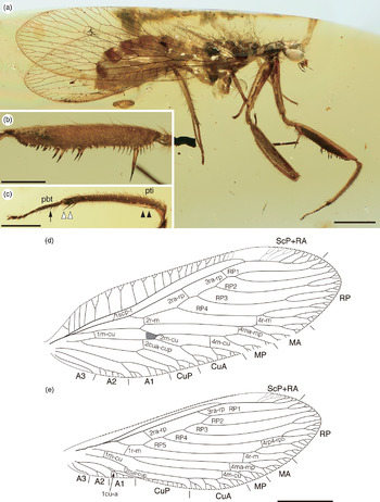
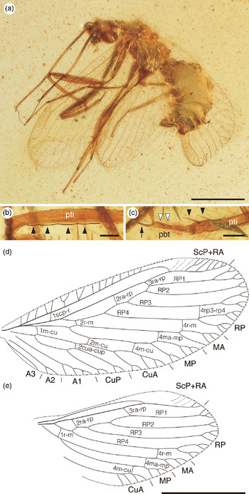
Habitus, forelegs and wing venation of Astioberotha falcipes gen. et sp. nov. (holotype, AMNH Bu-SY17) in mid-Cretaceous amber from northern Myanmar: (a) right lateral view; (b) left profemur, left lateral (outer) view; (c) left protibia, arrows showing slight protorusion, open arrows showing bearing spines, and linearrow showing small spine on middle ventral ridge in probasitarsus; (d) right forewing; and (e) right hindwing. Abbreviations as for Figures 1 and 2. Scale bars (a, d, e) 1.0 mm; and (b, c) 0.5 mm.

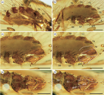
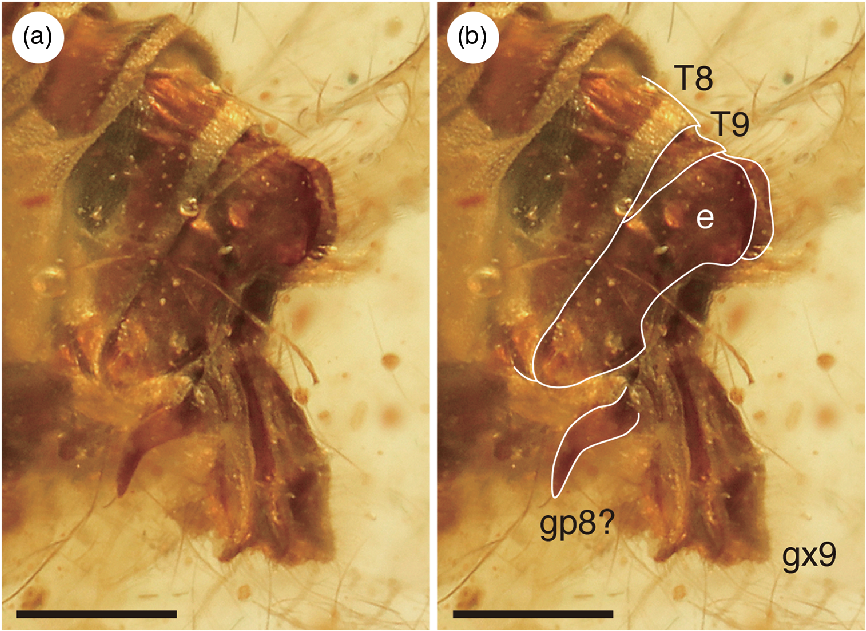
Abdomen of Astioberotha falcipes gen. et sp. nov. (holotype, AMNH Bu-SY17) in mid-Cretaceous amber from northern Myanmar: (a) latero-dorsal view; (b) latero-ventral view; (c) genital segments, latero-ventral view; (d) genital segments, latero-ventral view with white line indicating each segment and gonocoxite; (e) genital segments, ventral view; and (f) genital segments, ventral view with white line indicating each segment, gonapophysis and gonocoxite. e – ectoproct; gp8 – gonapophyses 8; gx9 – gonocoxites 9; S6–S7 – sternite 6–7; T3–T9 – tergite 3–9. Scale bars represent 0.5 mm.

Zoobank LSID. urn:lsid:zoobank.org:act:E18A6729-11F0-4615-8E69-D98656B31789
Derivation of name. Combination of the Latin ‘falcis’, meaning ‘the sickle’ and ‘pes’, meaning ‘a leg’.
Holotype. Well-preserved, incomplete female adult (Fig. 4a), antennae and left fore- and hindwing nearly completely lost due to over polishing, preserved in a c. 19.2 mm × 7.3 mm × 5.2 mm narrowly elongate, clear yellow, transparent amber piece; specimen accession number AMNH Bu-SY17, deposited in AMNH.
Diagnosis. As for the genus (vide supra).
Description. Body. Length c. 5.1 mm as preserved (measured from vertex to apex of abdomen).
Head. Vertex with dense fine setae. Eyes large and protruding laterally; postocular lobe not inflated. Antennae nearly lost with only 8 flagellomeres preserved on right antenna; scapus slightly elongate, c. 3 times as long as width; pedicellus short, c. 0.3 length of scape; both with fine setae; flagellum moniliform, with scattered fine setae on each segment. Maxillary palpi 5 segmented; labial palpi 3 segmented.
Thorax. Pronotum length c. 0.6 mm, elongate, narrower than width of head between compound eyes, scattered with fine setae. Mesothorax and metathorax not well visible due to milky substance on surface; scattered fine setae.
Legs. Foreleg (Fig. 4b, c). Procoxa elongate, c. 1.4 mm length, shorter than profemur, slightly curved, distally narrowed. Protrochanter elongate, slightly curved. Profemur long, c. 1.8 mm, slightly broadened, covered with dense fine setae on surface, with spines of variable lengths forming two rows on ventral ridge, only slightly curved towards distal, one markedly elongate spine proximal area, eight spines on the distal area of outer-ventral ridge. Protibia long, c. 1.3 mm, slender, covered with fine setae on surface; only slightly two protrusion on the proximal area of ventral ridge; bearing two or three spines in right protibial distally ventral ridge. Protarsus: all protarsomeres covered with fine setae; probasitarsus longest, relative length of tarsomeres 6.8–1.6–1.3–1–2, with one small spine on middle ventral ridge and one short spine distally; second to forth protarsomere with two short spines distally; claws curved.
Midleg. Mesocoxa stout and conical. Mesotrochanter elongate, slightly curved. Mesofemur long, c. 1.1 mm, slender, covered with dense long setae. Mesotibia long, c. 1.0 mm, slender, covered with dense long setae and several long, stronger almost perpendicular, setae. Mesotarsus: all mesotarsomeres covered with fine setae; first tarsomere longest, relative length of tarsomeres 6.7–2.3–1.5–1–1.3; claws small, curved.
Hindleg. Metacoxa similar to mesocoxa. Metatrochanter elongate, slightly curved. Metafemur long, c. 1.2 mm, slender, covered with dense long setae. Metatibia long, c. 1.8 mm, slender, covered with dense long setae, several long stronger almost perpendicular setae. Metatarsus: all metatarsomeres covered with fine setae; first tarsomere longest, relative length of tarsomeres 7.2–2.5–1.5–1–1.8; claws small, curved.
Wings (Fig. 4d, e). Forewing hyaline, elongate-ovoid, c. 5.4 mm length, c. 1.7 mm width. Trichosors present along the whole wing margin; marginal portions of veins and trichosors with tufts of fine setae; fine setae sparsely present on dorsal and ventral surfaces of longitudinal veins, but absent from crossveins. Costal space relatively broad; most subcostal veinlets once forked. ScP fused with RA at apical third. Subcostal space, proximal crossvein (1scp-r) located between origins of RP and M. RP divided into four branches; RP1 deeply forked. Two ra-rp crossveins present; 2ra-rp located between origin of RP3 and RP4; 3ra-rp located distally stem of RP1. M divided into MA and MP; one crossvein (4ma-mp) present. Two r-m crossveins present; 2r-m located slightly distal to RP and MA origin; 4r-m located between RP4 and MA. Cu divided into CuA and CuP; one crossvein (2cua-cup) present. Three m-cu crossveins present; 1m-cu located slightly distal to M and CuA origin; 2m-cu between distal to stem MP and CuA, maculation spot at narrowly margin; 4m-cu between MP and CuA. Three anal veins present: A1, A2 and A3; A3 simple.
Hindwing hyaline, elongate-ovoid, c. 4.7 mm length, c. 1.6 mm width, possibly an artefact between the terminal of CuP and A1 dimple. Trichosors present on wing margin; marginal portions of veins and trichosors with tufts of fine setae; fine setae sparsely present on dorsal and ventral surfaces of longitudinal veins, but absent from crossveins. Costal space narrowed, subcostal veinlets poorly preserved. ScP fused with RA at apical third. Subcostal space no crossvein. RP divided into five branches, intra-RP crossvein (4rp3-rp4) present. Two ra-rp crossveins present; 2ra-rp located between RP3 and RP4 origin; 3ra-rp located stem of RP1. M divided into MA and MP, one crossvein (4ma-mp) present. Two r-m crossveins present; 1r-m located near stems of RP and M; 4r-m located between RP5 and MA. Origin of Cu unclear; CuP pectinately branched with 11 simple branches; one crossvein (2cua-cup) present. Two m-cu crossveins present; 1m-cu located stems of M and Cu; 4m-cu located between MP and CuA. Three anal veins present: A1, A2 and A3; A3 simple. One short crossvein (1cu-a) present between CuP and A1.
Abdomen (Fig. 5). Abdomen well preserved, partly obscured due to milky layer on body surface; elongate-ovoid, each tergites well visible; tergites with scattered fine long setae; sternites with moderately scattered fine setae, gradually slightly longer towards distal. Genital segments well preserved and clear. Falciform structures considered as paired gonapophysis 8 (gp8). Tergite 9 ventroposterior part protruded; ectoproct covered with thin setae, dorsoventrally elongate. Gonocoxites 9 (gx9) paired, not developed.
Remarks. This specimen is one of the best-preserved paraberothines known to date in terms of the female genital segments. In this aspect, Astioberotha falcipes sp. nov. is similar to that of the holotype of Micromantispa cristata (see Shi et al. Reference Shi, Ohl, Wunderrich and Ren2015, figs 4, 5), extant Mantispinae and Drepanicinae (Mantispidae) (see Aspöck & Aspöck, Reference Aspöck and Aspöck2008, figs 123, 124; Liu et al. Reference Liu, Winterton, Wu, Piper and Ohl2015, figs 6, 10).
Genus Stygioberotha gen. nov.
Zoobank LSID. urn:lsid:zoobank.org:act:CFB453A6-D55E-485A-A30B-74C0262695A9
Type species. Stygioberotha siculifera sp. nov., here designated.
Derivation of name. Combination of the Greek ‘Stygios’ [Στγιος], meaning ‘the netherworld’, and the generic name Berotha. Gender feminine.
Diagnosis. Antennae: scapus markedly long, c. 10 times as long as width; flagellum thin moniliform. Profemur bearing two long spines each on the proximal and distal position of ventral edge. Protibia bearing four long slightly curved hooked spines towards apex on the distal area of ventral ridge. Protarsus: all tarsomeres lack spine or spine-like setae; probasitarsus slightly dilate, elongate, c. 7 times as long as width, longer than combined length of second to fifth tarsomeres. Forewing hyaline, elongate-ovoid; RP divided into five branches, intra-RP crossvein between RP4 and RP5 present; only one anal vein present. Hindwing hyaline, ovoid; RP divided into four branches, intra-RP crossvein between RP3 and RP4 present; anal vein absent.
Differential diagnosis. Stygioberotha gen. nov. can be distinguished from eight paraberothine genera (Acanthoberotha gen. nov., Astioberotha gen. nov., Creagroparaberotha, Eorhachiberotha, Micromantispa, Rhachibermissa, Scoloberotha and Uranoberotha gen. nov.) by length of scape (in Stygioberotha, 10 times as long as maximum width, while in other genera, 2–5 times width). Stygioberotha can be separated from Kujiberotha based on the numbers of flagellomeres (Stygioberotha has 37 flagellomeres, whereas at least 50 in Kujiberotha). Stygioberotha can be discriminated from six paraberothine genera (Albertoberotha, Alboberotha, Chimerhachiberotha, Paraberotha, Raptorapax and Spinoberotha) based on the numbers of spines on the protibia (Stygioberotha has 4 spines, whereas at least 15 in Acanthoberotha, 15–19 in Albertoberotha, 24 in Alboberotha, 12 in Paraberotha, 42 in Raptorapax, and numerous in Chimerhachiberotha and Spinoberotha). Furthermore, Stygioberotha can be distinguished from Retinoberotha by the hindwing venation (Stygioberotha has basal 1r-m crossvein present, while absent in Retinoberotha) (see details in Table 2).
Stygioberotha siculifera sp. nov.
(Fig. 6)
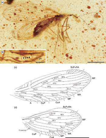
Habitus, foreleg and wing venation of Stygioberotha siculifera gen. et sp. nov., right lateral view (holotype, AMNH Bu-SY18) in mid-Cretaceous amber from northern Myanmar: (a) right lateral view; (b) left protibia and protarsus, right lateral (inner) view, arrows showing slightly curved four spines; (c) left forewing; and (d) right hindwing. Abbreviations as for Figures 1 and 2. Scale bars (a, c, d) 1.0 mm and (b) 0.2 mm.

Zoobank LSID. urn:lsid:zoobank.org:act:8ADE329D-3134-420A-BB32-4DEA81045DC4
Derivation of name. Combination of the Latin ‘sicula’ meaning ‘short sword or dagger’ and ‘-fer’ meaning ‘bearing’, in reference to short sword-shaped probasitarsus.
Holotype. Incomplete specimen of adult, sex undetermined (Fig. 6a), mostly part of right forewing and right side of abdomen compressed and deformed, preserved in a c. 17.1 mm × 11.2 mm × 3.1 mm broadly oval, flattened, clear yellow, transparent amber piece; specimen accession number AMNH Bu-SY18, deposited in AMNH.
Diagnosis. As for the genus (vide supra).
Description. Body. Length c. 2.7 mm as preserved (measured from vertex to apex of abdomen).
Head. Vertex with scarce setae. Eyes large, protruding laterally; postocular lobe not visible. Antenna: scapus elongate, c. 10 times as long as maximum width; pedicellus c. 0.6 times as length of scape; both covered with dense fine setae; flagellum thin and moniliform, relatively long, comprising 37 flagellomeres, with scattered fine setae on each segment.
Thorax. Pronotum length c. 0.4 mm, elongate, slightly narrower than width of head between compound eyes, covered with long setae. Mesothorax and metathorax with scattered long setae; detailed structures not visible due to milky substance on body surface.
Legs. Foreleg (Fig. 6b). Procoxa elongate, c. 0.7 mm length, only shorter than profemur, narrowed distally. Protrochanter elongate, slightly curved. Profemur long c. 0.7 mm, slightly broadened, covered with dense fine setae on surface, with spines of variable lengths forming two rows on ventral edge, markedly elongate two spines in proximal third and distal third, only slightly curved towards distal. Protibia long, c. 0.5 mm, slender, bearing four long slightly curved hooked spines towards apex on the distal area of ventral ridge. Protarsus: all protarsomeres covered with fine setae; probasitarsus slightly curved and dilate, longest, relative length of tarsomeres 7.4–1–1–1–2.4; claws not visible.
Midleg. Mesocoxa stout and conical. Mesotrochanter elongate, slightly curved. Mesofemur long, c. 0.6 mm, slender, covered with dense long setae. Mesotibia long, c. 0.6 mm, slender, covered with dense long setae, several long fine almost perpendicular setae. Mesotarsus: all mesotarsomeres covered with fine setae; first tarsomere longest, relative length of tarsomeres 9–2–1.5–1–1.2; claws small, curved.
Hindleg. Metacoxa similar to mesocoxa. Metatrochanter elongate, slightly curved. Metafemur long, c. 0.6 mm, slender, covered with dense long setae. Metatibia long, c. 1.0 mm, slender, covered with dense long setae and several long fine, almost perpendicular, setae. Metatarsus: all metatarsomeres covered with fine setae; first tarsomere longest, relative length of tarsomeres 7.5–2–1.3–1–1; claws small, curved.
Wings (Fig. 6c, d). Forewing hyaline, elongate-ovoid, c. 3.4 mm length, c. 1.1 mm width. Trichosors present along entire wing margin; marginal portions of veins and trichosors with tufts of fine setae; fine setae sparsely present on dorsal and ventral surfaces of longitudinal veins, but absent on crossveins. Costal space slightly broad; subcostal veinlets poorly preserved, rather simple. ScP fused with RA at apical third. Subcostal space broad; all paraberothine genera have crossveins (1scp-r) each at subcostal space; however, this is not visible due to some bubbles and debris on the subcostal space in basal third. RP divided into four branches; RP1 deeply forked; intra-RP crossvein (4rp3-rp4) present. Two ra-rp crossveins present; 2ra-rp located between origin of RP3 and RP4; 3ra-rp located at slightly distal of RP2 origin. M basally fused with R, divided into MA and MP; one crossvein (4ma-mp) present. Two r-m crossveins present; 2r-m located slightly distal to RP and MA; 4r-m located between RP4 and MA. Cu divided into CuA and CuP; one crossvein (2cua-cup) present. Three m-cu crossveins present; 1m-cu located slightly distal to origin of M and CuA; 2m-cu between slightly distal to stem of MP and CuA; 4m-cu between MP and CuA. One anal vein present. One crossvein (1cu-a) present between stem of CuP and A.
Hindwing hyaline, elongate-ovoid, c. 2.5 mm length, c. 1.1 mm width. Trichosors present on wing margin; marginal portions of veins and trichosors with tufts of fine setae; fine setae sparsely present on dorsal and ventral surfaces of longitudinal veins, but absent on crossveins. Costal space narrowed; subcostal veinlets poorly preserved. ScP fused with RA at middle slightly distal. Subcostal space no crossvein. RP divided into four branches; intra-RP crossvein (4rp3-rp4) present. Two ra-rp crossveins present; 2ra-rp located between RP3 and near RP4 origin; 3ra-rp located near stem of RP1. M divided into MA and MP; one crossvein (4ma-mp) present. Two r-m crossveins present; 1r-m located near stems of RP and M; 4r-m located between RP4 and MA. CuA pectinately branched with seven simple branches; CuP simple; one crossvein (2cua-cup) present. One crossvein (4m-cu) located between MP and CuA. Anal vein absent.
Abdomen. Not clearly visible due to milky substance on body surface; elongate-ovoid, covered with dense setae. Segmentation and structure of terminal segments unclear.
Genus Uranoberotha gen. nov.
Zoobank LSID. urn:lsid:zoobank.org:act:8B32FC0E-2C01-42CB-BDFD-F4D9B72FCB447
Type species. Uranoberotha chariessa sp. nov., here designated.
Derivation of name. Combination of the Greek ‘ouranos’ [θρανός], meaning ‘firmament’, and the generic name Berotha. Gender feminine.
Diagnosis. Antenna: scapus elongate, c. 5 times as long as width. Protibia with single row of 22 proximally inclined spines regularly spaced on external ventral edge. Probasitarsus: 5–7 small spine-like setae on external ventral edge and one long curved spine distally. Forewing hyaline, elongate-ovoid; costal space slightly broad; subcostal veinlets simple, not forked; intra-A crossvein between A1 and A2 present. Hindwing hyaline, elongate-ovoid; anal vein absent.
Differential diagnosis. Uranoberotha gen. nov. can be distinguished from seven paraberothine genera (Acanthoberotha gen. nov., Astioberotha gen. nov., Micromantispa, Raptorapax, Rhachibermissa, Scoloberotha and Stygioberotha gen. nov.) by length of scape (in Uranoberotha, c. 5 times as long as maximum width, while 2–3 times width in Acanthoberotha, Astioberotha, Micromantispa, Rhachibermissa and Scoloberotha or 8–10 times width in Raptorapax and Stygioberotha). Uranoberotha can be discriminated from four paraberothine genera (Creagroparaberotha, Eorhachiberotha, Kujiberotha and Paraberotha) based on the numbers of flagellomeres (Uranoberotha has 42 flagellomeres, whereas there are 32 in Creagroparaberotha, 30 in Eorhachiberotha, at least 50 in Kujiberotha and 20 in Paraberotha). Uranoberotha can be separated from two paraberothine genera (Chimerhachiberotha and Spinoberotha) based on the numbers of spines on protibia (Uranoberotha has 22 spines, while these are numerous in Chimerhachiberotha and Spinoberotha). Finally, Uranoberotha can be distinguished from Retinoberotha by the hindwing venation (Uranoberotha has basal 1r-m crossvein, while absent in Retinoberotha). Uranoberotha is most similar to Albertoberotha. However, Uranoberotha can be distinguished from Albertoberotha by a combination of the following character states: Antennae, scapus elongate, 5 times maximum width (8 times width in Albertoberotha), 42 flagellomeres (27 in Albertoberotha). Protibia with 22 spines ventral edge (at least 17 in Albertoberotha); probasitarsus longest (markedly short in Albertoberotha). Forewing, crossveins between MP and CuP (1m-cu and 4m-cu) and crossvein between CuA and CuP (2cua-cup) present (these crossveins absent in Albertoberotha) (see details in Table 2).
Uranoberotha chariessa sp. nov.
Habitus, forelegs and wing venation of Uranoberotha chariessa gen. et sp. nov. (holotype, AMNH Bu-SY19) in mid-Cretaceous amber from northern Myanmar: (a) left latero-dorsal view; (b) forelegs, left lateral view; (c) forewing; and (d) hindwing. pc – procoxa. Other abbreviations as Figures 1 and 2. Scale bars (a, c, d) 1.0 mm and (b) 0.5 mm.

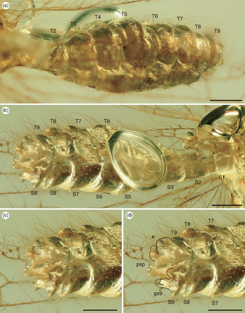
Abdomen of Uranoberotha chariessa gen. et sp. nov. (holotype, AMNH Bu-SY19) in mid-Cretaceous amber from northern Myanmar: (a) latero-dorsal view; (b) latero-ventral view; (c) genital segments, latero-ventral view; and (d) genital segments, latero-ventral view with line indicating ectoproct and gonocoxite 9. psp – pseudopenis. Other abbreviations as for Figure 5. Scale bars 0.2 mm.

Zoobank LSID. urn:lsid:zoobank.org:act:629F3334-AEDA-4843-943F-56FCFEBC0021
Derivation of name. From the Greek ‘charieis’ [χαρεις], meaning ‘graceful’.
Holotype. Exceptionally well preserved, nearly complete adult male (Fig. 7a), except distal part of the scraped right forewing, preserved in a c. 12.0 mm × 7.4 mm × 3.9 mm flattened oval, clear yellow, transparent amber piece; specimen accession number AMNH Bu-SY19, deposited in AMNH.
Diagnosis. As for the genus (vide supra).
Description. Body. Length c. 2.4 mm as preserved (measured from vertex to apex of abdomen).
Head. Vertex slightly protuberance, with long scarce setae. Compound eyes large, protruding laterally; postocular lobe slightly inflated. Antennae: scapus elongate, c. 5 times as long as width, slightly dilated toward apex; pedicellus one-half length of scape; both covered with dense fine setae; flagellum moniliform, relatively long, composed of 42 flagellomeres, with scattered fine setae on each segment. Maxillary palpi 5 segmented; labial palpi 3 segmented.
Thorax. Pronotum length c. 1.6 mm, elongate, slightly narrower than width of head between compound eyes, covered with thin and long setae. Mesothorax and metathorax with scattered long setae. Further details not completely visible.
Legs. Foreleg (Fig. 7b). Procoxa elongate, c. 0.6 mm length, only shorter than profemur, narrowed distally. Protrochanter elongate, slightly curved. Profemur long, c. 0.7 mm, broadened towards proximally, only slightly curved distally, with basal protuberance bearing slightly expanded circlet, covered with dense fine setae on surface, together with spines of variable lengths, forming two rows on ventral edge. Protibia long, c. 0.6 mm, slender, covered with fine setae on dorsal and lateral edges, single row of 22 proximally inclined spines regularly spaced on external ventral edge. Protarsus: all protarsomeres covered with fine setae; probasitarsus longest, relative length of tarsomeres 2.3–1–1.3–1–1.2, with 5–7 small spine-like setae on external ventral edge and one long curved spine distally; second protarsomere with at least two small spine-like setae on external ventral edge and one long curved spine distally; claws curved.
Midleg. Mesocoxa stout and conical. Mesotrochanter elongate, slightly curved. Mesofemur long, c. 0.7 mm, slender, covered with dense long setae. Mesotibia long, c. 1.2 mm, slender, covered with dense long setae. Mesotarsus: all mesotarsomeres covered with fine setae; first tarsomere longest, relative length of tarsomeres 10–2.8–1.6–1–1.6, all mesotarsomeres covered with fine setae; claws small, curved.
Hindleg. Metacoxa similar to mesocoxa. Metatrochanter elongate, slightly curved. Metafemur long, c. 0.6 mm, slender, covered with dense long setae. Metatibia long, c. 1.3 mm, slender, covered with dense long setae. Metatarsus: all metatarsomeres covered with fine setae; first tarsomere longest, relative length of tarsomeres 12–3–1.4–1–1.2; claws small, curved.
Wings (Fig. 7c, d). Forewing hyaline, elongate-ovoid, c. 3.2 mm length, c. 1.0 mm width. Trichosors present along entire wing margin; marginal portions of veins and trichosors with tufts of long fine setae; fine setae sparsely distributed on dorsal and ventral surfaces of longitudinal veins, but absent on crossveins. Costal space slightly broadened; subcostal veinlets simple, not forked; pterostigma area present at distal anterior margin, veinlets unclear. ScP fused with RA in pterostigma area. Subcostal space broad; proximal crossvein (1scp-r) located between origins of RP and M. RP divided into four branches. Two ra-rp crossveins present; 2ra-rp located between origin of RP3 and RP4; 3ra-rp located near stem of RP1. M basally fused with R, divided into MA and MP; one crossvein (4ma-mp) present. Two r-m crossveins present; 2r-m located slightly distal to RP and MA; 4r-m located between RP4 and MA. Cu basally fused at origin of ScP and R, divided into CuA and CuP; one crossvein (2cua-cup) present. Three m-cu crossveins present; 1m-cu located between slightly distal to origin of M and CuA; 2m-cu between slightly distal to stem MP and CuA; 4m-cu between MP and CuA. Three anal veins present: A1, A2 and A3; A3 simple; one crossvein (1a1-a2) present between stem of A1 and A2. One cu-a crossvein (1cu-a) present between stem of CuP and A1.
Hindwing hyaline, elongate-ovoid, c. 2.5 mm length, c. 0.9 mm width. Trichosors present on wing margin; marginal portions of veins and trichosors with tufts of long fine setae; fine setae sparsely distributed on dorsal and ventral surfaces of longitudinal veins, but absent on crossveins. Costal space strongly narrowed and absent medially. Pterostigma area located in distal anterior margin, veinlets unclear. ScP fused with RA at pterostigma area. Subcostal space without crossvein. RP divided into four branches. Two ra-rp crossveins present; 2ra-rp located near RP4 origin; 3ra-rp located stem of RP1. M divided into MA and MP; one crossvein (4ma-mp) present. Two r-m crossveins present; 1r-m located near each stem of RP and M; 4r-m located between RP4 and MA. Origin of Cu unclear; CuA modified with seven simple branches; CuP simple; one crossvein (2cua-cup) present between CuA and CuP. One crossvein (4m-cu) located between MP and CuA.
Abdomen (Fig. 8). Abdomen well preserved, elongate-ovoid; tergites and sternites with moderately scattered thin setae. Genital segments well preserved and clearly visible; gonocoxite 9 protruded, notched at centre; ectoproct covered with thin setae; abdominal apex with talon-shaped pseudopenis (paramere-mediuncus).
Remarks. This specimen is one of the best-preserved specimens among Paraberothinae, allowing detailed observations of the male genital segments. The structures of the genital segments of Uranoberotha are more similar to the extant mantispid genus Mantispa (e.g. Aspöck & Aspöck, Reference Aspöck and Aspöck2008, figs 40, 41) than the extant rhachiberothid genus Rhachiberotha (e.g. Aspöck & Mansell, Reference Aspöck and Mansell1994, figs 7, 8; Aspöck & Aspöck, Reference Aspöck and Aspöck1997, fig. 6; Aspöck & Aspöck, Reference Aspöck and Aspöck2008, figs 37, 38). For example, the unpaired pseudopenis (paramere-mediuncus) is located on the dorsal side of gonocoxite 9, loosely connected by membranes (in Rhachiberotha, the pseudopenis is located on the ventral side of gonocoxite 9). Furthermore, although the morphology of the pseudopenis of Uranoberotha is similar to the extinct paraberothine genus Albertoberotha, this genus has two (paired) talon-shaped pseudopenises (paramere-mediunci) (McKellar & Engel, Reference Mc Kellar and Engel2009, fig. 1).
Genus Creagroparaberotha Makarkin, Reference Makarkin2015a
Type species. Creagroparaberotha groehni Makarkin, Reference Makarkin2015a, p. 329.
Species composition. C. groehni Makarkin, Reference Makarkin2015a and C. cuneata sp. nov.
Revised diagnosis. Antenna with scapes long, c. 5–6 times as long as wide. Protibia with numerous (>12) ventral spine-like setae, covering entire length, together with slightly curved spine on dorsal edge of probasitarsus. Forewing with dark maculation; costal space broad; most subcostal veinlets forked. See details in Makarkin (Reference Makarkin2015a).
Remarks. We have deleted the two diagnostic characters mentioned in the original description, namely, (1) probasitarsus with six ventral spine-like setae; and (2) CuP deeply forked. This amendment of the diagnosis is based on the lack of these features in C. cuneata sp. nov.
Creagroparaberotha cuneata sp. nov.
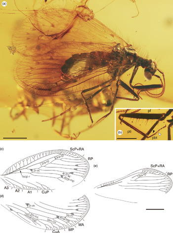
Habitus, foreleg and wing venation of Creagroparaberotha cuneata sp. nov. (holotype, AMNH Bu-SY20) in mid-Cretaceous amber from northern Myanmar: (a) right lateral view; (b) left foreleg, left lateral (outer) view, arrow showing spine-like setae on external ventral edge and open arrow showing long curved spine distally in the probasitsarsus; (c) right forewing; (d) left forewing; and (e) right hindwing. Abbreviations as for Figures 1 and 2. Scale bars (a, c–e) 1.0 mm; and (b) 0.5 mm.

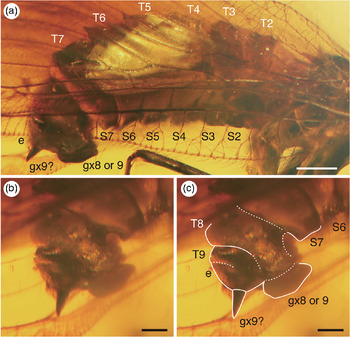
Abdomen of Creagroparaberotha cuneata sp. nov. (holotype, AMNH Bu-SY20) in mid-Cretaceous amber from northern Myanmar: (a) right lateral view; (b) genital segments, latero-caudal view; and (c) genital segments, latero-caudal view with white line and doted line indicating each segment and gonocoxite. Abbreviations as for Figure 5. Scale bars (a) 0.5 mm; and (b, c) 0.2 mm.

Zoobank LSID. urn:lsid:zoobank.org:act:1C533896-CACA-4757-AEDE-8557557F1A61
Derivation of name. From the Latin ‘cuneatus’, meaning ‘wedge-shaped’.
Holotype. A nearly completely preserved female adult (Fig. 9a), left hindleg originally being lost, preserved in a c. 29.0 mm × 9.2 mm × 8.4 mm narrowly cylindrical, yellow, transparent amber piece; specimen accession number AMNH Bu-SY20, deposited in AMNH.
Species diagnosis. Antennae with 37 flagellomeres (32 in C. groehni). Procoxa elongate, slightly longer than profemur (only shorter than profemur in C. groehni); protibia with at least 19 spine-like setae on ventral edge (at least 12 in C. groehni); probasitarsus with at least one ventral spine-like setae (six in C. groehni); slightly curved spine located on dorsal edge of probasitarsus only (two proximal protarsomeres present in C. groehni); one dark brown maculation spot located at primary fork inside of stem of MA (completely absent in C. groehni).
Description. Body. Length c. 4.0 mm as preserved (measured from vertex to apex of abdomen).
Head. Dorsum with slightly protuberance on vertex, with long scarce setae. Eyes large, protruding laterally; postocular lobe not inflated. Antenna: scapus elongate, c. 5 times as long as width, slightly dilated toward apex; pedicellus one-half length of scape; both covered with fine setae; flagellum moniliform comprising 37 flagellomeres, with scattered fine setae on each segment. Maxillary palpi 5 segmented; labial palpi 3 segmented.
Thorax. Well preserved. Pronotum length c. 0.6 mm, elongate, slightly expanded in anterior third, weakly wider than width of head between compound eyes, covered with long fine setae. Mesothorax: mesoprescutum with scattered fine setae along antero-lateral edge; mesoscutum and mesoscutellum visible; tegula with scattered fine setae. Metathorax structures not visible.
Legs. Foreleg (Fig. 9b). Procoxa elongate, c. 1.0 mm length, slightly longer than profemur, narrowed distally. Protrochanter elongate, slightly curved. Profemur long, c. 1.2 mm, broadened towards proximally, covered with dense fine setae, which become gradually longer towards apex on dorsal and lateral edges, with spines of variable lengths forming two rows on ventral edge, only slightly curved distally. Protibia long, c. 1.0 mm, slender, covered with fine setae on dorsal and lateral edges; several pairs of long setae on dorsal edge; single row of at least 19 proximally inclined spine-like setae, regularly spaced on external ventral edge. Protarsus: all protarsomeres covered with fine setae; probasitarsus longest, relative length of tarsomeres 3–1–1.3–1–1.5, with one spine-like setae on external ventral edge and one long curved spine distally; claws curved.
Midleg. Mesocoxa stout and conical. Mesotrochanter elongate, slightly curved. Mesofemur long, c. 1.0 mm, slender, covered with dense long setae. Mesotibia long, c. 1.3 mm, slender, covered with dense long setae and several long, stronger, almost perpendicular setae. Mesotarsus: all mesotarsomeres covered with fine setae; first tarsomere longest, relative length of tarsomeres 6.3–1.8–1.5–1–1.5; claws small, curved.
Hindleg. Left hindleg almost lost except metacoxa and matatrochanter. Metacoxa similar to mesocoxa. Metatrochanter elongate, slightly curved. Metafemur long, c. 1.2 mm, slender, covered with dense long setae. Metatibia long, c. 1.8 mm, slender, covered with dense long setae, several long stronger almost perpendicular setae. Metatarsus: second to fifth tarsomeres and claws not visible.
Wings (Fig. 9c–e). Forewing hyaline, broadly oval, c. 5.0 mm length, c. 2.2 mm width, wing venation partly obscured due to hindwing overlapping abdomen. Trichosors present along entire wing margin; marginal portions of veins and trichosors with tufts of fine setae; fine setae sparsely present on dorsal and ventral surfaces of longitudinal veins, but absent on crossveins. Costal space relatively broad, most subcostal veinlets once forked. ScP fused with RA at apical third. Subcostal space broad; proximal crossvein (1scp-r) located proximal forth. RP divided into four branches; RP1 deeply forked (right forewing, Fig. 9c). Two ra-rp crossveins present; 2ra-rp located between origin of RP3 and RP4; 3ra-rp located distally stem of RP2. M divided into MA and MP; one crossvein (4ma-mp) present. Two r-m crossveins present; 2r-m located slightly distally to RP and MA; 4r-m located between RP4 and MA. Cu divided into CuA and CuP; one crossvein (2cua-cup) present. Two m-cu crossveins ovserved; 2m-cu between distal to stem MP and CuA; 4m-cu between MP and CuA. Three anal veins present: A1, A2 and A3; A3 simple. Wing membrane with following dark brown maculation: margined crossveins 3ra-rp and 4ma-mp; semicircular spots at anterior part of 2r-m and 1cu-a; several spots at primary forks inside of stem of RPs; and one spot at primary fork inside of stem of MA.
Hindwing hyaline, elongate-ovoid, c. 4.3 mm length, c. 1.9 mm width, wing venation partly obscured due to forewing overlapping abdomen. Trichosors present on wing margin; marginal portions of veins and trichosors with tufts of fine setae; fine setae sparsely present on dorsal and ventral surfaces of longitudinal veins, but absent on crossveins. Costal space narrowed. ScP fused with RA at apical third. Subcostal space without crossvein.
Abdomen (Fig. 10). Elongate ovoid, moderately scattered setae, partly invisible due to coverage of wings on abdomen. Tergite 5 and 6 developed each posterior margin. Terminal segment unclear; gonocoxites 9 (or 8) visible, scattered setae on surface; one acute projection (possibly gonocoxites 9) developed on posterior of ectoproct.
Remarks. C. cuneata sp. nov. has a remarkable acute projection at the posterior margin of the ectoproct (Fig. 10b, c). Among the members of extant Mantispoidea, the females of Anchieta, Plega and Trichoscelia (Mantispidae: Symphrasinae) have an elongated and developed gonocoxite 9 (e.g. Liu et al. Reference Liu, Winterton, Wu, Piper and Ohl2015, fig. 11; Ardila-Camacho & García, 2015, figs 2, 6, 13). However, in extant Berothidae and Rhachiberothidae, no species with such an elongated gonocoxite have been found. In the family Dilaridae, the females have an elongated gonocoxite 9 (ovipositor) (Aspöck & Aspöck, Reference Aspöck and Aspöck2008, figs 113, 114; Liu et al. Reference Liu, Aspöck, Winterton, Zhang and Aspöck2017, fig. 9). Dilaridae have been recognized as a sister taxon to Mantispoidea based on several morphological traits (Aspöck et al. Reference Aspöck, Plant and Nemeschkal2001, Reference Aspöck, Haring and Aspöck2012; Aspöck & Aspöck, Reference Aspöck and Aspöck2008). Nevertheless, recent molecular phylogenetic studies suggest that Dilaridae is actually a sister taxon to all lacewing families, except for Coniopterygidae, Nevrorthidae, Sisyridae and Osmylidae (Engel et al. Reference Engel, Winterton and Breitkreuz2018; Winterton et al. Reference Winterton, Lemmon, Gillung, Garzon, Badano, Bakkes, Breitkreuz, Engel, Lemmon, Liu, Machado, Skevington and Oswald2018; Song et al. Reference Song, Li, Zhai, Bozdoğan and Yin2019).
Genus Micromantispa Shi et al. Reference Shi, Ohl, Wunderrich and Ren2015
Type species. Micromantispa cristata Shi et al. Reference Shi, Ohl, Wunderrich and Ren2015, p. 418.
Species composition. M. cristata Shi et al. Reference Shi, Ohl, Wunderrich and Ren2015, M. galeata sp. nov. and M. spicata sp. nov.
Revised diagnosis. Vertex slightly inflated; antennae with scapus relatively short, c. 2.5 times as long as width. Protibia bearing two or three spines on the distal area of ventral ridge, covered with thick prostrate setae on dorsal edge. Probasitarsus elongate, with two spine-like setae on ventral edge and one long spine on the end; second protarsomere bearing one spine at the end. Forewing hyaline, elongate-oval; RP divided into four branches, intra-RP crossvein between RP3 and RP4 present. Hindwing hyaline, ovoid; basal crossvein between RP and M present.
Remarks. This genus was originally established within the subfamily Drepanicinae (Mantispidae) (Shi et al. Reference Shi, Ohl, Wunderrich and Ren2015). However, Makarkin (Reference Makarkin2015c) suggested that the genus Micromantispa should instead belong to the subfamily Paraberothinae (Rhachiberothidae). Micromantispa was formally treated as a member of Mantispidae in a review article of the fossil Mantispidae (Jepson, Reference Jepson2015). At present, the morphological traits of the two new species described in this study strongly support the conclusion that Micromantispa belongs to Rhachiberothidae, based on characters such as the elongation of pronotum anterior to forelegs (sensu Aspöck & Mansell, Reference Aspöck and Mansell1994).
Micromantispa galeata sp. nov.
Habitus, forelegs and wing venation of Micromantispa galeata sp. nov. (holotype, AMNH Bu-SY21) in mid-Cretaceous amber from northern Myanmar: (a) left lateral view; (b) proximal area of left protibia, line and arrows showing four slight protrusions; (c) right protibia and probasitarsus, arrows showing two spines distally ventral ridge, open arrows showing two spines on probasitarsus, and linearrow showing one long curved spine distally on probasitarsus; (d) right forewing; and (e) right hindwing. Abbreviations as for Figures 1 and 2. Scale bars (a, d, e) 1.0 mm; and (b, c) 0.1 mm.

Genital segments of Micromantispa galeata sp. nov. (holotype, AMNH Bu-SY21) in mid-Cretaceous amber from northern Myanmar: (a) genital segments, left lateral view; and (b) genital segments, left lateral view with white line indicating each segment, gonapophysis and gonocoxite. Abbreviations as for Figure 5. Scale bars 0.2 mm.

Zoobank LSID. urn:lsid:zoobank.org:act:327E6B28-B590-4991-B2D2-E8FA5DA6364F
Derivation of name. From the Latin ‘galeatus’, meaning ‘helmeted’.
Holotype. A nearly completely preserved female adult (Fig. 11a), partly obscured due to cracks, preserved in a c. 13.1 mm × 9.3 mm × 3.0 mm flattened oval, clear yellow, transparent amber piece; specimen accession number AMNH Bu-SY21, deposited in AMNH.
Species diagnosis. Protibia with at least four only slightly developed protrusions on the proximal area of ventral ridge, bearing two spines on the distal area of ventral ridge (three in M. cristata). Hindwing RP divided into four branches; intra-RP crossvein absent (one intra-RP crossvein present in M. spicata).
Description. Body. Length c. 2.6 mm as preserved (measured from vertex to apex of abdomen).
Head. Vertex slightly inflated with long scarce setae. Compound eyes large. Antennae with scapus slightly elongate, c. 2.5 times as long as width; pedicellus short, two-fifths length of scape; both with fine setae; flagellum moniliform composed of 37 flagellomeres, with scattered fine setae on each segment. Segmentation of maxillary palpi and labial palpi unclear, each terminal segment acute.
Thorax. Pronotum length c. 0.4 mm, elongate anterior to forelegs, with scattered fine setae. Mesothorax with scattered long fine setae. Metathorax with slightly scattered fine setae.
Legs. Foreleg (Fig. 11b, c). Procoxa elongate, c. 0.9 mm length, shorter than profemur, bearing 2–3 fine setae on outer lateral surface. Protrochanter elongate, slightly curved. Profemur long, c. 1.0 mm, slightly broadened, covered with dense fine setae on surface, with spines of variable lengths forming two rows on ventral edge, only slightly curved towards distal, prural spines with rounded tip on the distal area of ventral ridge. Protibia long, c. 0.8 mm, slender, covered with fine setae on surface, at least four slight protrusions on the proximal area of ventral ridge, bearing two spines on distally ventral ridge. Protarsus: probasitarsus with two small spines on ventral ridge and one long curved spine distally; second to fifth tarsomere unclear.
Midleg. Mesocoxa stout and conical. Mesotrochanter elongate, slightly curved. Mesofemur long, c. 0.6 mm, slender, covered with dense setae. Mesotibia long, c. 0.9 mm, slender, covered with dense long setae. Mesotarsus: all mesotarsomeres covered with fine setae; first tarsomere longest, relative length of tarsomeres 6.5–2.3–1.5–1–1.5; claws small, curved.
Hindleg. Metacoxa similar to mesocoxa. Metatrochanter elongate, slightly curved. Metafemur long, c. 0.7 mm, slender, covered with dense setae. Metatibia long, c. 1.1 mm, slender, covered with dense long setae. Metatarsus: all metatarsomeres covered with fine setae; first tarsomere longest, relative length of tarsomeres 5.8–2.1–1.3–1–1.2; claws small, curved.
Wings (Fig. 11d, e). Forewing hyaline, elongate-ovoid, c. 3.0 mm length, c. 1.2 mm width. Trichosors present along the whole wing margin; marginal portions of veins and trichosors with tufts fine setae; fine setae sparsely present on dorsal and ventral surfaces of longitudinal veins, but absent on crossveins. Costal space slightly broad; middle two subcostal veinlets once forked. ScP fused with RA at apical third. Subcostal space broad; proximal crossvein (1scp-r) located between origins of RP and M. RP divided into four branches; one intra-RP crossvein (4rp3-rp4) present. Two ra-rp crossveins present; 2ra-rp located between origin of RP3 and RP4; 3ra-rp located stem of RP1. M divided into MA and MP; one crossvein (4ma-mp) present. Two r-m crossveins present; 2r-m located slightly distal to RP and MA origin; 4r-m located between RP4 and MA. Cu divided into CuA and CuP; one crossvein (2cua-cup) present. Three m-cu crossveins present; 1m-cu located slightly distal to M and CuA origin; 2m-cu between distal to stem MP and CuA; 4m-cu between MP and CuA. Three anal veins present: A1, A2 and A3; A3 simple.
Hindwing hyaline, elongate-ovoid, c. 2.3 mm length, c. 1.0 mm width. Trichosors present wing margin; marginal portions of veins and trichosors with tufts fine setae; fine setae sparsely present on dorsal and ventral surfaces of longitudinal veins, but absent on crossveins. Costal space narrowed, subcostal veinlets poorly preserved. ScP fused with RA at apical third. Subcostal space no crossvein. RP divided into four branches; intra-RP crossvein absent. Two ra-rp crossveins present; 2ra-rp located between RP3 and near RP4 origin; 3ra-rp located distal to stem of RP1. M divided into MA and MP, one crossvein (4ma-mp) present. One r-m crossveins ovserved, 4r-m located between RP4 and MA. Two r-m crossveins present; 1r-m located near stems of RP and M; 4r-m located between RP4 and MA. CuA visible, CuP not visible. One crossvein (4m-cu) located between MP and CuA.
Abdomen (Fig. 12). Abdomen preserved, partly invisible due to milky layer on body surface; oval, slightly compressed laterally, slightly curled ventrally, moderately scattered setae, segments 3–7 swollen. Tergite 9 much narrower than tergite 8. Gonapophyses 8 (gp 8) paired, elongate falciform. Gonocoxites 9 (gx9) exposed and deformed, structure unclear. Ectoprocts paired, valvate, covered with thin setae.
Remarks. The morphological traits of the female genital segments in M. galeata closely resemble those of Micromantispa cristata Shi et al. Reference Shi, Ohl, Wunderrich and Ren2015. The sickle-shaped gonapophysis 8 in the genital segments was observed on both anterolateral sides of gonocoxite 9, as in M. cristata (see Shi et al. Reference Shi, Ohl, Wunderrich and Ren2015, fig. 4).
Micromantispa spicata sp. nov.
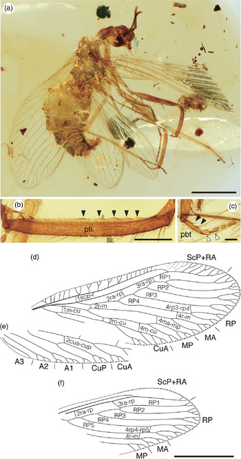
Habitus, forelegs and wing venation of Micromantispa spicata sp. nov. (holotype, AMNH Bu-SY22) in mid-Cretaceous amber from northern Myanmar: (a) right lateral view; (b) right protibia, arrows showing only slightly five protrusions; (c) left protarsus, arrows showing two spines on probastarsus and open arrows showing long curved spine distally on probasitarsus and second protarsomere; (d) left forewing; (e) right forewing; and (f) left hindwing. Abbreviations as for Figures 1 and 2. Scale bars (a, d–f) 1.0 mm; (b) 0.2 mm; and (c) 0.1 mm.

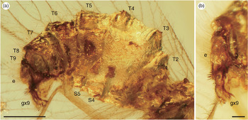
Abdomen of Micromantispa spicata sp. nov. (holotype, AMNH Bu-SY22) in mid-Cretaceous amber from northern Myanmar: (a) right lateral view; and (b) genital segments, latero-caudal view. Abbreviations as for Figure 5. Scale bars (a) 0.5 mm; and (b) 0.1 mm.

Zoobank LSID. urn:lsid:zoobank.org:act:E9C151B4-0D95-46FB-801D-29E38379F93F
Derivation of name. From the Latin ‘spica’, meaning ‘ear of grain’.
Holotype. Well-preserved, incomplete specimen of adult, sex undetermined (Fig. 13a), almost the whole antennae and part of the right forewing lost due to over polishing, preserved in a c. 11.0 mm × 7.3 mm × 3.3 mm in cabochon, clear yellow, transparent amber piece; specimen accession number AMNH Bu-SY22, deposited in AMNH.
Species diagnosis. Protibia five slight protrusions on the proximal area of ventral ridge, bearing two spines on the distal area of ventral ridge (three in M. cristata). Hindwing RP divided into five branches; one intra-RP crossvein (4rp4-rp5) present.
Description. Body. Length c. 3.1 mm (measured from vertex to apex of abdomen), slightly curled ventrally.
Head. Distorted anteroposterior direction. Compound eyes large. Antennae nearly completely lost, with only 14 flagellomeres preserved of left antenna; scapus slightly elongate, 2.5 times as long as width; pedicellus short, three-fifths length of scape; both with fine setae; flagellum moniliform, with scattered fine setae on each segment. Segmentation of maxillary palpi and labial palpi unclear, but each terminal segment acute.
Thorax. Pronotum elongate, distorted anteroposteriorly, slightly broken in posterior end. Mesothorax: mesopresctum with spine towards anterior; mesosctum projected, scattered with fine setae. Metathorax not completely visible.
Legs. Foreleg (Fig. 13b, c). Procoxa elongate, c. 1.1 mm length, shorter than profemur, slightly curved, bearing at least single row of six fine setae on outer lateral surface. Protrochanter elongate, slightly curved. Profemur long, c. 1.1 mm, slightly broadened, covered with dense fine setae on surface, with spines of variable lengths forming two rows on ventral edge, only slightly curved towards distal, prural spines with rounded tip on the distal area of ventral ridge. Protibia long, c. 0.9 mm, slender, covered with fine setae on surface, five slight protrusions on the proximal area of ventral ridge, bearing two spines distally on ventral ridge. Protarsus: all protarsomeres covered with fine setae; probasitarsus longest, relative length of tarsomeres 2.7–1.3–1.3–1–1.5, with two small spines on ventral ridge and one long curved spine distally; second protarsomere with one long curved spine distally; claws curved.
Midleg. Mesocoxa and mesotrochanter distorted. Mesofemur long, c. 0.8 mm, slender, covered with dense long setae. Mesotibia long, c. 1.0 mm, slender, covered with dense long setae. Mesotarsus: all mesotarsomeres covered with fine setae; first tarsomere longest, relative length of tarsomeres 6–2.2–1.3–1–1; claws small, curved.
Hindleg. Metacoxa and metatrochanter distorted. Metafemur long, c. 0.8 mm, slender, densely covered with long setae. Metatibia long, c. 1.4 mm, slender, densely covered with long setae. Metatarsus: all metatarsomeres covered with fine setae; first tarsomere longest, relative length of tarsomeres 5.8–2–1.3–1–1.3; claws small, curved.
Wings (Fig. 13d–f). Forewing hyaline, partly invisible due to curvature, elongate-ovoid, c. 3.6 mm length, c. 1.3 mm width. Trichosors present along the whole wing margin; marginal portions of veins and trichosors with tufts fine setae; fine setae sparsely present on dorsal and ventral surfaces of longitudinal veins, but absent on crossveins. Costal space slightly broad, middle five subcostal veinlets once forked. ScP fused with RA with strong curvature inwards at apical third. Subcostal space proximal crossvein (1scp-r) located nearly at origin of RP. RP divided into four branches; RP1 deeply forked; one intra-RP crossvein (4rp3-rp4) present. Two ra-rp crossveins present; 2ra-rp located between origin of RP3 and RP4; 3ra-rp located at stem of RP1. M divided into MA and MP; one crossvein (4ma-mp) present. Two r-m crossveins present; 2r-m located slightly distal to RP and MA; 4r-m located between RP4 and MA. Cu divided into CuA and CuP; one crossvein (2cua-cup) present. Three m-cu crossveins present; 1m-cu located slightly distal to M and CuA; 2m-cu between distal to stem MP and CuA; 4m-cu between MP and CuA. Three anal veins present: A1, A2 and A3; A3 simple.
Hindwing hyaline, elongate-ovoid, c. 2.8 mm length, 1.0 mm width, wing venation partly obscured due to wings overlapping abdomen. Trichosors present on wing margin; marginal portions of veins and trichosors with tufts of fine setae; fine setae sparsely present on dorsal and ventral surfaces of longitudinal veins, but absent on crossveins. Costal space narrowed, subcostal veinlets poorly preserved. ScP fused with RA at middle slightly distal. Subcostal space no crossvein. RP divided into five branches; one intra-RP crossvein (4rp4-rp5) present. Two ra-rp crossveins present; 2ra-rp located between RP3 and near RP4 origin; 3ra-rp located at stem of RP1. M divided into MA and MP. One r-m crossveins observed, 4r-m located between RP4 and MA.
Abdomen (Fig. 14). Abdomen preserved, part of segments unclear; oval, slightly compressed laterally, moderately scattered setae; segments partly unclear. Genital segments unclear; gonocoxites 9 paired developed finger-like projections, bearing with strong setae on surface; ectoproct covered with thin setae.
Comments. Although some females of extant Rhachiberothidae have developed gonocoxites (Aspöck & Mansell, Reference Aspöck and Mansell1994; Aspöck & Aspöck, Reference Aspöck and Aspöck1997, Reference Aspöck and Aspöck2008, figs 119–122), the holotypes of Micromantispa cristata and Astioberotha falcipes sp. nov. with undeveloped gonocoxites can be confidently interpreted as females (Shi et al. Reference Shi, Ohl, Wunderrich and Ren2015, figs 4, 5a, 11). The holotype of Spinoberotha mickaelacrai Nel et al. Reference Nel, Perrichot, Azar and Néraudeau2005a was originally considered female based on the presence of developed gonocoxites (referred to as ‘hypocauda’ in the original description). However, Makarkin (Reference Makarkin2015a) mentioned that the true female gonocoxites (hypocaudae) are usually directed anteriorly or ventrally, but those of Spinoberotha are caudally directed; he therefore suggested that the holotype may actually be a male. Although we confirmed a structure that may be considered a gonocoxite in the holotype of M. spicata, it was difficult to confidently determine the sex as we failed to observe the details of the genital segments.
4. Discussion
4.a. Morphological diversity of forelegs
This study revealed a remarkable diversity of paraberothines in the mid-Cretaceous Burmese amber from northern Myanmar. Previously, four species in four genera, together with an undescribed taxon, were known from Burmese amber (Engel, Reference Engel2004; Engel & Grimaldi, Reference Engel and Grimaldi2008; Shi et al. Reference Shi, Ohl, Wunderrich and Ren2015; Makarkin, Reference Makarkin2015a). In this study, we have added as many as four new fossil genera and seven new species, making Burmese amber by far the most species-rich deposit of Paraberothinae (Table 1). The recent explosion of palaeontological works based on Burmese amber now serves as an astonishing summary of the mid-Cretaceous biota (Ross, Reference Ross2019). Our study also supports the potentially diverse, but still undocumented, insect fauna in Burmese amber.
In this study, we have shown a surprising morphological diversity among the Burmese amber paraberothines. The newly described materials are generally well preserved, illuminating fine details of the foreleg structures that have not been previously addressed. Some important characteristics were clearly observable, and are sufficient for direct morphological comparisons among species, genera and even subfamilies. Our imaging efforts have revealed that morphological structures among the Cretaceous taxa were highly diverse, with a series of significant variations (e.g. length of scape, number of flagellomeres, foreleg structure and wing venation; summarized in Table 2).
The morphological diversity of the forelegs is particularly interesting, demonstrating great variation even within the Burmese amber taxa (Fig. 15). This morphological diversity is comparable to that of the entire subfamily. For example, the length of the profemur among Paraberothinae ranges from c. 0.5 mm in Spinoberotha to c. 1.9 mm in Kujiberotha; within the Burmese amber paraberothines it ranges from c. 0.7 mm in Stygioberotha gen. nov. and Uranoberotha gen. nov. to c. 1.8 mm in Astioberotha. Importantly, the number and shape of spines covering the protibiae varied (e.g. 2 spines in Astioberotha gen. nov. vs. 22 in Uranoberotha gen. nov.; spine shape is blade-like in Acanthoberotha, curved and hooked in Stygioberotha, and acicular in Creagroparaberotha cneata and Uranoberotha). Furthermore, the morphological structure of the probasitarsus varies greatly among taxa (e.g. remarkably elongated in Stygioberotha, bearing 9–10 spine-like setae in Acanthoberotha, 5–7 in Uranoberotha and 2 in Micromantispa). In addition, it is notable that the slight protrusions on the ventral ridges in the protibiae of Astioberotha and Micromantispa have only been found in Burmese amber (Figs 4c, 11b, 13b). These spines on the protibia and probasitarsus are completely lost in the remaining taxa of Rhachiberothidae. The presence of spines, or spine-like setae, on the inner edge of protibia is a synapomorphy of Paraberothinae, and it has been considered a plesiomorphy within Rhachiberothidae (Makarkin & Kupryjanowicz, Reference Makarkin and Kupryjanowicz2010; Makarkin, Reference Makarkin2015a). Indeed, the earliest fossil fauna of paraberothines from the Lower Cretaceous Lebanese amber had numerous spines, or spine-like setae, on the protibia (e.g. 12 in Paraberotha, 42 in Raptorapax, and numerous in Chimerhaberotha and Spinoberotha). The recently found Kujiberotha teruyukii from the Upper Cretaceous Kuji amber from Japan has nine spine-like setae on the external ventral ridge of the probasitarsus. Based on previous studies and our new material, the morphological diversity of forelegs is likely much greater than the extant Rhachiberothidae.
Reconstructed left forelegs of described paraberothine species in this paper: (a) Acanthoberotha cuspis gen. et sp. nov.; (b) Astioberotha falcipes gen. et sp. nov.; (c) Stygioberotha siculifera gen. et sp. nov.; (d) Uranoberotha chariessa gen. et sp. nov.; (e) Creagroparaberotha cuneata sp. nov.; and (f) Micromantispa spicata sp. nov. Scale bars 1.0 mm.

In the modern praying mantis, a completely different taxonomic group with similar raptorial forelegs, the diversity of foreleg morphology is thought to reflect the diversity of the habitat and predatory targets of the mantis (Wieland, Reference Wieland2013; Brannoch et al. Reference Brannoch, Wieland, Rivera, Klass, Béthoux and Svenson2017). The Austral Paraoxypilinae (Mantodea: Amorphoscelidae) have modified raptorial forelegs, possibly as an evolutionary adaptation to hunting ants (Wieland, Reference Wieland2013). In addition, the assassin bug (Heteroptera: Reduvioidea) also has various foreleg modifications that have evolved for prey capture (Zhang et al. Reference Zhang, Gordon, Forthman, Hwang, Walden, Swanson, Johnson, Meier and Weirauch2016). For example, in some species of Harpactorinae (Heteroptera: Reduviidae), it is suggested that differences in space and structure when the foretibia and forefemur are closed reflect the size and smoothness of the surface of the prey (Cai & Tomokuni, Reference Cai and Tomokuni2003; Zhao et al. Reference Zhao, Pham, Truong and Cai2014). Similarly, the diversity of foreleg morphology of Paraberothinae may also reflect ecological diversity. As mentioned in Section 3 above, the morphological diversity of forelegs of the extant Rhaciberothidae and Mantispidae is not necessarily high; that of the Mesozoic Paraberothinae is much higher. Based on these results, it is suggested that the morphological diversity of the forelegs of Paraberothinae reflects the diversity of prey and behaviour, as is the case in modern praying mantises and assassin bugs.
4.b. Biogeographical implications
The distribution of the extant rhachiberothids is limited only to sub-Saharan Africa (Aspöck & Aspöck, Reference Aspöck and Aspöck1997). In contrast, the extinct members of Rhachiberothidae have been found in various Lower Cretaceous – mid-Eocene amber deposits in the Northern Hemisphere, with different geological backgrounds (Table 1; Fig. 16). Most of the fossil localities of rhachiberothids are of Laurasian origin, whereas Lebanese amber is of Gondwanan origin (e.g. Azar et al. Reference Azar, Gèze, Acra and Penney2010); the Burmese amber was also recently suggested to have originated from Gondwana (Poinar, Reference Poinar2019; Westerweel et al. Reference Westerweel, Roperch, Licht, Dupont-Nivet, Win, Poblete, Ruffet, Swe, Thi and Aung2019). These backgrounds suggest a possible wider distribution on Pangea before the continental break-up; if so, this supports an early (probably Jurassic) origin of this group, as implied by recent studies (Engel et al. Reference Engel, Winterton and Breitkreuz2018). In addition, Khramov (Reference Khramov2013) mentioned that there are many as-yet undescribed fossils of Rhachiberothidae in the Upper Jurassic deposits in Karatau, Kazakhstan. Finally, our contribution adds a hidden assemblage to the mid-Cretaceous rhachiberothid fauna. This incredible mid-Cretaceous diversity also supports the earlier origin of Rhachiberothidae, at least for the subfamily Paraberothinae.
Global palaeogeographic reconstruction of the mid-Cretaceous (100 Ma) period, showing the distributions of the extant and extinct Rhachiberothidae; modified from Global Paleogeography and Tectonics in Deep Time © 2016 Colorado Plateau Geosystems Inc. The locality of amber-producing forests is based on Westerweel et al. (Reference Westerweel, Roperch, Licht, Dupont-Nivet, Win, Poblete, Ruffet, Swe, Thi and Aung2019).

Rhachiberothidae is currently considered to be closely related to the families Mantispidae and Berothidae, which constitute the superfamily Mantispoidea (Winterton et al. Reference Winterton, Hardy and Wiegmann2010, Reference Winterton, Lemmon, Gillung, Garzon, Badano, Bakkes, Breitkreuz, Engel, Lemmon, Liu, Machado, Skevington and Oswald2018; Engel et al. Reference Engel, Winterton and Breitkreuz2018; Song et al. Reference Song, Li, Zhai, Bozdoğan and Yin2019). The modern distribution of these two families is cosmopolitan, spanning all biogeographical regions. However, they have not been found at high latitudes in the Northern Hemisphere (Ohl, Reference Ohl, Pantaleoni, Letardi and Corazza2005; Wedmann & Makarkin, Reference Wedmann and Makarkin2007; Aspöck & Randolf, Reference Aspöck and Randolf2014). Thus, only Rhachibetohidae has a restricted distribution within the superfamily. As mentioned in the text above, Paraberothinae was once widespread in the Cretaceous, but other extinct rhaciberothids have been restricted to the Eocene Baltic and Oise ambers, with three species in two genera (Whalley, Reference Whalley1983; Engel, Reference Engel2004; Nel et al. Reference Nel, Perrichot, Azar and Néraudeau2005a, b; Makarkin & Kupryjanowicz, Reference Makarkin and Kupryjanowicz2010). A similar pattern (i.e. significant depletion or extinction after the Eocene Epoch) is found in the families Nymphidae and Psychopsidae as neuropteran groups (Engel & Grimaldi, Reference Engel and Grimaldi2008). Both families have already been found in Burmese amber (Engel & Grimaldi, Reference Engel and Grimaldi2008; Ross, Reference Ross2019), whereas the recent taxa of Nymphidae are confined to Australia and New Guinea, and those of Psychopsidae are distributed in southern Africa, Southeast Asia and Australia (Bakkes et al. Reference Bakkes, Sole and Mansell2017; Oswald, Reference Oswald2019). Judging from their current distributional patterns, these two relict families may be considered to be of so-called Gondwanan origin. However, the past wider distribution of rhachiberothids strongly indicates the difficulty of accurately determining biogeographic origin based solely on modern taxa.
4.c. Evolution of Rhachiberothidae
Although our study helps to clarify the hidden palaeodiversity of Rhachiberothidae in the Mesozoic, the evolution of rhachiberothids still remains unclear. Based on their morphological diversity, they may have had a complex evolutionary history over a long geological timescale. In this study, we abandoned the possibility of constructing a phylogenetic tree using rhachiberothid fossils, because several previously described extinct taxa are poorly preserved and have vague original descriptions. This lack of morphological information prevents us from elucidating the relationships among species and genera within Rhachiberothidae. Considering the apparently high diversity of paraberothines, future studies will likely discover more well-preserved fossils. It would then be possible to carry out a proper phylogenetic analysis based on informative rhachiberothid fossils.
In Rhachiberothidae, Makarkin (Reference Makarkin2015a) has considered that Paraberothinae could be a stem group in comparison with Rhachiberothinae, based on the presence of several primitive features (e.g. protibia with spines or spine-like setae on the inner edge and the presence of absolute CuP in the hindwing). Although the presence of spines or spine-like setae on the protibia suggests a monophyly of Paraberthinae (Pérez-de la Fuente & Peñalver, Reference Pérez-de la Fuente and Peñalver2019), the phylogenetic relationship of this subfamily is not well resolved as no phylogenetic analysis has been made for this group and even the entire family; more evidence is needed to discuss phylogenetic relationships within the family. In fossil Paraberothinae, the venation of CuP in the hindwing is diverse, regardless of its geological age and deposits (Table 2). For example, Stygioberotha gen. nov. and Uranoberotha gen. nov. have simplified, but not reduced, CuP venation in the hindwing, while the venation of CuP in the hindwing in extant and extinct Rhachiberothinae is partly reduced (e.g. Aspöck & Mansell, Reference Aspöck and Mansell1994, figs 18, 30, 43; Aspöck & Aspöck, Reference Aspöck and Aspöck1997, fig. 36).
Oisea celinea (Nel et al. Reference Nel, Perrichot, Azar and Néraudeau2005a, b) was originally described from the earliest Eocene Oise amber of France as a member of Rhachiberothinae. However, a later study by Makarkin & Kupryjanowicz (Reference Makarkin and Kupryjanowicz2010) refuted this taxonomic placement and considered it as incertae sedis within Rhachiberothidae. Furthermore, Makarkin & Kupryjanowicz (Reference Makarkin and Kupryjanowicz2010) mentioned that O. celinea shares several morphological traits with both subfamilies, providing firm evidence that each is indeed a valid subfamily. For instance, this species has a foretibia without spines on the inner edge and intermediate subcostal crossvein 2scp-r in the forewing, as is also seen in Rhachiberothinae. However, this fossil taxon has a forewing of distally fused ScP and RA, as in Paraberothinae. This combination of characters is of particular interest for inferring the rhachiberothid evolution. However, the use of a poorly preserved adult of the single holotype makes the application of phylogenetic methods challenging.
As discussed in Section 4.b above, the post-Eocene decline of Rhachiberothinae and extinction of Paraberothinae are interesting subjects for further study. Although we did not address these phenomena in this paper, possible explanations include dramatic climate change during the Eocene Epoch (Brunke et al. Reference Brunke, Chatzimanolis, Metscher, Wolf-Schwenninger and Solodovnikov2017) or a failure to shift prey preference. Unfortunately, there is little ecological and behavioural information available for extant or extinct rhachiberothids. We look forward to future discoveries regarding the morphological evolution, diversification and decline of the thorny lacewings.
Acknowledgements
We thank Dr Munetoshi Maruyama (Kyushu University Museum, Fukuoka, Japan) for useful suggestions on nomenclature. We also would like to thank Dr Ron Blakey (Colorado Plateau Geosystems Inc., Phoenix, AZ, USA) for his kind permission to reproduce a palaeogeographic map. The second author (SY) was supported by the JSPS (Japan Society for the Promotion of Science, Tokyo, Japan) Overseas Research Fellowship (no. 29-212).
Supplementary material
To view supplementary material for this article, please visit https://doi.org/10.1017/S0016756820000205