Introduction
Maternal age dependency of gestation time in hamster (Soderwall et al., Reference Soderwall, Kent, Turbyfill and Britenbaker1960; Navarro et al., Reference Stoddart, Roudebush and Fleming2000) and in other mammals, is a well demonstrated fact. Soderwall et al. (Reference Soderwall, Kent, Turbyfill and Britenbaker1960) reported that 1-month-old hamster females have gestation periods of 373 h, 4-month-old females gave birth after 378 h, 8-month-old females after 386 h and 14-month-old females after 402 h, i.e. they showed 24–29 h longer gestation periods than young females. We have recently shown (Trejo et al., Reference Trejo, Navarro, Ambriz and Rosado2005) that adult (8-month-old) nulliparous and multiparous females show significant asynchrony and retard on early embryo development (from two blastomeres to morula stages) when compared with nulliparous young (2-month-old) females.
Our knowledge of the factors that contribute to in vivo embryo development and transport are still incomplete. Solid experimental evidence supports the hypothesis that hormonal state of the female regulates ovum transport in mammals. Significant differences, however, between the rate of oviduct transport of embryos of different ages and of fertilized and unfertilized eggs in several mammalian species (Ortiz et al., Reference Ortiz, Bedregal, Carvajal and Croxatto1986), support the idea that the conceptus itself may autoregulate its own transport. This fact may be due, in part, to the secretion of some embryo-derived factors (Tranguch et al., Reference Tranguch, Steuerwald and Huet-Hudson2003) or to the capacity of the embryo to regulate its own development (Stoddart et al., Reference Stoddart, Roudebush and Fleming2001). The ability of embryos to develop to term, is related to the number of cell–cell adhesions between blastomeres during early development; more contacts result in a higher number of inner cell mass cells and, subsequently, a higher number of live offspring (Suzuki et al., Reference Suzuki, Togashi, Adachi and Toyoda1995). Formation of the firm cell–cell adhesions, indispensable for compaction and posterior differentiation of the conceptus (Neganova et al., Reference Neganova, Sekirina and Eichenlaub-Ritter2000; Kan et al. Reference Kan, Stemmler, Junghans, Kanzler, de Vries, Dominis and Kemler2007), require the presence and specific distribution of E-cadherin, a calcium-dependent cell–cell adhesion molecule and of their intracellular partner, β-catenin.
We have recently shown that preimplantation hamster embryo development and transport are highly synchronous in nulliparous young females (NYF), but not in adults. Nulliparous adult (8-month-old) females (NAF) and multiparous females (MAF), show significant asynchrony and retardation in early embryo development. In NYF all 8-cell embryos reached the uterus by 62 h after coitus. In adult females, both nulliparous and multiparous, a considerable number of 8-cell embryos failed to migrate into the uterus even at 67 h after coitus. We have also shown that the presence and distribution of E-cadherin on 6- and 8-cell embryos vary according to the parity and age of the females (Trejo et al., Reference Trejo, Navarro-Maldonado, Jimenez and Rosado2006). These differences are particularly important at the 8-cell stage. In a significant number of adult females, particularly in multiparous adult females, E-cadherin failed to be localized on all the cell–cell contacts. We think that these modifications in embryo development and transport are caused by abnormal functioning of E-cadherin-dependent cell–cell adhesion, which will lead to a delayed embryo compaction. We decided, therefore, to study by indirect immunofluorescence the presence and distribution of E-cadherin in 4-cell embryos obtained from young and adult hamster females. Our results show that alterations in expression and distribution of E-cadherin may be liable for the retard and asynchrony of hamster embryo preimplantation development, previously reported in adult hamster females, both nulliparous and multiparous.
Materials and Methods
All utilized animals were born in our animal facilities and kept at 25 °C under controlled conditions, with a light: dark cycle of 14: 10 h and food and water ad libitum. Normally cycling hamster females (Trejo et al., Reference Trejo, Navarro, Ambriz and Rosado2005), were divided into three experimental groups: (1) nulliparous young (2-month-old) females (NYF) weighing 128 ± 15 g; (2) nulliparous adult (8-month-old) females (NAF) weighing 168 ± 18 g; and (3) multiparous (four pregnancies), adult (7–8-month-old) females (MAF).
Females were individually caged with a male of proven fertility and trained to mount quickly (Navarro et al., Reference Navarro-Maldonado, Ambriz, Mundo, Trejo, Hernandez and Rosado2000; Trejo et al., Reference Trejo, Navarro, Ambriz and Rosado2005). Fifteen minutes were allowed for mating and the occurrence of the ejaculation pattern was directly observed. Fertilization was considered successful if sperm were in the vaginal smear taken 60 min after the mount.
As we know that adult females tend to retain 4-cell embryos longer than young females (Trejo et al., Reference Trejo, Navarro, Ambriz and Rosado2005), care was taken to obtain the required embryos at the earliest possible time. The time of fertilization, therefore, was carefully registered and the fertilized females were sacrificed by neck dislocation 58 h after coitus. The fallopian tubes were dissected and perfused (Navarro et al., Reference Navarro-Maldonado, Ambriz, Mundo, Trejo, Hernandez and Rosado2000; Trejo et al., Reference Trejo, Navarro, Ambriz and Rosado2005) to obtain 4-cell embryos. Fourteen embryos were obtained and processed for each of the female groups and 2–3 embryos were processed per female.
Four blastomeres embryos were incubated 60 s in a Tyrode acid solution (pH 2.5) (Sigma, T1788) to eliminate the zona pellucida. After twice washing with phosphate-buffered saline (PBS), the zona-free embryos were immunostained following the method proposed by Kawai et al. (Reference Kawai, Yamaguchi, Yoden, Hanada and Miyake2002) with some modifications. In short, embryos were fixed in 4% paraformaldehyde for 30 min. After two washes with PBS embryos were permeabilized by 10 min treatment with 1% Triton X-100 (Sigma, T8787). The zona-free, fixed and permeabilized embryos were then incubated in 5% albumin (fraction V, Sigma, A4503) for 1 h. Afterward the embryos were incubated with anti-E-cadherin antibody (Santa Cruz Biotechnology Inc.) (1: 50) during 2 h at room temperature and then stained in the dark for one hour at room temperature with fluorescein-tagged anti-IgG (1: 50) (Sigma) second antibody. Staining with no anti-E-cadherin antibody was utilized as a control for non-specific staining by the second antibody.
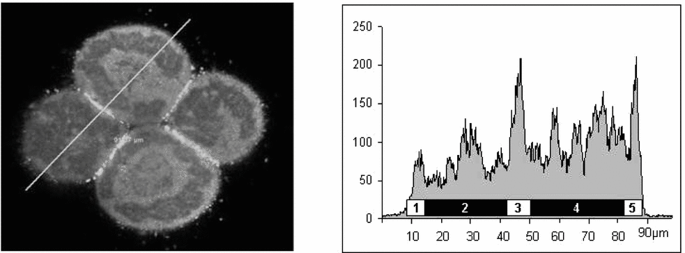
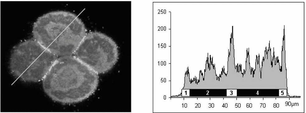
Immunoassayed embryos were mounted in Vecta shield (Vector Laboratories Inc.) and the intensity and distribution of fluorescence was observed and registered using a confocal scanning laser fluorescence microscope (Leica). In performing selected scans, the fluorescence recording was always set so that the observed fluorescence would totally fill the graph. Staining intensities for E-cadherin were quantified by computed densitometry, using the Number Cruncher Statistical Systems (NCSS, 2007). Independent integration of the area under the curve, divided in five sections (Fig. 1), allowed us to obtain the E-cadherin immune-concentration in: (i) the free membrane regions; (ii) the cytoplasm region (scanned twice following ∼perpendicular lines) (Fig. 2); and (iii) in the cell–cell adhesion regions (scanned transversally) of each embryo. In addition, fluorescence integration was obtained following longitudinally each of the four membrane-to-membrane adhesion zones of each embryo (Fig. 3). The collected data from each one of these regions per embryo were added. The respective means were obtained and these means were utilized in further statistical treatment as the amount per region and per embryo. In the case of the cytoplasm, the mean of the two perpendicular scans was similarly obtained. The data were analysed statistically by variance analysis. Differences were considered significant when p < 0.05.
Paraformaldehyde-fixed 4-cell hamster embryos were immunostained by incubation with anti-E-cadherin antibody. Intensity and distribution of fluorescence was observed and registered using confocal scanning laser fluorescence microscopy and the results of selected scans were submitted to computed integration. The figure shows the result of computed integration of a transverse confocal scanning passing through two adjacent blastomeres of a 4-cell embryo (left) obtained from a nulliparous young female and indicates (right) the five regions that were independently integrated. In the graph, x-axis indicates the distance in microns and y-axis fluorescence intensity in arbitrary units. Regions 1 and 5 indicate the amount of E-cadherin fluorescence observed in the free membrane surface of two adjacent blastomeres; 2 and 4 indicate the fluorescence found in the cytoplasm regions and 3 indicate the fluorescence found in the cell–cell adhesion zone integrated by the apposed membranes of two adjacent cells.
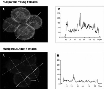
Representative images of immunostained 4-cell hamster embryos obtained from nulliparous young females (above) and multiparous adult females (below). Embryos obtained from nulliparous adult females are not included because no statistical differences were observed between them and multiparous adult females in any of the parameters studied. The intensity and distribution of fluorescence obtained by confocal scanning laser microscopy can be observed in the left side of the figure. (a) Together with an indication of the four paths followed during scanning and integration in each of the embryos studied. On the right side (b) we can observe the scans obtained by the confocal microscope following the path indicated at left with a continuous line. In the graphs, the x-axes indicate the distance in microns and the y-axes indicate fluorescence intensity in arbitrary units. We must stress that in the y-axes scale units are different.

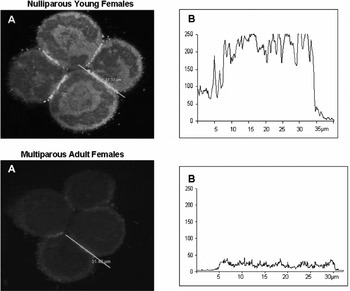
Representative images of immunostained 4-cell hamster embryos obtained from nulliparous young females (above) and multiparous adult females (below). Stress is made in the fluorescence observed in the adhesion zone between two adjacent blastomeres. The lines (a) indicate the traces followed by the confocal microscopy scans whose results are graphed in the right part (b) of each image. The same procedure was realized in each of the four cell–cell adhesion zones. In the graphs, the x-axes indicate the distance in microns and the y-axes indicate fluorescence intensity in arbitrary units. We must stress that in the y-axes scale units are different.

Results
Representative images of the results obtained from immunostained 4-cell embryos are exemplified in Fig. 2. As no statistical differences were observed between NAF and MAF, we present only the images obtained from NYF and from MAF. As indicated, each one of the studied embryos was scanned transversally four times. As E-cadherin fluorescence was irregularly distributed through the cytoplasm, we decided to measure it in both scanned directions, in order to obtain a more reasonable quantification of cytoplasmic E-cadherin. We indicate (right side) the results obtained by integration of confocal microscopy scans following the continuous line paths of the left side of Fig. 2. We must stress that the ordinate scales that indicate fluorescent intensity in arbitrary units, are different. Embryos obtained from young and adult females, both nulliparous and multiparous, show evident localization of E-cadherin fluorescence in all the cell–cell contact areas, but it was always clearly more intense in the embryos of NYF. Immunofluorescence is also observed in the free membrane surface and in the cytoplasm of all the observed embryos. Fluorescence intensity in these zones is also distinctly more intense in NYF.
Fluorescence intensity observed in the adhesion zone of the embryos obtained from NYF and from MAF females is indicated in Fig. 3. These type of scans would allow us to determine the number of cell–cell binding sites; however we preferred to use the total fluorescent intensity, obtained by integration of the scans, to estimate the amount of E-cadherin present in the cell–cell adhesion region in each group of embryos.
The quantitative data obtained from the integration of the transverse embryo scans are presented in Table 1. Immunostained E-cadherin in all the studied zones was significantly higher (p < 0.01) in NYF. No statistical difference was observed in any of the parameters studied between NAF and MAF; however it is possible to mention that a definite tendency to have higher values was consistently find in MAF.
Quantitative data obtained from the integration of the confocal microscope scans of immunostained 4-cell hamster embryos.

Data indicate the mean ± S.D. of 14 embryos per group of females.
Different letters indicate significant difference (p < 0.05) between data in the same column (ANOVA).
* Indicate the existence of a significant difference (p < 0.05) between the data in free membrane and adhesion zone (Student's t-test)
When the different studied zones were compared, the higher concentration of E-cadherin was found in the cytoplasm of the 4-cell embryos obtained from the three groups of females. The highly significant differences between young nulliparous females and adult females in the E-cadherin present in cell–cell adhesions is also signalled in Table 1.
Discussion
Cell–cell adhesion in early blastomeres is mainly caused by the extracellular, calcium dependent, homotypic interaction between E-cadherin molecules, but the strength of E-cadherin-mediated blastomere adhesion is regulated by intercellular anchorage of its intracellular dominion to the actin cytoskeleton through catenin molecules.
Components of the E-cadherin–catenin complexes are originally derived from maternal gene activity. Participation of the zygotic genome, however, is already important in the two blastomeres embryo. Unique gene expression at the 2-cell stage and clear differences between male and female embryos as early as the 4-cell stage (Minami et al., Reference Minami, Sasaki, Aizawa, Miyamoto and Imai2001), have indicated that in mammalian embryos the transition from a late 1-cell to a 4-cell embryo marks the change from control by the maternal genome to control by the zygotic genome (including degradation of maternal mRNA), arguing that differentiation in mammalian embryos may begin early. Additionally, the fact that mouse and human 2-cell embryos are already bilaterally polarized has been clearly demonstrated (Neganova et al., Reference Neganova, Sekirina and Eichenlaub-Ritter2000). Our data support this possibility: structure of 4-cell hamster embryos, particularly in embryos obtained from NYF, points to clear polarization in E-cadherin distribution between intercellular contacts and the rest of the plasma membrane. Blastomere flattening and good cell–cell adhesions, may indicate the existence of active intercellular communications.
Development and differentiation of multicellular organisms seems to be critically dependent of cell–cell adhesions. Cadherins were originally discovered as adhesion molecules functioning in mechanical linkage between adjacent cells. It has become apparent, however, that cadherins also participate in regulating cell functions via biochemical signalling pathways, both during embryo development and in adult tissue homeostasis (Yap & Kovacs, Reference Yap and Kovacs2003). In the mammalian embryo, E-cadherin cell–cell contacts pathways provide a key element in focusing the cellular activity of loosely associated cells to come together into more intimate contact so that proliferative signals, such as Rac-1 (Yap & Kovacs, Reference Yap and Kovacs2003), may elicit the necessary intracellular mechanisms to initiate differentiation to form a multicellular structure.
Numerous reports indicate that E-cadherin functions as an antiproliferative agent, but, in fact, E-cadherin cell–cell contacts can either enhance or inhibit proliferation (Liu et al., Reference Liu, Nelson, Pirone and Chen2006). The significant differences reported herein between NYF and both NAF and MAF, indicate that adequate distribution of E-cadherin, both to the cell–cell contacts and to the blastomere periphery in 4-cell embryos, may be an important factor in regulating and stimulating embryo cell multiplication. We may remember that one of the significant differences between NYF and MAF (Navarro et al., Reference Navarro-Maldonado, Ambriz, Mundo, Trejo, Hernandez and Rosado2000; Trejo et al., Reference Trejo, Navarro, Ambriz and Rosado2005) is the late appearance of morulae and the persistence of 4-cell embryos during unusually long periods. These results are similar to those of Blaha (Reference Blaha1964) who found <1% of 4-cell embryos at 63–68 h after ovulation in young animals, but reached nearly 18% in adult animals. The insufficient concentration, therefore and inadequate pattern of distribution of E-cadherin in embryos obtained from adult females, both nulliparous and multiparous, may be one of the factors that retard and desynchronize embryo development in these groups of females. These results will be in accord with other reports showing that enhancement of E-cadherin content and localization in the cell periphery, is associated with an increase in cell proliferation (Reddy et al., Reference Reddy, Liu, Ren, Lindgren, Boman, Shen, Lundin, Ottander, Rytinky and Liu2005; Liu et al., Reference Liu, Nelson, Pirone and Chen2006)
Embryo compaction, accompanied by blastomeres polarization of preimplantation mammalian embryos, is considered as the first step in differentiation and a process indispensable for the morphogenetic development that will form the trophectoderm and inner cell mass. It is then possible to postulate that synthesis, storage and distribution of E-cadherin in precompaction embryos must be of great importance for normal embryo development. Cadherin seems to function as both agent and subject in these processes. Interestingly, activation of Rac-1 by E-cadherin (Nakagawa et al., Reference Nakagawa, Fukata, Yamaga, Itoh and Kaibuchi2001; Noren et al., Reference Noren, Niessen, Gumbiner and Burridge2001), appears to lead to Actin recruitment and physical strengthening of adherents junctions (Chu et al., Reference Chu, Thomas, Eder, Pincet, Pérez, Thiery and Dufour2004). Moreover, in mice, in humans and probably also in the hamsters, compaction occurs in absence of protein synthesis and is characterized by the induction of after translational modifications of non-functional cytoplasm E-cadherin (Ohsugi et al., Reference Ohsugi, Butz and Kemler1996; Kawai et al., Reference Kawai, Yamaguchi, Yoden, Hanada and Miyake2002). These modifications induce E-cadherin to relocate into the lateral and basal cell surfaces to propitiate development and entail the conversion of limited, loose, cadherin contacts into broader zones of tight adhesion. Small GTPase proteins, such as Rac, appear to be critical for this process of contact-zone extension (Kovacs et al., Reference Kovacs, Ali, McCormack and Yap2002).
Ohsugi et al. (Reference Ohsugi, Butz and Kemler1996) have proposed that during preimplantation development E-cadherin is probably accumulated in the cytoplasm as a prefunctional molecule ready to be used. This accumulation is indispensable for the rapid occurrence of compaction at a precise developmental stage. The results of Goval et al. (Reference Goval, Van Cauwenberge and Alexandre2000) indicate that β-catenin is also redistributed to the apposing membranes as a prerequisite to physiological compaction. Adequate concentrations of E-cadherin seem to be necessary during the transition from morulae to blastocyst to ensure correct establishment of a functional trophectoderm (Kan et al., Reference Kan, Stemmler, Junghans, Kanzler, de Vries, Dominis and Kemler2007). Our results in the hamster support this conclusion and, in addition, show that retarded embryo development in adult females, may be partially caused by insufficient accumulation of E-cadherin in the blastomeres cytoplasm at the 4-cell stage of embryo development.
Enrichment of E-cadherin at contacts between sister blastomeres, contributes to organize preimplantation embryos from the 2-cell stage throughout cleavage, defining a microvillus-free area of the membrane (Neganova et al., Reference Neganova, Sekirina and Eichenlaub-Ritter2000). This early polarization of mammalian embryos is important in maintaining a harmoniously dividing preimplantation embryo and for allocation of cell fate (Antczak & Blerkom, Reference Antczak and Van Blerkom1999). Development of functionally specialized, polarized domains, in the plasma membrane are indispensable for the working of numerous important cellular processes (Neganova et al., Reference Neganova, Sekirina and Eichenlaub-Ritter2000; Kan et al., Reference Kan, Stemmler, Junghans, Kanzler, de Vries, Dominis and Kemler2007), including differentiation. Reduction in the concentration of membrane-associated E-cadherin, as we observed in the 4-cell embryos obtained from adult females, may, therefore, contribute to disturbances in embryo development, these results are similar to that reported, under different experimental circumstances by Barth et al. (Reference Barth, Nathke and Nelson1997).
In vitro studies suggest that a large proportion of embryonic loss occurs during preimplantation stages. Hardy (Reference Hardy and Bavister1993) showed that while 95% of human zygotes successfully undergo the first two cleavage divisions following IVF, ∼60% of preimplantation embryonic, arrest in vitro after the 8-cell stage, at the time of compaction and before blastocyst formation. More recently it has been confirmed that successful pregnancy outcome, can be correlated with adequate preimplantation embryo morphology and that only embryos destined to survive display an organized pattern of intercellular junctions during preimplantation development (Bloor et al., Reference Bloor, Metcalfe, Rutherford, Brison and Kimber2002; Kan et al. Reference Kan, Stemmler, Junghans, Kanzler, de Vries, Dominis and Kemler2007). In this context, our results seem to indicate that changes in the reproductive behavior related to age or multiparity (Van Kooij et al., Reference Van Kooij, Looman, Habbema, Dorland and te Velde1996) may also be correlated with changes in the processes related to intercellular adhesions during early cleavage.
Acknowledgements
We deeply appreciate M. En C Eduardo Carrillo of Laboratory of Microscopy Confocal of the Genomic Sciences Postgraduate of Universidad Autonoma de la Ciudad de Mexico for his invaluable assistance. This work was supported in part by grants Nos. 153054 and 35118 from CONACyT, Mexico.