Introduction
Chrysodeixis includens (Walker) (Lepidoptera: Noctuidae: Plusiinae) is a characteristic species from the Western Hemisphere that is distributed from northern United States of America to southern South America (Eichlin and Cunningham Reference Eichlin and Cunningham1978; Herzog Reference Herzog1980; Kitching and Rawlins Reference Kitching and Rawlins1987; Jost and Pitre Reference Jost and Pitre2002; Pogue Reference Pogue2005; Betancourt and Scatoni Reference Betancourt and Scatoni2006; Navarro et al. Reference Navarro, Saini and Leiva2009). Adults are small to average sized moth, with robust body, small tufts of scales in the dorsal part of thorax and anterior abdominal region, and characteristic silver marking near the center of the forewing. The larva is commonly known as semi looper or measuring worm (Eichlin and Cunningham Reference Eichlin and Cunningham1978; Lafontaine and Poole Reference Lafontaine and Poole1991) and it is considered a serious pest of economically important plants such as soybean (Glycine max (Linnaeus) Merrill (Fabaceae)), alfalfa (Medicago sativa Linnaeus (Fabaceae)), cotton (Gossypium hirsutum Linnaeus (Malvaceae)), bean (Phaseolus vulgaris Linnaeus (Fabaceae)), flax (Linum usitatissimum Linnaeus (Linaceae)), tobacco (Nicotiana tabacum Linnaeus (Solanaceae)), and several vegetables (Canerday and Arant Reference Canerday and Arant1967; Eichlin and Cunningham Reference Eichlin and Cunningham1978; Herzog Reference Herzog1980; Lafontaine and Poole Reference Lafontaine and Poole1991; Artigas Reference Artigas1994; Pastrana Reference Pastrana2004; Pastrana et al. Reference Pastrana, Di Iorio, Navarro, Chalup and Villagran2004; Betancourt and Scatoni Reference Betancourt and Scatoni2006; Navarro et al. Reference Navarro, Saini and Leiva2009). In the northwest and northeast of Argentina, Chrysodeixis includens is considered one of the major soybean pests (Berta et al. Reference Berta, Colomo, Valverde, Romero Sueldo and Dode2009; Casmuz et al. Reference Casmuz, Zaia, Socias and De La Vega2009; Valverde et al. Reference Valverde, Colomo, Berta, Romero Sueldo and Dode2010; Sosa Gomez Rolim et al. Reference Sosa Gomez Rolim, Akimi Cavaguchi Yano, Specht, Tardelli de Jesus Andrade and Sosa-Gómez2013).
Soybean is the most important crop in Argentina; for 2013–2014, the soybean planted area in Argentina was over 20 million ha and reaching an annual production of more than 53 million tonnes (Aizen et al. Reference Aizen, Garibaldi and Dondo2009; Bolsa de Cereales 2014; Ministerio de Agricultura, Ganadería y Pesca 2015). Occurrence of this species could cause significant economic losses in this region. In addition, some studies indicate that C. includens shows tolerance to various insecticides (Portillo et al. Reference Portillo, Felland, Pitre and Porter1993; Mascarenhas and Boethel Reference Mascarenhas and Boethel2000; Walker et al. Reference Walker, All, McPherson, Boerma and Parrott2000; Freitas Bueno et al. Reference Freitas Bueno, Corrêa-Ferreira, Hoffmann-Campo, Sosa-Gomes, Hirose and Roggia2012), suggesting higher doses than those recommended for other lepidopteran defoliators to control this pest (Igarzábal Reference Igarzábal2009).
Several aspects of this species have been studied as: diagnosis of preimaginal stages (Angulo and Weigert Reference Angulo and Weigert1975; Igarzábal et al. Reference Igarzábal, Fichetti and Tognelli1994), vital statistics (Canerday and Arant Reference Canerday and Arant1967; Jensen et al. Reference Jensen, Newsom and Gibbens1974; Combe and Pérez Reference Combe and Pérez1978; Shourt and Sparks Reference Shourt and Sparks1981; Barrionuevo et al. Reference Barrionuevo, Murúa, Goane, Meagher and Navarro2012), hosts (Silveira and Ruffinelli Reference Silveira and Ruffinelli1956; Pastrana Reference Pastrana2004; Pastrana et al. Reference Pastrana, Di Iorio, Navarro, Chalup and Villagran2004; Quimbayo et al. Reference Quimbayo, Serna, Olivares and Angulo2010), natural enemies (Margheritis and Rizzo Reference Margheritis and Rizzo1965; Harper and Carner Reference Harper and Carner1973; Edelstein and Leucuona Reference Edelstein and Leucuona2003; Valverde and Virla Reference Valverde and Virla2007; Valverde et al. Reference Valverde, Colomo, Berta, Romero Sueldo and Dode2010; Bueno et al. Reference Bueno, Parra and Bueno2012), bioecology and damage (Boldt et al. Reference Boldt, Biever and Ignoffo1975; Rizzo and Saini Reference Rizzo and Saini1990; Kitching et al. Reference Kitching, Orr, Thalib, Mitchell, Hopkins and Graham2000; Ashfaq et al. Reference Ashfaq, Young and McNew2001; Kidd and Orr Reference Kidd and Orr2001; Valverde and Virla Reference Valverde and Virla2007; De Freitas Bueno et al. Reference De Freitas Bueno, De Freitas Bueno, Moscardi, Postali Parra and Hoffmann-Campo2011), last instar identification keys (Angulo and Weigert Reference Angulo and Weigert1975; Godfrey Reference Godfrey1987; Lafontaine and Poole Reference Lafontaine and Poole1991; Igarzábal et al. Reference Igarzábal, Fichetti and Tognelli1994), and comparative diagnosis of adults (Eichlin and Cunningham Reference Eichlin and Cunningham1978; Rizzo and Saini Reference Rizzo and Saini1990; Lafontaine and Poole Reference Lafontaine and Poole1991; Pogue Reference Pogue2005).
The last instar and the adult of this species have been described several times. However, the information available for immature stages is outdated and superficial. For this reason, we provide a formal and detailed redescription of all preimaginal stages (egg, larva, and pupa) of Chrysodeixis includens, according to current standards of descriptive patterns. Our contribution will be useful for the correct identification of early-late larval stages and differentiation from other similar species as Rachiplusia nu (Guenée) (Lepidoptera: Noctuidae), with which it coexists in soybean crops and with which it is usually mistaken.
Materials and methods
Immature stages of C. includens were obtained from laboratory breeding under controlled conditions of 27±2 °C of temperature, 14:10 (light/dark) photoperiod, and 70–75% relative humidity. Adults were fed with a solution of 10% honey placed on glass jars of 5 mL with hydrophilic cotton. Eggs were procured from adults kept on oviposition cylindrical cages of polyethylene terephthalate (25 cm high by 10 cm diameter). After oviposition, eggs were taken with a moistened brush, individually placed on Petri dishes covered with filter paper, and daily moistened with distilled water until larval hatching. Ten eggs were used to evaluate height and diameter. For micropilar area description, a stereoscopic microscope was used and Angulo and Weigert (Reference Angulo and Weigert1975) method and Salkeld (Reference Salkeld1984) nomenclature were followed.
After hatching, 200 larvae were individually placed on Petri dishes and fed with artificial diet (Osores et al. Reference Osores, Willink and Costilla1982). Larval growth and stages differentiation were evaluated through cephalic capsule measurement and body total length for 20 specimens of each instar. Cephalic capsule width was measured following Dyar’s (Reference Dyar1890) method, according to Specht et al. (Reference Specht, Bogt and Corseuil2007), for 20 specimens of each instar. Larval colour pattern was described with specimens fed with natural diet (soybeans leaves) and using terminology proposed by Lafontaine and Poole (Reference Lafontaine and Poole1991). Chaetotaxy terminology employed corresponds to Hinton (Reference Hinton1946), with modifications added by Stehr (Reference Stehr1987).
Pupa were individually placed on Petri dishes covered with filter paper and daily moistened with distilled water until adult emergence. Total body length and maximum width were measured for 20 pupae. The terminology used follows Mosher (Reference Mosher1916).
Measurements were calculated in millimeters and are expressed as ranges or averages and standard deviations.
Results
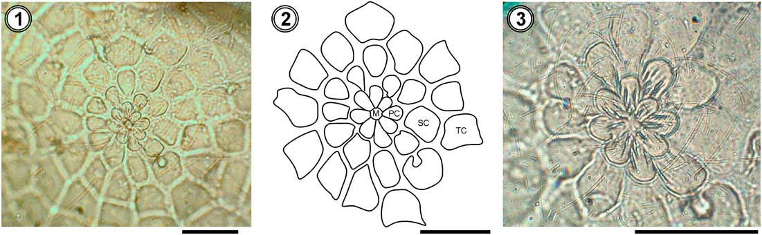
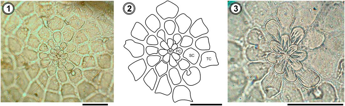
Egg (Figs. 1–3). Diameter: 0.58±0.01 mm, height: 0.42±0.02 mm. Individually deposited on the plant. Colouration: bright pearl-white after oviposition, becoming pale yellow with the advance of embryonic development, at the end of incubation with two dark points at polar area (corresponding to larva head and prothoracic shield). Morphology: straight, suboval, with flat and smooth base. Corion thin, transparent, pattern slightly marked. Sculptury consisting on 30–32 strong longitudinal ribs connected by thin irregular groves weakly marked, only a few ribs reach mycropilar area (Fig. 1). Mycropilar rosette 0.015 mm wide; located on centre of the superior polar region; more or less rounded; with 8–9 elongate, petal-like primary cells; distally rounded and juxtaposed at base; arranged as a conspicuous complete cycle of irregular shape and size (Figs. 2–3). Two cycles outside primary cells: first cycle formed by 11–12 secondary cells, bigger than primary cells, arranged in a close complete circle and the second cycle is formed by 12–26 irregular tertiary cells; with rounded, pointed, or truncate distal margin and arranged in an incomplete open cycle (Figs. 2–3); weak on some eggs. Primary and secondary cells on different levels.
Egg of Chrysodeixis includens: 1. superior polar region; 2. schematic drawing of mycropilar rosette; 3. close up photo of primary and secondary mycropilar cells. Scale bars=0.02 mm.
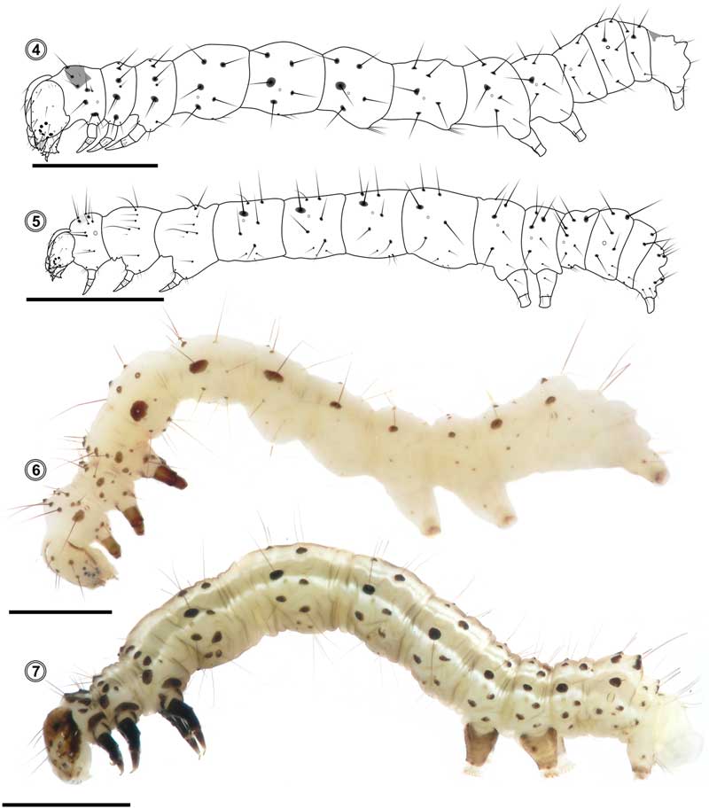
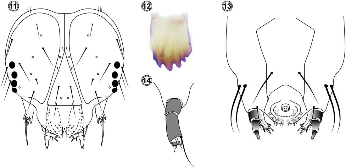
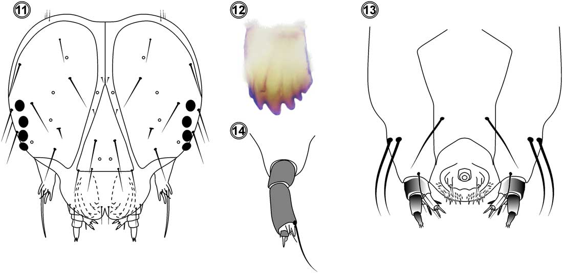
First instar (Figs. 4, 11–14). Head width 0.14±0.05 mm, body length 2.65±0.12 mm. Colour pattern: hyaline to white (artificial diet) and dark green (natural diet); head, legs, and pinaculae from light to dark brown; prothoracic and anal shields, spiracle peritreme, and leg plates weakly sclerotised, light brown; head and body setae same colour as shields or darker; epicraneal and adfrontal sutures and mandibles apex dark brown; stemmatal area black; antenna, labrum, labial and maxillary palpi, and hypopharynx from light brown to white; abdominal prolegs with subquadrate plates and anal prolegs with triangular plates; crochets from light brown to orange. Morphology: body thin, slightly narrow on thoracic segments. Head (Fig. 11). MD1–MD3 very shorts, posterior-lateral to P2. MD2 bristle closer to MD1 than to MD3. MDa puncture between MD2 and MD3. P1 and P2 similar in size, gap P2–P2 bigger than gap P1–P1. Pb puncture halfway between P1–P2. AF1 and AF2 similar in size, AF2 above frons apex, closer to P1 than to AF1. AFa puncture between AF1–AF2, closer to AF2 than to AF1. Fa puncture below F1, slightly separated. C1 and C2 bristles equal in length, C1 above frontclypeum superior vertex, C2 on ventrolateral vertex of frontclypeum. L1 bristle and La puncture dorsal and lateral to A3; A3 close to stemma 2 and similar in length to A2, A1 above A2, A2 closer to A3 than to A1. Aa puncture close to A2. S1–S3 equidistant and similar in length, S1 posterior and close to stemma 4, S2 posterior and ventral to stemma 1, S3 posterior and ventral to S2, below or very close to stemmatal band. Sb puncture anterior and medial to stemmata 3 and 4; Sa puncture between stemma 6 and S3, closer to stemma 6 than to S3. SS-group setae subequals and in triangular pattern, SS1 anterior and ventral to stemma 5, SS2 posterior and ventral to stemma 5, SS3 posterior and ventral to SS2. SSa puncture between SS2 and SS3, closer to SS2 than to SS3. MGa puncture posterior and dorsal to S3; MG1 puncture posterior and ventral to MGa. Stemmata subcirculars, placed lateral and anteriorly, stemmata 1–2 paired but separated, stemmata 3–4 nearly touching, stemma 5 slightly displaced to head ventral area, close to antennal base, stemma 6 posterior and dorsal to stemma 5 (Fig. 11). Antenna cylindrical, enlarged, with three antennomeres, with four sensilla; basal antennomere wider than long, middle antennomere larger than basal and distal antennomeres (Fig. 14). Maxillary palpus subconic, enlarged, with three palpomeres, without setae (Fig. 13). Labial palpus subconic, with two palpomeres, with two sensilla (Fig. 13); both palpi with sclerotised apex, varying from light brown to dark brown; spinneret conic, more than twice as long as labial palpus (Fig. 13). Hypopharynx with distal region without setae, medial proximal region with small setae, and proximal lateral region with several big and small setae (Fig. 13). Mandible (Fig. 12) with a raw of six teeth; first tooth short, placed above second; second, third, and fourth teeth enlarged, similar in size; fifth and sixth teeth smaller than the others; teeth with sharp edges and upward curved except sixth tooth, which is short with wide and blunt edge; second, third, and fourth teeth with internal processes, strongly marked and interrupted before reaching the cutting edge on second and third teeth. Labrum bilobed, with a medial and ventral indent (Fig. 11). Thorax (Fig. 4). Prothoracic shield subtrapezoidal; XD setae placed along anterior margin, slightly longer and ticker than those of D group, XD2 slightly behind XD1; D2 close to posterior margin, D1 close to body middle line; SD2 shorter and thicker than SD1, both setae closely spaced, separated from the prothoracic shield and arranged on the same pinnacle; L-group anterior to spiracle, on the same pinnacle, setae very close to each other, L1 anterior, dorsal, and longer than L2, L2 thin, slightly above or in line with spiracle ventral border, L3 absent; SV-group arranged as a horizontal line below spiracle, on the same pinnacle, SV2 longer and thicker than SV1; MV1 slightly before coxa anterior margin; V1s close to each other, slightly after coxa posterior margin. T2–T3 with similar chaetotaxy; D1, D2, SD2, SD1 arranged on a vertical line; D2 and SD1 with pinnacles and setae bigger than those of D1 and SD2; L1 and L2 close, on same pinnacle, close to anterior margin, L1 dorsal and posterior to L2, longer and thicker than L2, L3 absent; SV1 close to ventral region, SV2 setae absent; MV1 anterior to coxa and V1s slightly posterior to coxa, more separated than V1s on T1. Abdomen (Fig. 4). Pinnacles on A1–A9 of different size, even on same segment, with a progressive reduction of pinnacles size towards posterior segments, segments A1 to A4 with SD1 pinnacle twice as big as segments A5–A9; D1 and D2 similar in size, on A1–A7 D1 close to dorsal medial line, dorsal and anterior to D2, on A8 D1 and D2 at same level, segment A9 with D1 anterior and ventral to D2, A10 with D1, SD1, and SD2 setae similar in size and equidistant, D2 closer to dorsal medial line than D1; A1 with SD1 vertically lined with spiracle, A2–A6 and A8 with SD1 anterior and dorsal to spiracle, A9 with setae SD1 and D1 only, on an apparent vertical line; L2 anterior and ventral to spiracle and L1 posterior and ventral on A2–A6 and A8, L1–L2 close to each other on A10, placed towards posterior margin of anal proleg, L3 absent on all abdominal segments; A1 with SV1 only, SV2 missing, V1 shorter than SV1, SV-group bisetose on A2–A4, A2 with SV1, SV2, and V1 on separate pinnacles, SV1 closer to SV2 than to V1, A3–A4 with SV1, SV2, and V1 on one triangular pinnacle, A5–A9 with SV1 and V1 setae only, SV-group bisetose on A10, SV1 and SV2 placed towards anal proleg anterior margin, V1 ventral to them; SV3 absent on all abdominal segments. Anal shield subtriangular. Abdominal prolegs on A5–A6 with seven to nine crochets in mesoseries and anal proleg with eight to nine crochets in mesoseries, proleg absent on A4. Spiracles rounded, prothoracic and A8 spiracles bigger than A1–A7 spiracles.
First to fourth instar: 4. first instar; 5. second instar; 6. third instar; 7. fourth instar. Scale bars=0.5 mm (Fig. 4), 1 mm (Figs. 5–6) and 2 mm (Fig. 7).

Second instar (Fig. 5). Head width 0.40±0.03 mm; body length 4.65±0.26 mm. Colour pattern: white (artificial diet) to light green (natural diet); head from light brown to pale yellow; epicraneal and adfrontal sutures from light to dark brown; pinnacles and setae from dark brown to black; stemmata black; antenna, labrum, mandible apex, hypopharynx, maxillary apex, and labial palpus sclerotised, from orange to dark brown; prothoracic and anal shields, legs and prolegs shields from light brown to same colour as body; legs from dark brown to black; dorsal, lateral, subventral, and ventral pinnacles from light to dark brown; subdorsal pinnacles black; setae thin and black; spiracles peritreme, abdominal and anal prolegs crochets from light brown to orange. Morphology (Fig. 5) similar to first instar, except for the following features. Head with A3 seta more than twice as long as A2. Thorax T1 with SD1–SD2, L1–L2, and SV1–SV2 setae on separated pinnacles; segments T2–T3 with L1 and L2 setae as in T1; L3 present, posterior and in horizontal line with L1. Abdomen with D2 setae slightly longer on A8–A9 than the other abdominal D2 setae; L3 present, on A1–A6 and A8 placed posterior and ventral to L2, on A7 in a vertical line with SD1, spiracle, and L2; A10 with L1, L2, and L3 setae close to each other, located towards anal prolegs posterior margin; SV-group trisetose on A1–A4, SV1, SV2, and SV3 equidistant and on separate pinnacles, SV3 on straight line or slightly posterior to L2; V1 shorter than SV1 and SV3. Abdominal prolegs on A5–A6 with 8–10 crochets in mesoserie and anal prolegs with 9–12 crochets in mesoserie.
Third instar (Fig. 6). Head width 0.53±0.05 mm; body length 7.42±0.99 mm. Colour pattern: white (artificial diet) or from light green to yellowish green (natural diet), with two pairs of white continuous slightly conspicuous lines, subdorsal line surrounds D2 by dorsal margin, lateral line ventral to SD1; head from light brown to yellowish green; some specimens with dendritic patches, from light brown to black, these patches can cover the head partial or totally and they can remain on successive instars; epicraneal and adfrontal sutures, pinnacles, and setae from dark brown to black; stemmata black; middle and distal antennomeres, labrum, maxillary and labial palpi last antennomere, and mandible apex from orange to light brown; prothoracic and anal shields slightly noticeable, concolour with body; pinnacles and setae black; spiracles peritreme from dark brown to black, inner area light brown or same colour as tegument; legs brightly black, light brown, or same colour as tegument; legs, abdominal, and anal prolegs plates inconspicuous, same colour as tegument; abdominal and anal crochets sclerotised, from light brown to orange. Morphology (Fig. 6) similar to second instar, except for the following features. Body with tiny black spicules, conspicuous at sides of middle dorsal line and over supraspiracular area. Thorax T1 with SD1 and SD2 setae on the same or different pinnacles; L1 and L2 on the same pinnacle, bigger than prothoracic spiracle; SV1 and SV2 setae on different pinnacles; T2–T3 with L1 and L2 setae on separated pinnacles. Abdomen with D1–D2 and SD1 pinnacles protruding as cones from tegument on A1–A4; segments A8–A9 with D2 setae longer than the other abdominal D2 setae; SD1 pinnacles twice or thrice as big on A1–A4 as those on A5–A7. There is a progressive size reduction of pinnacles towards posterior segments, on A8 increases size again. Abdominal prolegs on A5–A6 with 14–16 crochets in mesoserie and anal proleg with 13–16 crochets in mesoserie.
Fourth instar (Fig. 7). Head width 0.76±0.05 mm; body length 11.70±1.55 mm. Colour pattern (Fig. 7): variable, more frequently from brightly light green to yellowish green, some specimens olive green or with two colours: dark green over supraspiracular area and light green below it; with four pairs of white continuous conspicuous lines of variable size: dorsal, addorsal, subdorsal, and lateral; head from light green to yellowish green; epicraneal and adfrontal sutures light brown, black, or concolour with head; pinnacles black or same colour as head; setae light brown or black; some specimens with black diagonal bands on genae at stemmata 5 and 6, from stemma 6 to head posterior region; stemmata black; labrum, middle and distal antennomeres, maxillary and labial palpi from black to same colour as tegument; thorax with prothoracic shield same colour as tegument, lateral margins with addorsal and subdorsal lines fused in one solid line, from T2 lines separate; dorsal line inconspicuous, lateral line at same level as L-group and spiracle; T2–T3 with dorsal line thinner than the other lines and closer to middle dorsal line; addorsal line of similar wide as lateral line, at same level as D1, surrounding it and partially enclosing dorsal side of D2 seta, subdorsal line narrower than addorsal line, placed below D2, partially enclosing its ventral margin; lateral line more than twice as wide as dorsal line, running through spiracular area; abdomen with lines similar to T2–T3, except for the following features. Segment A10 with dorsal, addorsal, and subdorsal lines reaching anal plate and fused on medial region; lateral line ventral to SD1 and SD2, extending to ventral region; spiracles peritreme dark or light brown to black, inner area light brown to same colour as tegument. Morphology (Fig. 7) similar to third instar, except for the following features. Thorax T2 with interspace D1–D2 smaller than interspace D2–SD2; spiracles oval, T1 and A8 spiracles bigger than the others. Tegument densely covered dorsally with light and dark, tiny spines. The pinnacles and diagonal bands, if present, remain on later stages. Dendritic patches like the ones described for third instar. Small outlines of vestigial prolegs become visible on A3–A4; A5 proleg with 16–19 crochets; A6 with 18–20 crochets; and anal proleg with 17 crochets; all crochets in mesoserie.
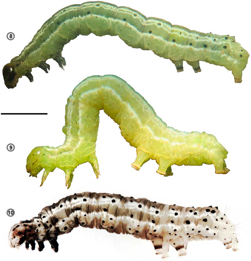
Fifth instar (Figs. 8–10). Head width 1.04±0.06 mm; body length 20.00±1.51 mm. Colour pattern (Figs. 8–10) similar to fourth instar, except for the following features. Head from light green to yellowish green; P1, P2, A3, and L1 setae generally surrounding by black pinnacles, twice or thrice as big as the other pinnacles; antenna with all antennomeres or only middle and distal antennomeres light to dark brown or as tegument, basal antennomere short and wide, middle antennomere shorter than distal, distal antennomere long, with four setae, one of those setae longer than antenna itself. Thorax T1 with XD, D, SD, L, and SV on black pinnacles, T2–T3 with D1 and D2 on pinnacles black, white, or as tegument; SD1 and SD2 on pinnacles black or white; L1–L3 on pinnacles black, white, or as tegument. Abdomen covered by black small spicules, similar in size; abundant over dorsal line sides, over supraspiracular area, and on ventral region; abdominal A1–A9 with subdorsal pinnacles generally black and dorsal and lateral pinnacles white, black, or as tegument. Setae from light brown to translucent. Ventral and subventral pinnacles concolour with tegument. Body slightly uniform in wide. Vestigial prolegs conspicuous on A3–A4, reduced to small button-like structures, without crochets; A5–A6 prolegs with 20–24 crochets, anal prolegs with 22–23 crochets.
Fifth instar colour pattern variation and morphology. Scale bar=4 mm.

First instar head: 11. head, anterior view; 12. mandible, median view; 13. hypopharyngeal complex, posterior view; 14. antenna, anterior view.
Sixth instar. Head width 1.54±0.05 mm; body length 27.45±2.74 mm. Larva conserve the same characters as fifth instar. Two or three days before start to weave the cocoon, larva lose colouration and patterns intensity and activity decrease (feeding and mobility).
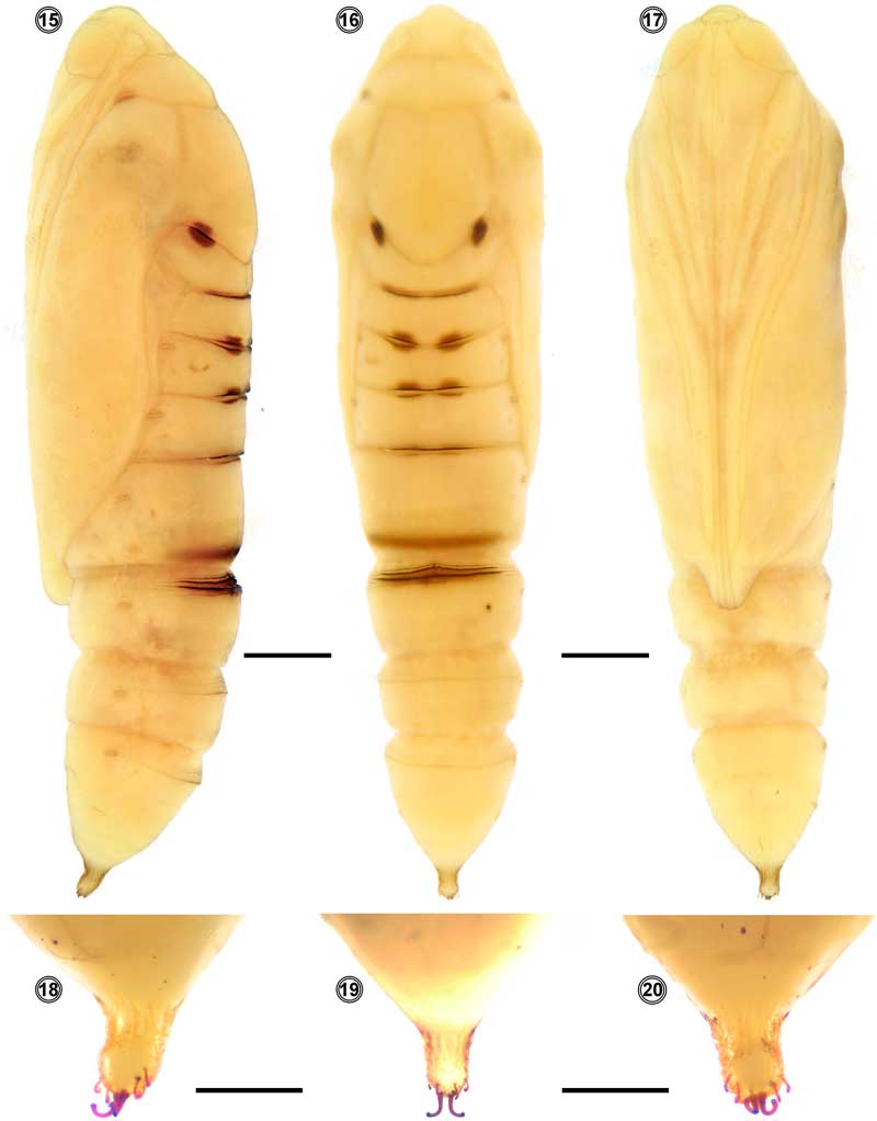
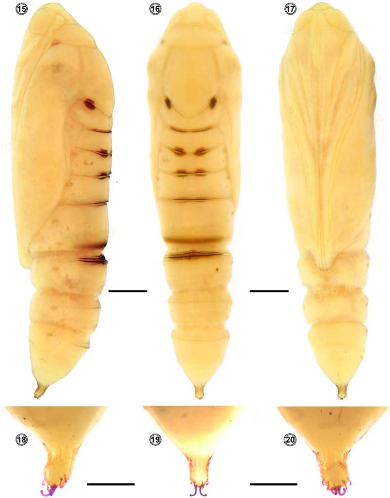
Pupa (Figs. 15–20). Female: length 18.2±0.1 mm, width 3.5±0.1 mm. Male: length 18.6±1.9 mm, width 3.6±0.1 mm. Obtect, adectic, with delicate glaucous cocoon. Brightly green on firsts days turning light brown when it is close to adult emergence. Thorax and abdomen slightly rugous. Spots from dark brown to orange in (Figs. 15–16): circular lateral spots between posterior margin of T2 and anterior margin of T3 and between A1 and A3, the latter close each other on medial region; posterior margin of T3 and posterior margins of A3–A5 spots as transversal lines along dorsal area. Anterior margin of A5–A7 sculptured with a dorsal transversal line of big spines, followed by sparse transversal grooves with same colour as tegument, light brown, or orange (Figs. 15–16). Spiracle same colour as tegument, reduced on T1; segments A2–A7 with oval spiracles; A8 with spiracle bigger than the others. Galea and forewings extended to A5 posterior margin; gena and eyes from dark brown to orange; labial palpus extended from clypeus to middle of profemur or beyond it; profemur short, placed between anterior part of maxilla and foreleg; maxilla long, extended to A4 posterior margin, expounding distal apex of metaleg; mesoleg slightly shorter than metaleg; antenna widely arched anteriorly, posteriorly convergent on medial ventral region, slightly shorter than mesoleg; forewing with differentiated veins; hindwing borders almost disappear on ventral view (Fig. 17). Cremaster (Figs. 18–20) longer than wide, rugous, light yellow or same colour as tegument; ventrally (Fig. 20) with a central pair of double spines joined at the base, thick, twice as long as adjacent spines; dorsally (Fig. 19) with three pairs of small single spines, thin, sclerotised, dark brown, with curved hook-like tip.
Pupa: 15–17 entire body, 18–20 cremaster. 15, 18, lateral view; 16, 19, dorsal view, 17, 20, ventral view. Scales bars=2 mm (Figs. 15–17) and 1 mm (Figs. 18–20).
Discussion and conclusion
Morphological studies of Chrysodeixis includens (Walker) carried out on this work allow to find dependable characters to differentiate their stages/instars. Among proposed characters, more useful ones to recognise preimaginal stages are:
Egg with rosette as elongate, petal-like cells, with two to three cells cycles, irregulars in size and shape, only first cycle cells are juxtaposed at the base, and primary and secondary cells are not on the same level.
First instar with sclerotised head, mandible differentiated by internal processes on second, third, and fourth teeth and conic spinneret. Abdomen with SV1, SV2, and V1 setae on A3–A4 arranged on a triangular pinnacle, this arrangement remain stable until last instar and it is typical of the tribe Argyrogrammatini (Eichlin and Cunningham Reference Eichlin and Cunningham1978). Absence of L3 and SV3 setae on all abdominal segments, this character separate first instar from the others.
Second and third instar are characterised by SD1 pinnacles on A1–A4 bigger than on the other segments and D2 setae on A8–A9 longer than the other D2 setae. Third instar also presents pinnacles SD1 and D1–D2 of abdominal segments protruding from tegument as cones.
Fourth instar head may have a black band on gena, not covering stemmata 5 and 6; and P1, P2, A3, and L1 setae pinnacles differentiated by their bigger size. T2 with interspace D1–D2 smaller than interspace D2–SD2. Thorax and abdomen with four pairs of white continuous conspicuous lines of variable size; small outlines of vestigial button-like prolegs on A3–A4 (Argyrogrammatini) become visible.
On fifth and sixth instar colouration pattern is unhelpful since it is highly variable. The tiny black spicules with the characters mentioned in the fourth stages are relevant to identify the last two stages.
Pupa brightly green; eyes stand out by their brown to orange colour; dorsally, thorax and abdomen with conspicuous spots, circular and as transversal lines, dark brown to orange and posterior margin of segments A4–A6 with a band of big spines, cremaster longer than wide.
In this works, we recorded six instars for Chrysodeixis includens. Supernumerary larval stages for C. includens have been reported depending on diet used (Navarro et al. Reference Navarro, Saini and Leiva2009). Furthermore, Boldt et al. (Reference Boldt, Biever and Ignoffo1975), Herzog (Reference Herzog1980), Shourt and Sparks (Reference Shourt and Sparks1981), and Barrionuevo et al. (Reference Barrionuevo, Murúa, Goane, Meagher and Navarro2012) recorded six instars in larva fed with artificial diet or soybean leaves. Other authors, found five instars in larva fed with leaves of Brassica oleracea Linnaeus (Brassicaceae) or artificial diet (Combe and Pérez Reference Combe and Pérez1978; Shourt and Sparks Reference Shourt and Sparks1981) and as many as seven instars in larva reared only with artificial diet (Shourt and Sparks Reference Shourt and Sparks1981).
To determine larval stage, we used a standard method consisting of measuring cephalic capsule width. Dyar (Reference Dyar1890) measured the cephalic capsule width of each instar of 28 species of caterpillars and found that the progression was more or less constant and expected from a geometric progression. This is known as Dyar’s law, with a mean rate value of progression of 1.5, fluctuating between 1.3–1.7 (Berg and Merritt Reference Berg and Merritt2003). In this study, the ratios of progression of cephalic capsules width for each instar were comprised in Dyar’s law except for the first instar, with a ratio of 2.86 compared to second instar. Berg and Merritt (Reference Berg and Merritt2003) stated that contradiction with this law could occur when number on instars is not constant. As mentioned before, larval instars of C. includens vary between five and seven and maybe this could be an explanation for the discrepancy observed.
Measurements of pupa revealed that male are larger than female pupae, contrary to pupae of other lepidopteran species. This is the first time that pupa are sexed and measured independently for C. includens. Same discrepancy was observed by Barrionuevo (Reference Barrionuevo2011) with Rachiplusia nu pupa.
The effect of insecticides on the insects may vary from instar to instar and the proper timing of insecticide applications depends primarily on the predominance of a particular instar. Head size has been successfully used for larval instar determination on Lepidoptera (Hsia and Kao Reference Hsia and Kao1987; Iannacone and Alvariño Reference Iannacone and Alvariño2007; Specht et al. Reference Specht, Bogt and Corseuil2007; Zenker et al. Reference Zenker, Specht and Corseuil2007; Barrionuevo Reference Barrionuevo2011). Larval instar determination for this species may be carried out taking into account morphological characters mentioned along with cephalic capsule width measurements and identification key provided below.
We consider that opportune detection of preimaginal stages of Chrysodeixis includens will help to control this pest species, especially at admissible economical levels (Angulo Reference Angulo1987). Eggs, larvae, and pupae of C. includens coexist and it is commonly misidentified with Rachiplusia nu. Thus, we present below a key to differentiate immature stages of both species:

Acknowledgements
The authors thank Dr. Fernando Navarro for valuable suggestions and advice during the preparation of the manuscript. Andrew Smith and an anonymous reviewer offered many comments that substantially improved the manuscript. The Instituto Superior Dr. Abraham Willink (INSUE) and Consultora Rurais provided laboratory, chambers, and equipment. This study was supported by the Consejo Nacional de Investigaciones Científicas y Técnicas (CONICET).