Introduction
The role of the intra-uterine environment in influencing developmental trajectories and biological outcomes in later life has been gaining in scientific importance.Reference Talge, Neal and Glover 1 The ontogenic plasticity of the foetus confers it with the ability to adapt to changes in the intra-uterine environment, in preparation for its survival and optimal functioning in the environment in to which it will eventually be born.Reference Gluckman, Hanson and Pinal 2 However, recent research suggests that this programmability of the foetus may function as a double-edged sword. In situations where optimal matching of intra- and extra-uterine environments occur, the foetus maybe conferred with developmental and evolutionary advantages such as increased vigilance and greater willingness to explore new environments.Reference Glover 3 However, perturbations in the intra-uterine environment, may result in the foetus being conditioned in a manner that is no longer conducive to optimal performance in the postnatal environment, thus leading to an increased risk of neurodevelopmental and health problems during childhood and later life.Reference Glover 3
A growing body of evidence has revealed maternal psychological stress, depression and anxiety, to be a perturbation in the intra-uterine environment capable of exerting programming effects on the foetus.Reference Talge, Neal and Glover 1 The neuronal substrates reported to be most vulnerable to the programming influences of maternal prenatal stress are the hypothalamic–pituitary–adrenal (HPA) axis, the hippocampus and the parahippocampal regions of the foetal brain.Reference Glover, O’Connor and O’Donnell 4 These glucocorticoid receptor rich areas are responsible for the regulation of stress responses, emotional responses, sleep, memory and learning.Reference Talge, Neal and Glover 1
The human evidence most frequently cited in support of the association between prenatal maternal depression and foetal responses to a novel, potentially stressful stimulus, have emanated from four cohorts in Baltimore, New York, California and Rhode Island in the United States.Reference DiPietro, Costigan and Gurewitsch 5 – Reference Allister 12 These studies employ either foetal exposure to a vibroacoustic stimulation (VAS) or maternal exposure to a challenging mental task to study foetal responses to a potential stressor. An overview of this literature points to reports of poorer foetal performance on markers of neurological maturity [such as greater increases in mean foetal heart rate (FHR) and delayed habituation to VAS] in the foetuses of depressed mothers compared with controls.Reference DiPietro, Costigan and Gurewitsch 5 , Reference Monk, Fifer and Myers 8 , Reference Monk, Myers, Sloan, Ellman and Fifer 9 , Reference Allister 12 However the cohorts in Baltimore and California also report higher levels of foetal neuromaturity (measured through concordance in foetal physiological responses) in foetuses exposed to moderate levels of maternal prenatal stress.Reference DiPietro, Costigan and Gurewitsch 5 , Reference Sandman, Glynn and Wadhwa 10 , Reference Sandman, Wadhwa, Chicz-DeMet, Dunkel-Schetter and Porto 11 , Reference DiPietro, Kivlighan and Costigan 13 These findings are in agreement with a recent extension to the foetal programming hypothesis which contends that both dangerous and very safe environments promote high HPA and autonomic responsivity giving rise to U-shaped relationships between environment quality and stress responsivity.Reference Del Giudice 14
The goal of the current study is to add to the literature examining the relationship between maternal prenatal depression and foetal responses to a potentially stressful stimulus by exploring this association in a socio-economically disadvantaged cohort of mothers from South India. To date, no literature exists on this subject from the developing world where both, total fertility rates and the prevalence of maternal depression, are higher than those in western countriesReference Bennett, Einarson, Taddio, Koren and Einarson 15 (rates of prenatal depression are found to range from 42.7% in Pakistan and 42.06% in India to 33% in BangladeshReference Imran and Haider 16 – Reference Gausia, Fisher, Ali and Oosthuizen 18 ). A rural South Indian sample was selected to address this gap in the literature. Additionally, the sample provided an opportunity to examine the association independent of the confounding influences of maternal smoking and alcohol consumption – common high risk behaviours observed in depressed western mothers with independent effects on foetal neurodevelopment.Reference Goldenberg 19
Methods
Participants
Women were recruited from the prenatal clinic of an obstetric hospital in Solur, Karnataka, India. Eligibility was restricted to pregnant women with singleton foetuses at 28 or more weeks of gestation. Women with past or existing severe psychiatric conditions (e.g. psychotic disorders and schizophrenia), and severe medical conditions (e.g. type I diabetes, heart disease, primary hypertension) were excluded. Women with chronic depression and those in whom the onset of the current depressive episode occurred before pregnancy were also excluded from participation. Pregnancy dating was based on the last menstrual period and early confirmation by ultrasound (M gestation age at assessment=35.5 weeks, s.d.=2.9 weeks). Of the 133 women enrolled, 131 women were retained in the current analysis. Two mothers declined foetal stimulation by the VAS following baseline recordings. The sample represents a population of young (M maternal age=21.5 years, s.d.=2.6), moderately educated (M years of education=9.3, s.d.=3.1), and married (98%) women. Most women were expecting their first child (62%) and were not employed outside their homes (84%). The women lived in families where the annual income was slightly below the 75th percentile for the national rural average (Median annual income in USD currency equivalent=657, interquartile range=$ 438–1095).Reference Desai, Dubey and Joshi 20 A small proportion of the women reported experiencing hunger due to insufficient money to purchase food (17%). None of the women received any psychotropic medication at the time of participation. None of them smoked or consumed alcohol before or during their pregnancies. Data on foetal sex was obtained retrospectively following birth, as foetal sex determination is prohibited by law in India. Of the 108 babies who were delivered at the study location, 39% were male. The study was approved by the local Institutional Ethics Review Board and participants provided written informed consent in the regional language, Kannada.
Procedure
Women reported on their experience of symptoms of depression and psychological stress during pregnancy. Following this, they underwent a period of foetal heart rate monitoring using a conventional foetal monitor (the Philips Avalon FM20). FHR was recorded continuously for a total of 30 min under baseline resting conditions (duration=10 min), during foetal exposure to a VAS (duration=10 min) and post-stimulation (duration=10 min). All recordings took place in a quiet, gently lit hospital room, 20–30 min after the mother had consumed a snack. During the stimulation period, a vibroacoustic foetal stimulator, emitting a stimulus at a frequency of 65 Hz±7%, and amplitude of 68 dB, was placed on the maternal abdominal wall in close proximity to the foetal head and one stimulus, 3 s in duration, was applied at the beginning of every minute from minute 10:00 to minute 19:00.
Measures
Maternal characteristics
Maternal age and socio-demographic characteristics were obtained using demographic questionnaires. Medical and obstetric information were extracted from hospital records.
Maternal psychological stress
Maternal depression and psychological stress were assessed using the regional language translations of the Edinburgh Postnatal Depression Scale (EPDS) and the Kessler 10 Scale (K10). Both measures were used in this study as there is no one established measure for assessing perinatal maternal stress and depression in rural India. The EPDS is a 10-item self-report instrument based on a 7-day recall, specifically designed to assess perinatal depression. It has been found to be a well-accepted screening measure for pre- and postnatal maternal depression in both western and developing settings with a sensitivity and specificity of 100% and 87% at a threshold score of 13 and above.Reference Murray and Cox 21 The K10 is an established measure of general psychological distress, measuring symptoms of both anxiety and depression. It is an effective screening instrument for maternal postnatal depression in the developing world with a sensitivity and specificity of 59% and 91% at a cut-off score of 4 and above.Reference Baggaley, Ganaba and Filippi 22
Foetal variables
Information on the mean FHR and mean heart rate variability was computed for each foetus during the baseline, stimulation and post-stimulation periods. During the stimulation period, FHR data was analysed to determine the number of times the foetus responded to the VAS by an acceleration in its heart rate. An acceleration in FHR is defined as an abrupt increase in the heart rate of 15 or more beats per minute above baseline heart rate, and sustained for at least 15 s.Reference Parer and Nageotte 23 Two variables pertaining to foetal response and one variable pertaining to foetal recovery to stimulation were then computed in MATLAB as follows:
-
I. Foetal response variables
-
i. Total foetal response to stimulation (TFR): The area under the curve for every acceleration during the stimulation period was calculated and summed together for each foetus.
-
ii. Foetal habituation: Foetuses were classified as those who habituated to the VAS, that is stopped responding via accelerations in heart rate to the 9th and/or 10th stimulus and those that continued to respond to the VAS till the end of the stimulation period. Habituation was studied in 96 foetuses, as the remainder’s responses did not meet criteria for FHR accelerations.
-
-
II. Foetal recovery variables
-
i. Delta FHR: The change in mean FHR from baseline to post-stimulation levels was calculated for each foetus.
Data analysis plan
Two sets of statistical analyses were performed using the Statistical Package for Social Sciences v.10.0.0. First, a two-group analysis was carried out to compare response and recovery variables between the foetuses of women with and without prenatal stress and depression. Women scoring 13 and above on the EPDS or 4 and above on the K10 were classified as ‘depressed’, while those scoring below these thresholds on both scales were classified as ‘controls’. Total foetal response and delta FHR were compared between the groups using t tests and Mann–Whitney tests. Habituation was compared between the groups using the χ 2 test. Second, the pattern of association between maternal prenatal depression and foetal response variables was analysed by plotting the means of total foetal response and delta FHR in each quintile of maternal depression, and applying curve estimation techniques. The EPDS was selected for the quintile analysis over the K10 as it specific to perinatal maternal stress and depression, whereas the K10 is a measure of general psychological distress.
Results
Sixty seven of the 194 women (34.54%) screened in the study scored above the threshold for prenatal depression. Sixty seven depressed women and the first 66 controls underwent FHR monitoring (total n=133). Exploratory analysis revealed no associations between foetal baseline, response and recovery variables, and foetal age and sex (Table S1). Mean FHR and variability did not differ between the foetuses of depressed mothers and controls during the baseline, stimulation and post-stimulation phases of FHR monitoring.
In the first analysis, 31 (67%) foetuses in the control group habituated to the VAS compared with 28 (56%) foetuses in the depressed group. This difference was not significant (χ 2=1.31, P=0.27) and both groups of foetuses habituated after a mean exposure to 6.7 vibroacoustic trials (s.d.=2.3; z=−0.20, P=0.84). There were no differences (U=2193.0, P=0.83) in total foetal response between the foetuses of control mothers (Mean=88,374.0, s.d.=5769.0) and of stressed mothers (Mean=88,416.1, s.d.=5995.1).
The analysis of foetal recovery found no differences in delta FHR (t=−0.46, P=0.65) between the foetuses of stressed mothers (Mean=3.0, s.d.=7.7) and controls (Mean=2.3, s.d.=7.5).
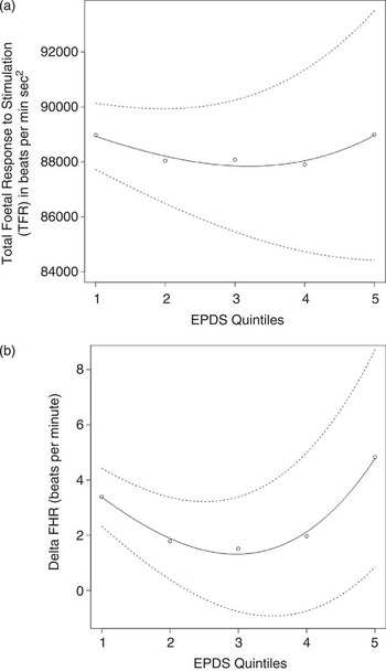
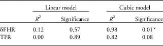
The second analysis is illustrated by Figure 1a and 1b. The foetuses of mothers in the first and fifth prenatal depression quintiles (corresponding to EPDS scores of 0–1 and 16–27, respectively) showed higher levels of total foetal response and increased delta FHR compared with the foetuses of mothers in the second, third and fourth quintiles (corresponding to EPDS scores of 2–15). Curve estimation techniques revealed that a cubic solution explained these associations better than a linear model (Table 1).
Curvi-linear association between maternal prenatal depression and foetal response and recovery variables. Note: The figures shows the curvilinear association between maternal prenatal depression and (a) mean total foetal stress responsivity to vibro-acoustic stimulation (solid line) and (b) the mean change in foetal heart rate (FHR) from baseline to post-stimulation levels (solid line) flanked by the upper and lower limits of standard deviation (broken line).

Curve estimation for maternal prenatal depression and foetal responses to stress

FHR, foetal heart rate; TFR, total foetal response to stimulation.
This table shows findings of curve-estimation for Fig. 1a and 1b. For both TFR and δFHR, a cubic model explained the association with continuous maternal prenatal depression scores better than a linear model.
*P<0.05
Discussion
In this study, the association between maternal prenatal depression, and foetal cardiac measures of response and recovery to a potentially stressful VAS, in a socio-economically disadvantaged population from rural India, is U-shaped. Lower levels of response and recovery were demonstrated by the foetuses of mothers with moderate levels of prenatal depressive symptoms compared with the foetuses of mothers with very high and very low prenatal depression scores. Given that previous literature has reported similar U-shaped associations between prenatal stress and offspring performance,Reference DiPietro, Costigan and Gurewitsch 5 , Reference Sandman, Glynn and Wadhwa 10 , Reference Sandman, Wadhwa, Chicz-DeMet, Dunkel-Schetter and Porto 11 , Reference DiPietro, Kivlighan and Costigan 13 our results suggest that, in certain environmental contexts, exposure to moderate amounts of stress may be important to condition foetuses towards the optimal regulation of their stress responses.Reference Del Giudice 14 These findings add to the body of evidence suggesting that exposure to maternal stress during intra-uterine life induces changes in foetal behaviour, and that these programming influences vary as a function of socio-environmental contexts.
Studies of humans and rodents show moderate intra-uterine exposure to stress to be conducive to foetal and infant neurodevelopment and performance.Reference DiPietro, Hodgson, Costigan, Hilton and Johnson 7 , Reference Meek, Burda and Paster 24 – Reference Fujioka, Fujioka, Ishida, Maekawa and Nakamura 26 Examples in support of this hypothesis include associations between moderate prenatal stress and enhanced foetal neuromaturity, low levels of maternal prenatal depression and higher infant cognitive scores, and moderate early life stress and lower cortisol reactivity in 10–12 year olds.Reference DiPietro, Kivlighan and Costigan 13 , Reference Gunnar, Frenn, Wewerka and Van Ryzin 27 , Reference DiPietro, Novak, Costigan, Atella and Reusing 28 We believe that our finding adds to the literature highlighting the potential adaptive function of intra-uterine exposure to moderate stress levels in increasing an organism’s capacity to function better in moderately stressful external environments.Reference Del Giudice 14 In our study, children from rural South India were exposed to under-nutrition, the threat of famine, infections, and poor levels of social and economic security. Compared to western settings, such rural environments in the developing world may be considered ‘stressful’ in the context of child development. As such, an optimal matching of intra-uterine and extra-uterine influences may confer the child with survival advantages and therefore intra-uterine exposure to moderate amounts of maternal stress may favourably adapt foetuses destined to be born into moderately stressful environments (via their stress responsivity mechanisms), thereby preparing them to cope better with adversity in later life. This hypothesis may afford an explanation of why better levels of foetal stress responsivity were observed among the foetuses of moderately stressed mothers in this study. However, larger prospective studies are needed to substantiate this theory and to elucidate the inter-play between the underlying mechanisms.
Our study was limited in that foetuses were assessed only at one time point during the third trimester and that foetal response patterns in 35 foetuses were not sufficient to assess habituation in this group, lowering the total sample size to 96. The ability of the VAS to be sufficiently stress-provoking has also been questioned, however it is important to keep in mind the ethical constraints in subjecting foetuses to stressors while designing such studies. Despite the relatively small sample size, and the use of indirect measures (i.e. foetal heart rate) to monitor foetal responses to a relatively mild stressor, the study revealed findings of a U shaped association, previously reported in larger western inquiries. The absolute difference in bpm between quintiles was small and may not be of clinical significance; nonetheless these differences may be of importance from a developmental perspective when detected early in life. Another limitation of our study is that while certain factors known to influence foetal HPA axis development such as foetal age and sex were assessed, we were unable to measure some biological factors (such as placental blood flow, maternal catecholamine levels and maternal cardiovascular activity) which have been reported, in previous work, to contribute to the association.Reference Glover, O’Connor and O’Donnell 4 , Reference DiPietro, Hodgson, Costigan, Hilton and Johnson 6 , Reference Monk, Myers, Sloan, Ellman and Fifer 9 – Reference Sandman, Wadhwa, Chicz-DeMet, Dunkel-Schetter and Porto 11 Of importance is that these results are the first evidence from the developing world, where more than 80% of the world’s pregnancies occur each year. 29 Furthermore, the findings of this study are not confounded by foetal exposure to alcohol, nicotine, recreational drugs or psychotropic medication.
We conclude that the association between maternal prenatal depression and foetal responses to a potentially stressful stimulus, in a socio-economically disadvantaged population from the developing world, is U-shaped. This raises the possibility that exposure to some amount of intra-uterine stress may favour offspring development in certain environmental contexts. The study represents the first attempt to investigate the programming of foetal responses in the context of maternal stress in the developing world and highlights the need for further research in this field in these regions.
Acknowledgements
The authors are grateful to the participating mothers and children of the Ramnagara District in Karnataka, India, and to the staff and doctors at Snehalaya Hospital, Solur for their diligence and co-operation without which this project would not be possible.
Financial Support
This research was supported by the Child Care Health and Development Research grant, the Ashok Ranganathan Bursary and the Oxford University Department of Psychiatry Doctoral Research Fund awarded to M. C. Fernandes.
Conflicts of Interest
None.