Introduction
Parasites belonging to the insect order Hymenoptera constitute a large part of biodiversity in terms of species richness, with an estimated number of several hundred thousand species (Rodriguez et al. Reference Rodriguez2013). This estimation is rising regularly with sustained taxonomic efforts helped by DNA barcoding and turbo-taxonomy initiatives (Butcher et al. Reference Butcher2012; Smith et al. Reference Smith2013), but also with the ecological characterization of species complexes (Muirhead et al. Reference Muirhead2012; Kaiser et al. Reference Kaiser2015). The larval stages of parasitic wasps exploit other arthropods, in particular insects of the orders Diptera and Lepidoptera. They are also known as ‘parasitoids’ because they kill their hosts, which place them as intermediates between parasites and predators (Godfray, Reference Godfray1994). Different types of parasitoids can be distinguished: the terms ectoparasitoid and endoparasitoid refer to species feeding respectively externally or internally during the larval stage of their hosts. In addition, koinobiont species that keep their hosts alive and developing during parasitism, are distinguished from idiobionts parasitoids that kill or paralyze permanently their host during oviposition and, as such, behave more like predators (Quicke, Reference Quicke2015). As idiobionts and koinobionts do not have the same relationships with their hosts, they use different factors to achieve parasitism success. Idiobionts usually rely on the injection of venom components to paralyze their host, while koinobionts preserve their host even if they may paralyze them transiently (Moreau et al. Reference Moreau2002). Moreover egg survival and larval development inside the host require finely tuned host physiology modifications to allow parasite development as well as healthy host growth for the duration of the interaction (Pennacchio and Strand, Reference Pennacchio and Strand2006). Koinobiont endoparasitoids are thus particularly exposed to the host immune system from which they should protect themselves, but which needs to remain functional to prevent infections that would kill the host by septicemia. The evolution of the koinobiont life history strategy has therefore led to various stratagems to precisely manipulate the host immune response.
As they lead to host death, many parasitoid species significantly impact host populations and are therefore used for biological control applications against insect pests (Hawkins, Reference Hawkins1994). In addition, the endoparasitoid lifestyle involves strong co-evolutionary interactions between parasitoids and hosts, which lead to host specialization, i.e. wasps of restricted host range. Specialist parasitoids are particularly well suited for biological control, as compared with generalists, since they have limited impact on non-targeted species, thus preserving local biodiversity (Bigler et al. Reference Bigler, Babendreier and Kuhlmann2006). However, some biological control agents have been shown to shift to non-target species (Henneman and Memmott, Reference Henneman and Memmott2001); therefore understanding the evolutionary factors involved in host range determination is essential in the context of biological control.
Endoparasitoids have intimate relationships with their hosts. This implies molecular interactions controlled by virulence and resistance genes. As the outcome of parasitism is obligatorily the death of at least one of the partners, strong selection pressures affect both host and parasitoid, which are locked in an arms race leading to specialization (van Valen, Reference van Valen1973; Henry et al. Reference Henry, Roitberg and Gillespie2008). The ability to overcome host immunity and modify host physiology represents two key traits involved in host–parasitoid coevolution. Accordingly, various strategies of host manipulation have been selected by hymenopteran parasites. The most original strategy is undoubtedly recurrent domestication of virus through stable viral genome integration into wasp genomes, a phenomenon referred to as endogenization. These viruses, grouped together in the Polydnaviridae family (PDV), are vertically transmitted and used by wasps to produce viral particles that can transfer virulence genes to host cells. These genes are collectively essential for parasitism success as they express diverse factors altering host physiology, thus making the host permissive to the development of wasp offspring (Dushay and Beckage, Reference Dushay and Beckage1993).
This review takes the standpoint of parasitoid wasps to examine the question of the convergent domestication of viruses. First, we describe a series of virus domestication events in several lineages of Ichneumonoidea. Then, we consider the evolutionary challenges associated with domesticating a virus for a wasp. Finally, we compare PDVs and the other virulence factors to discuss the advantages that virus domestication might provide to the wasp compared with other strategies to successfully parasitize their hosts.
Virus domestication by parasitoid wasps: an example of convergent evolution
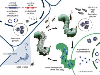
The first immunosuppressive virus-like particles (VLPs) found to be associated with parasitoid wasps were discovered in the late 1960s in the ovaries of Venturia canescens (Rotheram, Reference Rotheram1967). In the early 1970s, PDVs were described in different Hymenoptera lineages from the Ichneumonoidea superfamily (reviewed in Stoltz and Krell, Reference Stoltz, Krell, Beckage and Drezen2012). However the demonstration that these particles derived from conventional viruses awaited decades until advances in DNA sequencing technologies and mass spectrometry enabled the analyses of both host genomes and PDV/VLP protein components (Bézier et al. Reference Bézier2009a; Volkoff et al. Reference Volkoff2010; Wetterwald et al. Reference Wetterwald2010; Béliveau et al. Reference Béliveau2015; Pichon et al. Reference Pichon2015). In all cases, the particles produced (PDVs and VLPs) were shown to result from the stable integration of virus genomes within wasp genomes, allowing the production of viral particles specifically in the reproductive tract of female wasps. PDV particles contain double-stranded circular DNA, encoding virulence genes expressed in parasitized host and involved in parasitism success, while VLPs are devoid of DNA, but instead contain virulence proteins produced by the wasp. For both PDVs and VLPs, the virus genes are integrated in the wasp genome and vertically transmitted to the wasp progeny, like regular wasp genes. The DNA molecules packaged in PDV particles do not contain replication genes, consistent with the fact that viral particles do not produce a viral progeny in host cells, unlike regular viruses that replicate autonomously in infected cells (Fig. 1). This domestication, historically described as a symbiotic association between the wasp and the virus, involved gene loss or gene exchange between the genomes of the partners as observed in obligatory associations between prokaryotic symbionts and insects (Moran, Reference Moran2007; Husnik et al. Reference Husnik2013). However, PDVs could also be viewed as tools shaped during parasitoid wasp evolution to deliver virulence factors into the host.
Life cycle of the wasp Cotesia congregata and its BV. Within the wasp ovaries, in the nucleus of the calyx cells (i) proviral loci (in blue) are amplified, excised and circularized, (ii) the nudivirus-like genes (in red) are expressed to produce viral particles and (iii) viral circles are encapsidated in BV particles, which acquire an envelope upon exiting the nucleus. (iv) When the wasp oviposits in Manduca sexta caterpillars, BV particles are injected in the host along with the eggs. (v) BV particles enter host cells, where the virulence genes (in blue) are expressed to produce virulence factors. (vi) Virulence factors induce physiological modifications allowing the wasp larvae to develop inside the host body until their egress for pupation outside the host before emerging as adults. BV, Bracovirus.

Virus domestication independently occurred at least three times in the Ichneumonoidea. We might expect other examples to be discovered given the diversity and species richness of parasitoid wasps. Other examples of recurrent virus gene domestication by eukaryotes have been described, such as the retroviral syncitin genes involved in mammalian placenta development (Lavialle et al. Reference Lavialle2013), but to date parasitoid wasps are the only metazoans to have domesticated the production of complete viruses. We might however expect this strategy to occur in other, as yet unexplored, branches of the tree of life.
Bracoviruses (BVs) originated from a betanudivirus
The PDVs of braconid wasps are referred to as BVs. All braconid species that have a PDV share a common ancestor and form a monophyletic group named the ‘microgastroid complex’ composed of six subfamilies, the Microgastrinae, Cardiochilinae, Cheloninae, Miracinae, Mendesellinae and Khoikhoiiinae (Whitfield and O'Connor, Reference Whitfield, O’Connor, Beckage and Drezen2012). The divergence of this group from the other braconid subfamilies occurred 100 mya, as estimated from the age of wasp fossils in amber used to calibrate the molecular clock (Whitfield, Reference Whitfield2002; Murphy et al. Reference Murphy2008; Whitfield and O'Connor, Reference Whitfield, O’Connor, Beckage and Drezen2012). Based on this phylogeny, it was suggested BVs could all originate from the integration of a single viral genome into the genome of the ancestor of the microgastroid complex. This hypothesis was confirmed by sequencing cDNAs from the ovaries producing BV particles in several species, which revealed that numerous genes typical of nudiviruses were highly expressed in this tissue (Bézier et al. Reference Bézier2009a, Reference Bézierb; Burke and Strand, Reference Burke and Strand2012b). Nudiviruses are large DNA viruses infecting arthropods. They are a sister group to the better-known baculoviruses, used for biological control of lepidopteran pests. Proteomic analyses showed the nudivirus-like genes found in BV-carrying wasps encode viral particle components (Bézier et al. Reference Bézier2009a; Wetterwald et al. Reference Wetterwald2010; Burke and Strand, Reference Burke and Strand2012b). Functional analyses also confirmed the involvement of nudivirus genes in particle production (Burke et al. Reference Burke2013). Moreover homologues of nudiviral genes could be isolated by PCR in species from most subfamilies of BV-associated wasps as expected for a virus inherited from their common ancestor (Bézier et al. Reference Bézier2009a). Of note, these findings confirmed the initial prediction based on the typical shape of their nucleocapsids (nudiviruses and BVs are the only eukaryotic viruses with a tail on their capsid) that BVs might originate from nudiviruses (Stoltz and Vinson, Reference Stoltz and Vinson1979; Stoltz and Whitfield, Reference Stoltz and Whitfield2009). The phylogenomic analysis of the nudivirus-like genes in braconid wasp genomes showed the viral ancestor could be assigned to the genus Betanudivirus (Thézé et al. Reference Thézé2011). Since the ancestral nudivirus genome integration event, the parasitoid lineage has highly diversified as the Microgastrinae subfamily alone represents between 17 000 and 46 000 BV-associated species (Rodriguez et al. Reference Rodriguez2013).
Ichnoviruses (IVs) originated from viruses belonging to a new undescribed virus family
In ichneumonid wasps, PDVs are named IVs. However, the phylogenetic distribution of PDVs in the Ichneumonidae is more scattered than in the Braconidae. Wasps from the Campopleginae and Banchinae subfamilies harbour PDVs, but do not constitute a monophyletic group and are separated by other subfamilies that lack PDVs (Béliveau et al. Reference Béliveau2015). Two hypotheses have been formulated to explain this distribution: (1) a unique ancestral domestication occurred and PDVs were lost in several lineages, or (2) two independent domestications occurred, with different viruses, one in Banchinae, the other in Campopleginae. Recently, the viral machinery of PDVs present in banchine wasps was showed to derive from the same kind of viruses as that of IVs from campoplegine wasps (Volkoff et al. Reference Volkoff2010; Béliveau et al. Reference Béliveau2015). This points to the possibility of a single virus integration into the genome of the ancestors of these wasps. However the lack of data showing conserved integration sites does not allow the exclusion of a scenario whereby two viruses from the same family were independently captured in the Campopleginae and Banchinae. The genomic regions encoding genes involved in PDV particle production were named Ichnovirus Structural Protein Encoding Regions (IVSPER). A high density of genes lacking introns characterizes IVSPERs, suggesting an endogenized virus origin. Most IVSPER genes have no similarities with any other viral genes (Volkoff et al. Reference Volkoff2010). A few genes from banchine IVSPERs were found to be homologous to DNA replication genes of large DNA viruses belonging to the Megavirale order; however no phylogenetic link could be made with any particular viral family (Béliveau et al. Reference Béliveau2015). No pathogenic virus having homologues of IVSPER genes have yet been characterized. This is not so surprising as new viral families are regularly discovered (Abergel et al. Reference Abergel, Legendre and Claverie2015), especially as viruses of this potential new family may have remained unnoticed for lack of striking impact on insect health. Alternatively, the ancestral virus, in its exogenous form, could now be extinct (Volkoff et al. Reference Volkoff, Beckage and Drezen2012).
Symbiotic virus replacement in V. canescens
Venturia canescens is an ichneumonid wasp belonging to the Campopleginae subfamily. Its phylogenetic placement suggests it should harbour an IV. However V. canescens instead is associated with virus-like particles, devoid of nucleic acids (Rotheram, Reference Rotheram1967). Genome analyses revealed traces of IV sequences as expected for a Campoplegine species (Pichon et al. Reference Pichon2015). However these sequences are non-functional and thus do not encode the VLPs produced by V. canescens. Transcriptomic and proteomic analyses showed that the genes expressed in the wasp ovaries and producing VLPs derive from a nudivirus (like BVs). These genes are organized in six clusters in the wasp genome originating from a relatively recent virus endogenization event (Pichon et al. Reference Pichon2015). Although VLPs and PDVs derive from the same evolutionary processes, VLPs are not classified as viruses because they transfer virulence proteins and do not contain dsDNA, which is the hallmark of PDVs (named after the multiple DNA molecules contained in their particles). It is somewhat surprising that a strategy that appears a priori less sophisticated than PDV utilization has been selected in the lineage of V. canescens. However, the transfer of virulence factors in the form of proteins does ensure parasitic success. One might speculate that the original IV might have been lost because of the development of host resistance and that the selection of another tool was favoured as a means to enhance parasitic success in this lineage.
Diverse types of VLPs have been observed in wasps from several Hymenoptera families such as Microctonus aethiopoides (Braconidae: Euphorinae; Barratt et al. Reference Barratt1999), Opius concolor (Braconidae, Opiinae; Jacas et al. Reference Jacas1997), Meteorus pulchricornis (Braconidae: Euphorinae; Suzuki and Tanaka, Reference Suzuki and Tanaka2006) and several Leptopilina species (Figitidae; Dupas et al. Reference Dupas1996). However, their origins have yet to be elucidated, so whether or not endogenous viruses contribute to their production remains unknown.
Evolutionary success of virus-associated wasp lineages
The convergence in domestication processes through independent virus endogenization events and the maintenance of genetic elements from ancestral viruses in each wasp lineage over several million years required multiple evolutionary steps whereby the ancestral viruses were transformed into the gene-transfer agents, which we now know as PDVs. Several factors such as stinging behavior favouring encounters with viruses or the integrative properties of viruses may have promoted viral endogenization in wasp genomes. Because these events repeated themselves several times, the ability to produce PDVs must confer a strong evolutionary advantage to the wasp. The lineages of wasps with PDVs are indeed characterized by high species richness, e.g. Microgastrinae 17 000 to 46 000 species (Rodriguez et al. Reference Rodriguez2013), Banchinae 4000 species and Campopleginae 15 000 species (Gauld and Bolton, Reference Gauld and Bolton1988; Quicke, Reference Quicke2015). By their functions, PDVs have probably played a role in the diversifications of these subfamilies by providing a flexible tool, tailored to different host species and allowing adaptation to the hosts, specialization and speciation (Volkoff et al. Reference Volkoff2010; Herniou et al. Reference Herniou2013).
The evolutionary challenge of transforming a virus into a PDV
BVs and IVs originate from different viruses, but share common features such as particle production in the wasp calyx and packaging of double-stranded DNA circles encoding virulence genes in these particles. Numerous modifications of the originally integrated virus had to occur during parasitoid wasp evolution to build up the complex PDVs we observe today. Although we do not know the temporal order of these events, we propose a scenario based on the mechanisms of PDV production to highlight the complexity of the evolutionary process that might have happened. We will focus on BVs because we cannot make functional hypotheses for IVs since most IVSPERs genes do not display similarity with other virus or metazoan genes. In the absence of functional studies on BVs, gene functions have been inferred based on gene homologies with nudiviruses and baculoviruses with the assumption that sequence homology probably reflects similar functions during the viral cycle.
Nudivirus genome integration into the genome of the ancestor of microgastroid wasps
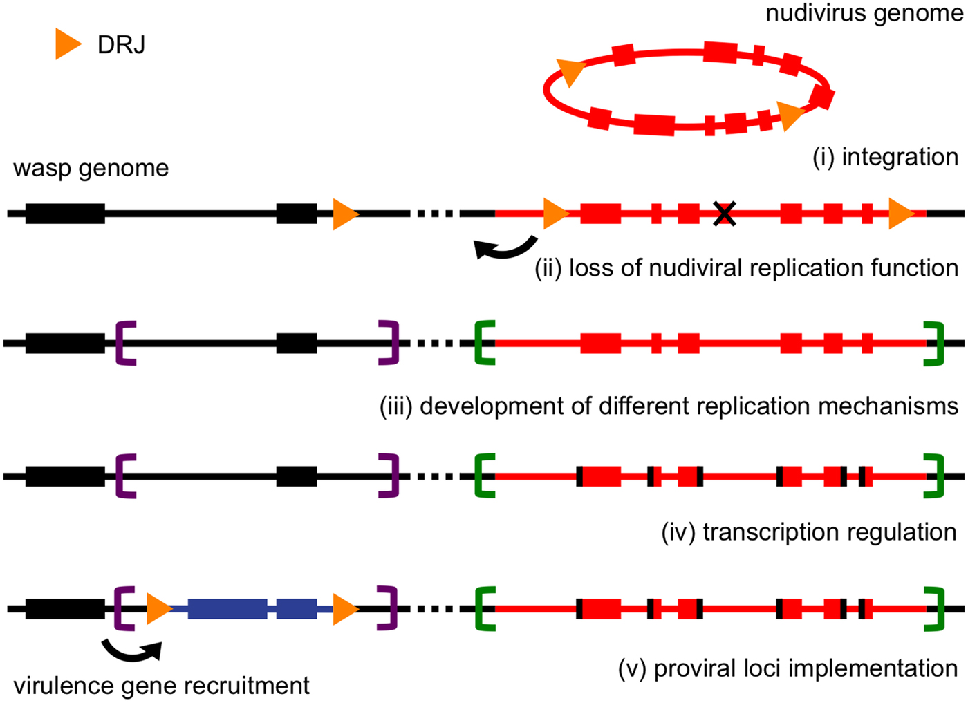
The first event of the domestication was the integration of a nudivirus genome into the genome of the ancestor of all microgastroid wasps (Fig. 2(i)). Based on extent nudiviruses, this genome was likely over 100 kb and contained about 130 genes (Wang et al. Reference Wang, Bininda-Emonds, Jehle, Garcia and Romanowski2012, Bézier et al. Reference Bézier2015). As expected if we assume a single genome integration event, the phylogenies of the nudivirus genes found in braconid wasps and their exogenous homologs are congruent (Thézé et al. Reference Thézé2011). Moreover, 17 nudivirus genes encoding major viral capsid components are still co-localized in the wasp genome, which could reflect the ancestral organization of the nudivirus genome (Bézier et al. Reference Bézier2009a, Reference Bézierb; Burke et al. Reference Burke2014). Since this cluster is amplified during particle production (although not packaged in the particles) its organization may have been maintained to enable synchronous gene amplification (Louis et al. Reference Louis2013). This amplification may allow more efficient transcription, resulting in high levels of particle production. The dispersion of the other nudivirus genes over the wasp genome could be attributed to regular genome shuffling as a result of recombination.
Schematic scenario of the genomic events leading to the evolution of BVs from the domestication of a nudivirus. Rectangles represent nudivirus-like genes in red, wasp genes in black and BV virulence genes in blue. Yellow arrowheads represent DRJ sequences necessary for the viral DNA circularization and encapsidation in BV particles. Purple and green brackets represent the boundaries of different RU. Note that the temporal order is hypothetical especially between steps (ii) and (iii). BV, bracovirus; DRJ, direct repeat junction; RU, replication units.

Unique features of the nudivirus life cycle may have facilitated the ancestral integration. The nudiviruses are arthropod dsDNA viruses and can infect insects from different orders such as Coleoptera (OrNV, Wang et al. Reference Wang2011), Orthoptera (GbNV, (Wang et al. Reference Wang2007b), Diptera (ToNV, Bézier et al. Reference Bézier2015) and Lepidoptera (HzNV, Burand et al. Reference Burand2012) and they comprise two genera Alphanudivirus and Betanudivirus. The betanudivirus HzNV, which infects the corn earworm moth (Helicoverpa zea), is the closest known relative of the BV ancestor (Thézé et al. Reference Thézé2011). Interestingly, the cycle of HzNV may comprise a latent phase of infection. During this phase the virus genome is found both as free circular DNA and integrated into the host genome (Lin et al. Reference Lin1999). The nudivirus ancestor of BVs and HzNV may have shared this ability to integrate their genomes into the host DNA. More generally, the endoparasitoid life history leads to proximity between the wasp larvae and the viruses infecting the lepidopteran host. Some of these viruses may infect both the host and the parasite, such as ascoviruses (Bigot et al. Reference Bigot1997). A virus, morphologically similar to nudiviruses, was reported to replicate in both the wasp Microplitis croceipes and its lepidopteran host Heliothis virescens and to be transmitted vertically in the wasp (Agra Gothama et al. Reference Agra Gothama, Sikorowski and McLaughlin1998). Similar interactions could have occurred 100 mya and favoured the nudivirus ancestor integration in the wasp genome. Moreover, for maintenance in the wasp genome and vertical transmission, the integration needs to have occurred in germ cells. Interestingly, HzNV replicates specifically in the gonads (Raina et al. Reference Raina2000). The nudivirus ancestor of BVs may have similarly infected wasp ovaries. This proximity increases the likelihood of integration event in germ cells, which is a prerequisite to vertical transmission. As illustration, DNA of the symbiotic prokaryote Wolbachia, known to infect germ cells, was also found integrated into several arthropod genomes (Dunning Hotopp et al. Reference Dunning Hotopp2007; Drezen et al. Reference Drezen2016; Leclercq et al. Reference Leclercq2016).
The BV is no longer autonomous for its replication
Another step in the evolution of the integrated viral genome towards the genesis of PDVs is the loss of self-replication (Fig. 2(ii)). A large proportion (18/33) of nudivirus core genes (conserved in the whole family Nudiviridae) has been conserved in the wasp genomes, where they are presumed to be required for most key viral functions, such as gene transcription, DNA packaging into nucleocapsids and particle entry into infected cells. However, most DNA replication genes have been lost, including the nudiviral DNA polymerase (Burke et al. Reference Burke2014). The amplification of DNA packaged in particles within ovarian calyx cells (Fig. 2(iii)) is a complex, yet incompletely elucidated process (Pasquier-Barre et al. Reference Pasquier-Barre2002). Specific genomic regions, named replication units (RUs), are amplified but only some are packaged in the particles (Louis et al. Reference Louis2013). The mechanisms enabling RU amplification and the proteins involved in this process have not been identified yet. However based on the structure of the amplified molecules, three different molecular mechanisms have been proposed. The proviral loci correspond to the endogenous form of the sequences clustered in the genome that will be packaged in the particles as DNA circles (Desjardins et al. Reference Desjardins2008; Bézier et al. Reference Bézier2013). They are amplified either as head-to-tail concatemers or head-to-head/tail-to-tail concatemers (Louis et al. Reference Louis2013; Burke et al. Reference Burke2015) depending on the RU and from precise borders containing specific regulatory sequences. These concatemers likely correspond to the products of two types of replication intermediates known to be produced by baculoviruses for head-to-tail type and poxviruses for the tail-to-tail/head-to-head type (Du and Traktman, Reference Du and Traktman1996; Kornberg and Baker, Reference Kornberg and Baker2005; Weller and Coen, Reference Weller and Coen2012). Although not packaged in the particles the nudiviral cluster is also amplified (Louis et al. Reference Louis2013; Burke et al. Reference Burke2015), but the amplification pattern resembles that described for chromosomal amplification of chorion genes in Drosophila following the ‘onion skin’ model where the DNA is amplified at the highest level at the centre of the RU, amplification decreasing progressively towards the periphery (Kim and Orr-Weaver, Reference Kim and Orr-Weaver2011). This RU differs by having no clear boundaries (Burke et al. Reference Burke2015) suggesting a third mechanism is involved in the amplification. It is possible that the integration of the nudivirus occurred in a region that was already amplified in the wasp ovaries (as it is the case for chorion genes in Drosophila nurse cells) providing the basis for viral DNA amplification without nudiviral DNA polymerase contribution. Amplification of the nudiviral cluster is probably needed to increase concentration of the DNA template for the transcription of these genes and to produce a large amount of particles by the limited number of calyx cells in the ovaries. This amplification induces genomic constraints and, accordingly, a comparison of the nudiviral cluster in two distantly related species within the microgastroid complex, Microplitis demolitor and Cotesia congregata, revealed a strong conservation of the genomic architecture in this region (Burke et al. Reference Burke2014). Altogether, the absence of nudivirus DNA polymerase, the use of a eukaryotic replication system for nudivirus-like gene amplification and the absence of nudiviral sequences in BV particles suggest the wasp has to keep control of virus replication, possibly to avoid the induction of a pathogenic cycle in wasp tissues.
Particle production is developmentally regulated and probably controlled by the wasp
BV particle production takes place specifically in calyx cells within the wasp ovaries (Fig. 2(iv)). This tissue specificity may be promoter-dependent and could have been inherited from the nudivirus ancestor, especially if it displayed gonad tropism such as HzNV (Raina et al. Reference Raina2000). Viral particle production is also temporally tuned with wasp metamorphosis (Pasquier-Barre et al. Reference Pasquier-Barre2002) but the mechanism that initiates the expression of the first set of nudivirus-like genes early during wasp pupal development remains unknown. Another set of genes coding for particle components is expressed coincidently with the onset of particle replication, which begins at day 4 of pupal development as observed using TEM analysis of calyx cells and confirmed using molecular assays detecting the amplification of viral sequences and circle production (Pasquier-Barre et al. Reference Pasquier-Barre2002; Bézier et al. Reference Bézier2009a). A series of nudivirus-like genes are homologous to baculovirus genes expressed during the different stage of infection, classified as early, late and very late genes. In baculoviruses, the early genes encoding the RNA polymerase subunits regulate the transcription of ‘late’ and ‘very late’ genes coding for structural components of the nucleocapsid and proteins included in the viral envelope (Rohrmann, Reference Rohrmann2013). Similarly, it has been proposed that the nudiviral RNA polymerase may control the expression of the other nudivirus-like genes (Bézier et al. Reference Bézier2009a). This hypothesis is supported by a functional analysis using RNA interference targeting an RNA polymerase subunit that resulted in the reduced production of BV particles (Burke et al. Reference Burke2013). A detailed transcriptomic analysis of gene expression at different pupal stages would be necessary to determine whether nudivirus-like genes could be classified as early, late and very late genes, or whether their temporal expression pattern is different in the context of the wasp genome.
After viral DNA amplification in calyx cells, the molecules produced are further processed. They are circularized using specific motifs [named direct repeat junctions (DRJs) or Wasp Integration Motifs (WIMs) ] through a recombination process that yields a single motif in each circle (Savary et al. Reference Savary1997, Reference Savary1999; Bézier et al. Reference Bézier2013; Burke et al. Reference Burke2014). Finally, the circles are packaged inside the viral particles. The circularization involves the integrases/recombinases VLF-1 and INT-1 derived from the ancestral nudivirus (Rohrmann, Reference Rohrmann2013) as shown by functional analyses (Burke et al. Reference Burke2013).
Possible impact of particle production on wasp immunity
The large production of viral particles in ovarian calyx cells (Beckage, Reference Beckage1998) raises the question of whether the wasp immune system might be induced upon virus release by cell lysis into the ovary lumen. Indeed in the case of symbiotic bacteria a specialized structure, the bacteriocyte, keeps symbiotic bacteria isolated (Login et al. Reference Login2011). Moreover this structure is maintained by local regulation of the immune system, involving antimicrobial peptide synthesis to control the number of bacterial cells in the bacteriocyte, in a context of global immune repression (Masson et al. Reference Masson2015). This type of repression has also been reported in Glossina sp. (Diptera) to maintain the bacteria Wigglesworthia (Wang and Aksoy, Reference Wang and Aksoy2012). Repression of immune pathways, targeting in particular antiviral pathways, such as RNA interference (van Rij and Berezikov, Reference van Rij and Berezikov2009), might similarly operate in the wasp ovaries during PDV particle production. This hypothesis could be tested by comparing the level of immune gene expression at different steps of ovaries development.
During oviposition, the wasp injects BV particles in the host body within the haemocoel. Then, the particles enter into host cells. Immune cells, such as hemocytes (Beck et al. Reference Beck, Inman and Strand2007) are the first targeted, but PDV particles enter virtually all tissues including the gut and nerve cells (Provost et al. Reference Provost2004; Beck et al. Reference Beck, Inman and Strand2007). The mechanism involved in binding to and entering inside host cells remains unknown. However, the BV envelope contains eight proteins homologous to ‘per os infectivity factors’, which form a complex allowing baculovirus entry into midgut cells during primary infection (Peng et al. Reference Peng2010). This suggests a similar PIF protein complex might be involved in BV entry into host cells, with a broader cellular entry capacity than observed for occlusion-derived virions of baculoviruses. This would not be unexpected as pif genes are also shared with nudiviruses and hytrosaviruses infecting cells other than those of midgut epithelium (Wang et al. Reference Wang, Burand and Jehle2007a) and are indeed present on viral particles (Kariithi et al. Reference Kariithi2010; Bézier et al. Reference Bézier2016). The inactivation of two pif genes by RNA interference was shown to result in a dramatic decrease in the number of infected host cells (Burke et al. Reference Burke2013). It is not known whether infections by the nudivirus ancestor of BVs were tissue specific, as is the case for most nudiviruses (Wang et al. Reference Wang, Burand and Jehle2007a), or of broad cellular spectrum as observed for the virus of M. croceipes, which infects fat body, midgut and haemocytes (Hamm et al. Reference Hamm, Styer and Lewis1988). However, tissue tropism of BVs is unusually broad and may have been conferred by a particular adaptation of pif genes, in particular the p74 gene, which binds target cells in baculoviruses (Peng et al. Reference Peng2010). Once into host cells, BV particles move to the nucleus where they release dsDNA circles. These circles may integrate in the host genome, using a specific mechanism involving a Host Integration Motif (HIM) (Beck et al. Reference Beck2011; Burke et al. Reference Burke2013). This integration mechanism of BV circles into the DNA of host cells could derive from a property of the ancestral nudivirus, as HzNV can integrate into host DNA in vitro (Lin et al. Reference Lin1999).
The mechanisms described above reveal the complexity of the BV life cycle. The main mechanisms involved in virulence gene delivery were probably inherited from the nudivirus ancestor, but have been transformed through recycling. The virus cycle follows a different order, as it is split between two organisms: the particle-producing cells of the wasps and the infected cells of the lepidopteran host (Fig. 1). The regulatory sequences involved, e.g. RU boundaries, Direct Repeat Junctions at the circle extremities or the HIMs, are conserved among microgastrine wasps. However, so far, no homology has been detected with sequences of nudivirus genomes (Burke et al. Reference Burke2014). These regulatory sequences may have diverged since the integration of the ancestral genome. Alternatively, it may be that among the few nudivirus genomes currently available, none is close enough to the nudivirus ancestor to enable the detection of homologous regulatory sequences.
Origin and evolution of virulence factors
Bracovirus dsDNA circles contain virulence genes expressed by the host cellular machinery (Chevignon et al. Reference Chevignon2014) and manipulating the physiology of the parasitized host (Strand and Burke, Reference Strand and Burke2014). However, BV gene content is highly variable among wasp species, both in number and type of genes (Dupuy et al. Reference Dupuy, Gundersen-Rindal, Cusson, Beckage and Drezen2012; Burke and Strand, Reference Burke and Strand2012a; Bézier et al. Reference Bézier2013). Notably, the gene content of the BV carried by the egg-larval parasitoid Chelonus inanitus (Cheloninae), parasitizing its host at the egg stage (Weber et al. Reference Weber, Annaheim and Lanzrein2007), is completely different from those of the larval parasitoid wasps (Microgastrinae and Cardiochilinae), ovipositing in larvae. This highlights that the main function conferred by the BVs is the ability to transfer genes, but that the set of genes maintained in the circles and transferred to the host varies as a function of the host–parasite interactions. Therefore, BV circle contents evolved after the initial domestication and became lineage specific as a result of diverse evolutionary constraints including co-evolutionary arm-race with the hosts (Bézier et al. Reference Bézier2008) (Fig. 2(v)). Phylogenetic analyses clearly showed that some BV circle genes originate from the wasp genomes, e.g. sugar transporters (Desjardins et al. Reference Desjardins2008), C-type lectins (Gasmi et al. Reference Gasmi2015), Egf (Burke and Strand, Reference Burke and Strand2014) or Ankyrins (Falabella et al. Reference Falabella2007). Many BV genes harbour conserved metazoan domains but are so divergent that their phylogenetic origin is unclear, although they most likely originate from the wasp gene set. In addition, a series of BV-specific genes do not contain any conserved domains, such as many protein components of wasp venoms (Vincent et al. Reference Vincent2010). This suggests they may have specifically evolved after the domestication. Strikingly although some virus genes are used to manipulate infected hosts (Hoover et al. Reference Hoover2011; van Houte et al. Reference van Houte, Ros and van Oers2014; Ros et al. Reference Ros2015) not a single gene of detectable nudiviral origin has been found to date in a BV packaged circle. In IVs, the dichotomy between the viral machinery producing the particles and the virulence genes is less pronounced. Indeed some gene families (N genes, p12) are present both in the packaged genome and in the IVSPERs (Volkoff et al. Reference Volkoff2010; Béliveau et al. Reference Béliveau2015).
Surprisingly, several virulence gene families (encoding Cysteine-rich, Ankyrin (ank), Protein Tyrosine Phosphatase) are shared between BVs and IVs despite the independent origins of these PDVs (Burke and Strand, Reference Burke and Strand2012a; Dupuy et al. Reference Dupuy, Gundersen-Rindal, Cusson, Beckage and Drezen2012). This convergence suggests similar evolutionary constraints led to the selection of the same genes in different lineages. The genes probably originated from cellular wasp genes, which should be quite similar in braconid and ichneumonid wasps (superfamily Ichneumonoidea) (Whitfield and O'Connor, Reference Whitfield, O’Connor, Beckage and Drezen2012).
Curiously, the ank genes of IVs and BVs share specific molecular signatures, suggesting non-independent evolution. This might be explained by an ancient horizontal gene transfer event between the two groups of PDVs. This transfer could have been facilitated by the integrative properties of BV circles in parasitized host and wasp DNA (Beck et al. Reference Beck2011; Serbielle et al. Reference Serbielle2012), as attested by the presence of BV sequences in several lepidopteran genomes (Serbielle et al. Reference Serbielle2012; Gasmi et al. Reference Gasmi2015). Multiparasitism, whereby two wasps species parasitize the same host, may have promoted direct transfer between BV and IV. This may have resulted in the integration of some BV sequences into the proviral form of an ichnovirus resulting in ank gene acquisition. Alternatively, the transfer could have occurred in the opposite direction since IV circles might have similar integrative properties (Doucet et al. Reference Doucet2007).
A specific feature of BV virulence proteins, such as PTPs and ANK, is their simple organizations, consisting solely of the conserved domain, while homologous wasp proteins generally contain several associated domains. This suggests the proteins have been truncated since their acquisition by PDVs. This simplified organization might be sufficient for PDV virulence factors to act as constitutive inhibitors of host regulatory pathways.
Many PDV virulence genes belong to multigenic families (Serbielle et al. Reference Serbielle2012; Bézier et al. Reference Bézier2013). For BVs, most variants evolved through large genomic duplications encompassing several segments (Bézier et al. Reference Bézier2013), but new gene copies also arise from cDNA reintegration (Serbielle et al. Reference Serbielle2012). After duplication, selection may drive the evolution of new copies in response to an arms race with the hosts (Dupas et al. Reference Dupas2008; Serbielle et al. Reference Serbielle2012).
Our current knowledge of PDV components and organization highlights the complexity of the system that enables virulence gene delivery and sheds light on the origin and evolution of virus domestication. Whole genome sequencing can provide a global view of the manner in which the domesticated virus is organized in the wasp genome. Additional sequencing efforts would allow a comparison of different species, wasps and other insects, with ancient or recently acquired endogenous viruses (Burke et al. Reference Burke2014; Cheng et al. Reference Cheng2014; Pichon et al. Reference Pichon2015) for a deeper understanding of virus domestication and its impact on insect genomes.
Polydnaviruses perform crucial functions
The transition to koinobiont endoparasitoid life history, involving more intimate relationships with the host, required the development of various virulence factors to enable development inside the developing host (Pennacchio and Strand, Reference Pennacchio and Strand2006). Comparison of the virulence arsenal of wasps with and without PDVs highlights the central role played by PDVs during parasitism, which could explain wasp evolutionary success.
The roles of PDVs
BVs encode virulence genes expressed in the host cells that interfere with the immune system and development (Stoltz and Cook, Reference Stoltz and Cook1983; Beckage et al. Reference Beckage1990). In particular, BVs were found to inhibit host metamorphosis through the modification of ecdysone and juvenile hormone levels (Beckage, Reference Beckage, Beckage and Drezen2012).
The arsenal of PDV virulence genes is very complex. For example, the BV of C. congregata contains 312 genes in 37 multigenic families (Bézier et al. Reference Bézier2013). A model, based on the gene-for-gene co-evolution, was proposed to explain such large gene expansion in BVs (Herniou et al. Reference Herniou2013). Furthermore, some BVs genes evolve under positive selection, and might be involved in adaptation (Serbielle et al. Reference Serbielle2008; Jancek et al. Reference Jancek2013). These genes would be good candidates for functional analyses. Indeed, given the large catalogue of BV genes, it appears difficult to determine the function of each gene, without prior hypotheses. Most functional studies to date have thus focused on genes containing conserved protein domains. For example, the cystatin gene family was found to inhibit cathepsin-like cysteine proteases involved in the immune response (Serbielle et al. Reference Serbielle2009). High throughput RNA-Seq analyses gave a global perspective on host–parasite interactions and, in particular which BV genes are overexpressed during parasitism (Chevignon et al. Reference Chevignon2014) and which host immunity genes are up- or downregulated (Chevignon et al. Reference Chevignon2015).
Other wasp virulence effectors and comparison with the BV
Beyond the strategies used to simply evade the host immune response (e.g., oviposition in tissues that are not accessed by hemocytes; Osman, Reference Osman1974; Kraaijeveld and Van Alphen, Reference Kraaijeveld and Van Alphen1995; Shaw and Quicke, Reference Shaw and Quicke2000) endoparasitoids employ various virulence molecules and agents to actively modify host physiology, including a depression of the host immune response (Burke and Strand, Reference Burke and Strand2014). Venom is a good example, as it is a common feature in the Hymenoptera where it performs diverse functions depending on its use (reviewed in Moreau and Asgari, Reference Moreau and Asgari2015). In ectoparasitoids, one of the primary function of venom is to induce host paralysis (Piek, Reference Piek1990). Endoparasitoids do not paralyze their host and their venoms have lost paralytic factors, such as neurotoxins (Pennacchio and Strand, Reference Pennacchio and Strand2006). Instead, endoparasitoid venoms are involved in host immune suppression. For example, in Pimpla hypochondriaca (Hymenoptera: Ichneumonidae), several venom proteins disrupt haemocyte spreading capacity and inhibit egg encapsulation (Dani and Richards, Reference Dani and Richards2010). Venoms could also play a role in developmental arrest, such as in Pteromalus puparum (Hymenoptera: Pteromalidae) where they disrupt the endocrine system of the lepidopteran host Pieris rapae (Lepidoptera: Pieridae) (Zhu et al. Reference Zhu2009). The venom of the parasitoid Aphidius ervi (Hymenoptera: Braconidae) induces developmental arrest (Digilio et al. Reference Digilio, Pennacchio and Tremblay1998) and castration (Digilio et al. Reference Digilio2000) of the pea aphid Acyrthosiphon pisum (Hemiptera: Aphididae). Lastly, venom composition analyses revealed antibacterial proteins in several endoparasitoid species (Dani et al. Reference Dani2003). This function, also known in social hymenoptera species, seems important to keep the host in good condition (Moreau, Reference Moreau2013).
Teratocytes are giant cells derived from the extraembryonic membrane surrounding the wasp embryo. Three functions are associated with teratocytes. Modification of host development and pupation disruption has been observed in several species, mainly through modifications of juvenile hormone and ecdysone metabolism (Strand and Wong, Reference Strand and Wong1991; Beckage and Buron, Reference Beckage and Buron1997; Dahlman et al. Reference Dahlman2003). Teratocytes also alter host immune defences by targeting haemocytes (Tanaka and Wago, Reference Tanaka and Wago1990; Andrew et al. Reference Andrew, Basio and Kim2006; Gao et al. Reference Gao2016), while secreting many proteins and antimicrobial peptides (Burke and Strand, Reference Burke and Strand2014). However, they could also contribute to larval nutrition, as a direct source of larval food (Kadono-Okuda et al. Reference Kadono-Okuda1995) or by helping the larvae to feed on host fat body through collagenase activity (Nakamatsu et al. Reference Nakamatsu, Fujii and Tanaka2002).
Advantages of PDVs
There is a certain degree of redundancy in the host functions targeted by venoms, teratocytes and PDVs. However their activities are partitioned (Burke and Strand, Reference Burke and Strand2014) to provide synergistic or additive effects for parasitism (Tanaka, Reference Tanaka1987; Strand and Dover, Reference Strand and Dover1991). In wasps associated with viruses, venom functions (e.g., altering immune defences, modifying host development and keeping the host edible) have most often been taken over by PDVs. Venoms are still produced though. They act in the very first stages of parasitism, before PDVs can protect the eggs through the expression of their genes. In Cotesia rubecula, a 50 kDa venom protein (Vn50) reduces egg melanization by inhibiting the phenol-oxidase cascade (Asgari et al. Reference Asgari2003). Furthermore, BV genes are not expressed in the absence of venom in C. rubecula (Zhang et al. Reference Zhang2004). Altogether, this shows that while some wasps can manage without PDV, the PDV on its own is not sufficient to ensure parasitic success.
However, PDVs do have several advantages compared with venom or teratocytes. Firstly, PDVs inherited from their viral ancestor the ability to target and enter specific cells. PDV virulence proteins are produced by the host against itself. This transfers the physiological cost of virulence protein production from the wasp to the host. The action of PDV virulence proteins on host physiology may also extend during the entire duration of parasitism. This prolonged action might be facilitated by the integration of viral DNA into the lepidopteran host genome (Beck et al. Reference Beck2011). The many virulence proteins encoded in the PDV circles provide a flexible, upgradable tool with a large set of virulence factor. The PDV arsenal could limit host resistance by targeting different host functions, but could also confer the ability to attack several host species (Herniou et al. Reference Herniou2013).
Concluding remarks
Most parasitoid wasps do not rely on the injection of virus particles, but the high diversification of the lineages of PDV-associated wasps suggests virus domestication leads to evolutionary success. Surprisingly evolution can also lead to the replacement of the sophisticated PDV-based strategy with that deployed by seemingly more rudimentary VLPs (such as in V. canescens), in the first reported example of symbiotic endogenous virus replacement.
The evolutionary history of PDVs since their integration remains highly speculative, in part because of the scarcity of data on the virus families from which they derive. Reconstruction of more accurate evolutionary scenarios would require additional studies of pathogenic exogenous viruses. One of the main challenges is now to identify pathogenic viruses with the same set of genes as IVS. More knowledge is available on nudiviruses, but the mechanisms of genome integration during their latent phase remain unexplained. Moreover it would be interesting to know how nudiviruses were hijacked to package other sequences instead of the virus genome during BV evolution, and how nudiviruses lost the ability to incorporate DNA in V. canescens VLPs. Knowledge on nudivirus replication could also help disentangle the different mechanisms at work during BV DNA amplification. On the wasp side, virus-associated clades with their very high number of species provide a basis for very detailed comparative genomics studies on the evolution of virus genes in wasp genomes. Decrease in the cost of sequencing and progress in genome assembly will soon allow undertaking genome-wide studies at the population and species levels that will uncover the evolutionary forces at play in parasitoid wasps.
Acknowledgments
The authors would like to thank Georges Periquet, Elisabeth Huguet, Annie Bézier, Germain Chevignon, Philippe Gayral, Sébastien Moreau and Thibaut Josse for numerous discussions on BV evolution, as well as anonymous reviewers for their constructive comments.
Financial support
This work was supported by the Agence Nationale de la Recherche grant ABC Papogen.