Introduction
Embryo implantation is a well defined and precise process in which various factors come into play one after the other, remaining in close collaboration. Significant pregnancy loss resulting from preimplantation embryonic death is common to many mammals and is considered to be a selection process that leads to the survival of superior embryos for implantation. However, dysregulation of the events before, during, or immediately after implantation also may often be a cause for poor pregnancy rates (Ma et al., Reference Ma, Song, Das, Paria and Dey2003). The foremost cause of reduced reproductive efficiency in mammal is preimplantation embryonic loss, which materializes because of intrauterine infections in pregnant women. Despite multiple strategies that attempt to impede preterm delivery, the rate has increased from 10 to 12.7% of all pregnancy (Martin et al., Reference Martin, Hamilton, Sutton, Ventura, Mathews, Kirmeyer and Osterman2010).
Progesterone (P4) and estrogen are the ovarian steroids that play a vital regulatory role in development and implantation of blastocyst. P4 regulates uterine receptivity for blastocyst attachment, induces stromal cell proliferation and differentiation, regulates epithelial cell proliferation, and coordinates uterine and embryonic interactions at a morphologic and molecular level (Lee et al., Reference Lee, Jeong, Tsai, Tsai, Lydon and DeMayo2006). The effects of P4 are mediated by the progesterone receptor (PR). 17β-Estradiol (E2) is the most potent estrogen and acts as a growth hormone for the tissues of the reproductive organs by supporting the lining of the vagina, cervical glands, endometrium and lining of the fallopian tubes. E2 enhances the growth of the myometrium. The rising estrogen levels during the first part of the reproductive cycle enhance endometrial cell proliferation. Following ovulation, progesterone (P4) secreted by the luteinized follicles lead to the differentiation of these cells (Stouffer, Reference Stouffer2003). At this point, the endometrium is mature and primed for embryo implantation. During pregnancy, E2 increases due to placental production (You et al., Reference You, Yang, Tang, Gao and Ni2006). E2 mediates its effect through two intracellular estrogen receptors (ER) i.e. ER-α and ER-β.
It has been established that Gram-negative bacteria, or their component, trigger pre-term labour in various animal models; many attempts have been made to clarify this mechanism (Koga & Mor, Reference Koga and Mor2010). We established a Gram-negative bacteria infection model in the mouse by intraperitoneal (i.p.) injection of lipopolysaccharide (LPS) (Deb et al., Reference Deb, Chaturvedi and Jaiswal2004) and showed that endotoxins derived from Gram-negative bacteria cause embryonic cell death (Jaiswal et al., Reference Jaiswal, Chaturvedi and Deb2006) and induce the DNA damage in embryonic and uterine cells (Jaiswal et al., Reference Jaiswal, Jaiswal, Agrawal and Chaturvedi2009).
We reported that LPS alters the serum level of P4 and E2 during the preimplantation days of pregnancy and elevates the E2/P4 ratio. A large infiltration of macrophages in the corpora lutea (CL) and appearance of Graafian follicles from day 3.5 of pregnancy after LPS treatment are responsible for ovarian dysfunction and altered E2/P4 ratio (Agrawal et al., Reference Agrawal, Jaiswal and Jaiswal2011). Other studies reported that LPS stimulates the macrophages to secrete cytokines, which in turn inhibit ovarian function (Sugino et al., Reference Sugino, Telleria and Gibori1998) and appears to alter the P4 and E2 (Fidel et al., Reference Fidel, Romero, Maymon and Hertelendy1998; Refojo et al., Reference Refojo, Arias, Moguilevsky and Feleder1998; Sugino et al., Reference Sugino, Telleria and Gibori1998; Williams et al., Reference Williams, Sibley, Miller, Lane, Fishwick, Nash, Herath, England, Dobson and Sheldon2008). The hormones mediate their effects via complement receptors. We hypothesize that PR and ER may be altered in response to LPS in embryos and uterus. The aim of this work was to unravel the effect of Gram-negative bacteria infection on the expression of PR and ER in embryos and uterus during preimplantation days of pregnancy by using Gram-negative bacteria infection model in mouse.
Materials and methods
Animal model
All animals were treated in accordance with institutional ethics committee guidelines for the care and use of animals in research. Park strain mice (6–8 weeks, 20–21 g) in proestrus were selected by measuring vaginal electrical resistance (Agrawal et al., Reference Agrawal, Jaiswal, Chaturvedi, Tiwari and Jaiswal2009) and impregnated naturally by males. Mating was confirmed by the presence of vaginal plug and morning of plugging was considered as day 0.5 of pregnancy.
Treatment and specimens
Conditions of Gram-negative bacteria infection were induced by LPS (250 μg/kg body weight i.e. 5 μg/animal, Salmonella enterica serotype Minnesota Re 595; Sigma, St. Louis, MO, USA) in pregnant animals on day 0.5 of pregnancy through i.p. route, in all experiments (Deb et al., Reference Deb, Chaturvedi and Jaiswal2004).
For measurement of PR and ER transcripts in embryos (n = 5 in each group), animals were superovulated with standard protocol (Deb et al., Reference Deb, Chaturvedi and Jaiswal2004) and, in uterus (n = 4 in each group) non-superovulated animals were used. Animals received either normal saline or LPS on day 0.5 of pregnancy were sacrificed on days 1.5, 2.5, 3.5, 4.0, 4.125, 4.33 and 4.42 of pregnancy and embryos or uterus were collected and processed for RNA extraction, immediately.
Measurement of PR and ER
RNA extraction
Embryos
In total, 100 embryos were collected on each day of pregnancy (days 1.5 to 4.42), washed and were re-suspended in 50 μl sterile PBS and used for extraction of total cellular RNA using the High Pure RNA Isolation Kit (Roche Diagnostics GmbH, Mannheim, Germany) according to manufacturer's instructions. Final washes of embryos were collected and used as a negative control. RNA was eluted in 60 μl of elution buffer and RNA equivalent to five embryos was used for each reverse-transcription polymerase chain reaction (RT-PCR).
Uterus
Total cellular RNA was purified from tissue specimens after homogenization in Trizol reagent (GIBCO-BRL, USA) according to the manufacturer's instructions. The quantity and quality of RNA were verified by spectrophotometry and 1.5% agarose gel electrophoresis, respectively.
Primer designing for RT-PCR
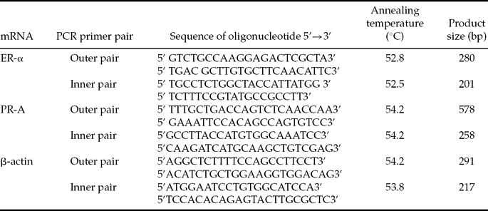
The RT-PCR primers for PR, ER and β-actin were designed using Primer Express software (Primer Express, Applied Biosystems, CA, USA). DNA and mRNA sequences of genes were taken from MGI 3.54 (The Jackson Laboratory, Bar Harbor, Maine, USA), World Wide Web (URL: http://www.informatics.jax.org). The approximate positions of primer pairs were checked by Spidey software (National Library of Medicine, USA). The oligonucleotide primers were designed to cross introns and exons boundaries for discrimination of the amplified products from genomic DNA or cDNA and were synthesized by Sigma Genosys (Sigma Aldrich Chemicals Pvt. Ltd., Bangalore, India). Sequences of primers, annealing temperature and amplicon lengths are shown in Table 1.
Primers sequences and sizes of the amplified products

RT-PCR
Expression of PR, ER and β-actin genes was analyzed by RT-PCR (with outer primer) followed by nested PCR (with inner primer) in embryos and by RT-PCR (with inner primer) in uterus samples. Reverse transcription followed by PCR was carried out using the ‘Titan One Tube RT-PCR System’ (Roche Diagnostics GmbH, Mannheim, Germany) according to manufacturer's instructions. Each RT-PCR reaction was performed in 50 μl mixtures that contained 2 μg RNA for uterus and for embryos RNA equivalent to five embryos with 0.4 μM of each primer, 0.2 μM dNTP mix, 5 mM dithiothreitol (DTT), 1 0 U RNase inhibitor and 1 U enzyme mix. Cycling conditions were followed according to manufacturer's instructions with annealing temperature mentioned in Table 1, by using a gradient in thermocycler (Bio-Rad Laboratories, Richmond, USA) and 35 cycles for each gene.
Nested PCR
The product of first round gene specific RT-PCR was used to carry out nested PCR for embryos. Nested PCR reaction was performed in 50-μl reaction mixtures containing 0.2 μl of the first round PCR product, 0.4 μM each primer, 0.2 μM dNTP mix, 0.5 U Taq DNA polymerase (MBI Fermentas, UK). Cycling conditions were followed initial denaturation at 94°C 2 min; denaturation at 94°C, 10 s; annealing temperature is as given in Table 1, 30 s; elongation 72°C, 45 s; final elongation at 72°C, 7 min using gradient in thermocycler and 25 cycles for each gene.
After amplification, PCR products were size fractionated by agarose gel electrophoresis (1.5%). Next, 15 μl of amplified product using inner primers from embryos and uterus was loaded onto the agarose gel and visualized by ethidium bromide staining under a UV transilluminator (Bio-Rad Laboratories, Richmond, USA) and photographed. The semi-quantitative analysis of PCR products was done by densitometric scanning using AlphaEaseFC™ software, version 3.0 (Alpha Innotech Corporation, San Leandro, CA, USA). The intensity of bands observed for different genes in both groups were normalized with the intensity of β-actin band observed under same experimental conditions. The identities of amplified PCR products for all cDNAs were verified by sequencing.
Statistical analysis
The results of each experiment were analyzed by using one-way analysis of variance (ANOVA) with Duncan's multiple range test for comparison of the significance level (P) between control and treated values. A P-value <0.05 value was considered as a significant difference between the values compared.
Results
Effect of LPS on PR and ER transcripts in embryos
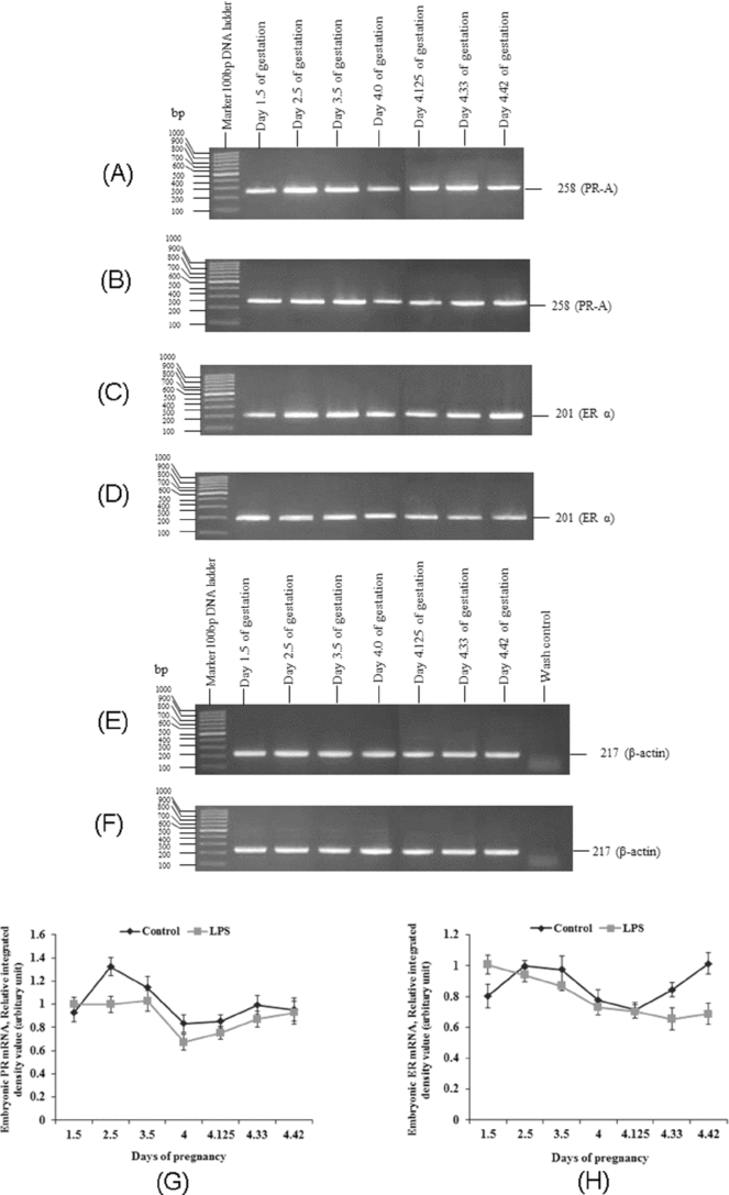
To determine whether transcription of PR and ER is affected by Gram-negative bacteria infection, semi-quantitative RT-PCR followed by nested PCR was performed in embryos obtained from days 1.5 to 4.5 of pregnancy from animals injected with normal saline or LPS (Fig. 1).
Expression of progesterone receptor (PR) and estradiol receptor (ER) mRNA in the mouse embryos collected from normal saline (control) or lipopolysaccharide (LPS)-treated animals on days 1.5, 2.5, 3.5, 4.0, 4.125, 4.33 or 4.42 of pregnancy. PR, ER and β-actin transcripts were detected by reverse-transcription polymerase chain reaction (RT-PCR) followed by nested PCR. PR (A) control (B) LPS-treated, ER (C) control (D) LPS-treated, β-actin (E) control (F) LPS-treated. A representative figure from among three repeat experiments is depicted. Ratios of the relative signal intensities of (G) PR/β-actin and (H) ER/ β-actin are depicted (n = 5 in each group).
The PR mRNA expression was detected on all studied preimplantation days of pregnancy in embryos recovered from both the groups. To the best of our knowledge, for the first time we observed the expression of PR mRNA in embryos from the onset of first zygotic cleavage i.e. from day 1.5 of pregnancy. An increase in PR mRNA expression was observed from day 1.5 to 2.5 with a maximum level on day 2.5 of pregnancy in embryos recovered from control animals. A sharp decrease in its expression was observed from days 3.5 to 4.0 and the decreased level was maintained until day 4.42 of pregnancy in embryos recovered from control animals (Fig. 1A,G). PR mRNA expression in embryos recovered from LPS-treated animals was same as the control on day 1.5 of pregnancy. This mRNA expression was markedly reduced on day 2.5 with a slight decrease from days 3.5 to 4.33 of pregnancy in the embryos recovered from LPS-treated animals as compared with the respective controls (Fig. 1B,G). The expression of PR mRNA was almost same on day 4.42 of pregnancy in embryos recovered from both the groups (Fig. 1A,B,G).
The ER-α mRNA expression was observed from day 1.5 of pregnancy until the day of implantation in embryos recovered from both the groups. An increase in its expression was observed on day 2.5 and the same increased level was maintained until day 3.5 of pregnancy in embryos collected from control animals. A sharp decrease in its expression was observed from day 4.0 to day 4.125 of pregnancy followed by a marked increase on day 4.33 to the day of implantation in embryos collected from control animals (Fig. 1C,H). However, an early rise in the ER-α mRNA expression was observed on day 1.5 of pregnancy in embryos collected from LPS-treated animals. ER-α mRNA expression was same from day 2.5 to day 4.125 of pregnancy in embryos collected from LPS-treated animals as compared with the respective controls. The ER-α mRNA expression was lower from day 4.33 to day 4.42 of pregnancy in embryos recovered from LPS-treated animals as compared with the respective controls (Fig. 1D,H).
The uniform expression of β-actin gene was observed in embryos collected at different stages of preimplantation period of pregnancy from control and LPS-treated animals (Fig. 1E,F).
Effect of LPS on PR and ER transcripts in the uterus
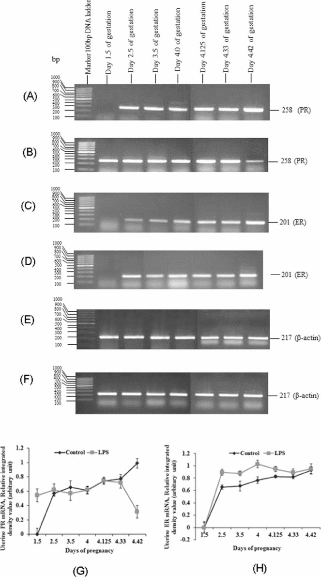
The expression of PR mRNA was examined in the uterus that was collected during different preimplantation days of pregnancy by one-step RT-PCR (Fig. 2).
Expression of progesterone receptor (PR) and estradiol receptor mRNA in the mouse uterine horns collected from normal saline (control) or lipopolysaccharide (LPS)-treated animals on days 1.5, 2.5, 3.5, 4.0, 4.125, 4.33 or 4.42 of pregnancy. The PR, ER and β-actin transcripts were detected by reverse-transcription polymerase chain reaction (RT-PCR). PR (A) control (B) LPS-treated, ER (C) control (D) LPS-treated, β-actin (E) control (F) LPS-treated, depicted is a representative figure from among three repeat experiments. Ratios of the relative signal intensities of (G) PR/β-actin and (H) ER/ β-actin are depicted (n = 4 in each group).
PR mRNA expression was not detected on day 1.5 of pregnancy; but was detected from day 2.5 with a constant expression until day 4.0 of pregnancy in uteri recovered from control animals. PR mRNA expression increased gradually from days 4.125 to 4.33 and peaked on day 4.42 of pregnancy in uteri recovered from control animals (Fig. 2A,G). PR mRNA expression was apparent from day 1.5 expression was equivalent to the controls levels until day 4.33, followed by a marked decrease on day 4.42 of pregnancy in uteri recovered from LPS-treated animals as compared with their respective controls (Fig. 2B,G).
ER-α mRNA expression was not observed on day 1.5 of pregnancy in uteri collected from both groups (Fig. 2C,D,H). ER-α mRNA expression was detected from day 2.5 of pregnancy, with gradual increase until the day of implantation in uteri collected from control animals (Fig. 2C,H). ER-α mRNA expression was greater from day 2.5 to day 4.125 of pregnancy in uteri recovered from LPS-treated animals as compared with their respective controls (Fig. 2D,H). ER-α mRNA expression on days 4.33 and 4.42 of pregnancy in uteri recovered from LPS-treated animals was the same as their respective controls (Fig. 2D,H).
Uniform expression of the β-actin gene was observed in the uteri of control and LPS-treated animals collected at different stages during the preimplantation period of pregnancy (Fig. 2E,F).
Discussion
In the present study, we report that the expression of PR and ER-α mRNA is altered during the preimplantation days of pregnancy in embryos and uteri recovered from a Gram-negative bacteria infection model in mouse. Previously we have reported that LPS significantly alters the serum level of P4 and E2 during the preimplantation days of pregnancy and elevates the E2/P4 ratio (Agrawal et al., Reference Agrawal, Jaiswal and Jaiswal2011). These steroid hormones regulate the levels of PR and ER.
It was reported that P4 is essential for implantation in mice (Hou and Gorski, Reference Hou and Gorski1993). The PR−/− mouse uterus fails to undergo decidualization, which shows that P4 is essential for the induction of this process (Tan et al., Reference Tan, Paria, Dey and Das1999). P4 is the best studied immunomodulator at the maternal–fetal interface (Peltier, Reference Peltier2003) and is considered as Nature's immunosuppressant because it plays a significant role in promoting the survival of the fetus (Peltier et al., Reference Peltier, Tee and Smulian2008). It possesses anti-inflammatory (Hardy et al., Reference Hardy, Janowski, Corey and Mendelson2006) and immunomodulatory properties at the maternal–fetal interface and supports the adhesion of the blastocyst (Peltier, Reference Peltier2003). The observed high PR mRNA expression throughout the preimplantation period of pregnancy in control embryos may support the function of P4 to respond and promote the development and adhesion of blastocysts. PR mRNA is down-regulated throughout the preimplantation period of pregnancy in embryos recovered from LPS-treated animals, except for at days 1.5 and 4.42 of pregnancy. The low expression of PR mRNA in embryos recovered from LPS-treated animals may not support the hypothesis that the embryos to respond to P4 and may inhibit the development and adhesion of blastocysts on uterine endometrium.
The down-regulation of PR mRNA expression may allow the initiation of proinflammatory cascades in embryos and uteri, and which otherwise remain suppressed, due to elevated circulating levels of P4. Abrogation of the anti-inflammatory effect of P4, due to down-regulation of PR mRNA expression in developing embryos in response to LPS may disrupt normal embryonic development during the preimplantation days of pregnancy, which may inhibit embryo implantation and ultimately lead to early pregnancy loss in mouse. Our report is in contrast with a previous in vitro study that showed that PR mRNA appears only at the blastocyst stage of embryonic development (Hou & Gorski, Reference Hou and Gorski1993).
We reported that LPS significantly elevates the serum level of E2 during the preimplantation days of pregnancy (Agrawal et al., Reference Agrawal, Jaiswal and Jaiswal2011). E2 activates the silent endogenous PR mRNA level, which is followed by expression of PR protein (Lee & Gorski, Reference Lee and Gorski1996; Das et al., Reference Das, Tan, Johnson and Dey1998; Paria et al., Reference Paria, Tan, Lubahn, Dey and Das1999; Tan et al., Reference Tan, Paria, Dey and Das1999; Murata & Higuchi, Reference Murata and Higuchi2003). The early expression of PR mRNA (i.e. on day 1.5 of pregnancy) in uteri recovered from LPS-treated animals may be due to the observed high serum level of E2 in response to LPS. Our observation of high expression of PR mRNA in uteri from days 2.5 to 4.42 of normal pregnancy is in agreement with an earlier report (Tan et al., Reference Tan, Paria, Dey and Das1999).
PR mRNA expression was very low on day 4.42 of gestation in uteri recovered from LPS-treated animals as compared with the control. Stromal expression of PR and adequate levels of P4 are necessary for optimal decidualization (Tan et al., Reference Tan, Paria, Dey and Das1999). In addition, expression of PR in the uterus is required to mediate the decidualization response to P4 (Mulac-Jericevic & Conneely, Reference Mulac-Jericevic and Conneely2004). P4 maintains uterine quiescence throughout pregnancy by acting through its nuclear receptor PR. In the majority of mammalian species, transition of the pregnant uterus from a state of near quiescence to an activated state of responsiveness to contractile stimuli is associated with decline in circulating levels of P4 (Hardy et al., Reference Hardy, Janowski, Corey and Mendelson2006). PR knockout mice result in pleiotropic reproductive abnormalities, including behavioral defects, failure to ovulate, failure of the uterus to support implantation, and defects in glandular development (Lee et al., Reference Lee, Jeong, Tsai, Tsai, Lydon and DeMayo2006). At extremely low PR mRNA expression, as observed in uterus recovered from LPS-treated animals on day 4.42 of pregnancy, uteri may not mediate the responses to P4. The absence of these feed-forward processes in uteri together may not allow the blastocyst to attach for implantation and may be responsible for LPS-induced implantation failure and early pregnancy loss in mouse.
In the present study, ER-α mRNA expression was detected from day 1.5 to the day of implantation in embryos recovered from control and LPS-treated animals. Embryo implants at the late blastocyst stage and estrogen stimulate the development of embryo and prepare it for implantation. Estrogen is essential for on-time uterine receptivity and blastocyst activation in mice (Paria et al., Reference Paria, Huet-Hudson and Dey1993). Ovariectomy of mice in the morning of day 4, i.e. before preimplantation estrogen secretion, results in implantation failure by initiating the state of blastocyst dormancy (Hamatani et al., Reference Hamatani, Daikoku, Wang, Matsumoto, Carter, Ko and Dey2004). It was reported that the expression of ER-α mRNA follows the changes in the level of E2 in peripheral blood (Wang et al., Reference Wang, Eriksson and Sahlin2000). This finding suggests that LPS-induced early expression of ER-α mRNA in preimplantation stage embryos may be due to the observed high serum level of E2 (Agrawal et al., Reference Agrawal, Jaiswal and Jaiswal2011) in LPS-treated animals. The increased expression of ER-α mRNA at the time of implantation (i.e. days 4.33 to 4.42 of pregnancy) in embryos recovered from control animals may mediate the required responses of E2 for the activation of the developing dormant blastocysts and make them adhesion competent for implantation. However, a sharp decrease in ER-α mRNA expression at the same time in embryos recovered from LPS-treated animals may not support the conversion of dormant blastocyst to the activated state and may not make them adhesion competent for implantation. The hang-up of this process may not bolster the blastocyst to attach for the implantation and responsible for LPS-induced implantation failure and early pregnancy loss in mouse. Our study in vivo is in contrast with the study in vitro by Hou & Gorski (Reference Hou and Gorski1993). The discrepancy in the results could be due to the difference in the sensitivity of the PCR methods.
There is no expression of ER-α mRNA on day 1.5 of pregnancy in the uterus recovered from animals of both the groups. However, other previous study reports an earlier observation of ER-α mRNA using northern blot analysis for uteri from days 1 to 8 of pregnancy (Tan et al., Reference Tan, Paria, Dey and Das1999). Our observation of increase in ER-α mRNA expression from day 2.5 to 4.42 of normal pregnancy with a high level at the time of implantation in uteri is in agreement with previous reports (Curtis Hewitt et al., Reference Curtis Hewitt, Goulding, Eddy and Korach2002; Minorics et al., Reference Minorics, Ducza, Marki, Paldy and Falkay2004). The elevated level of the ER-α mRNA on day 4.42 of pregnancy may be related to E2-induced blastocyst implantation and epithelial proliferation in the endometrium, which is necessary for blastocyst attachment to be manifested by the day 4.5 (Minorics et al., Reference Minorics, Ducza, Marki, Paldy and Falkay2004).
It has been reported that the expression of uterine genes contributing to blastocyst implantation is maintained at a lower E2 dose, whereas gene expression promptly becomes aberrant in the uterus, becoming refractory at higher E2 levels (Ma et al., Reference Ma, Song, Das, Paria and Dey2003). The greater level of ER-α mRNA expression from days 2.5 to 4.33 of pregnancy in uteri recovered from LPS-treated animals suggests that LPS increases the response to E2 via increasing ER-α mRNA levels in uteri and thereby converting it to refractory state. Greater ER-α mRNA expression in uteri, in the background of high serum level of E2 (Agrawal et al., Reference Agrawal, Jaiswal and Jaiswal2011) during preimplantation days of pregnancy may promptly reduce gene expression in the uterus and transforms it to the refractory state, which results in LPS-induced implantation failure in the mouse.
Disturbance in the expression of PR or ER in response to LPS may lead to early embryonic loss in mouse. However, the exact mechanism that underlies embryonic loss is not clearly understood. This mechanism may be induced by numerous factors that trigger different pathways and that may culminate in elimination of the developing embryos from the mother. The present study will contribute to our understanding of the mechanism of early pregnancy loss during Gram-negative bacteria infection in the pregnant mother. A thorough understanding of this mechanism may lead to the development of effective strategies that either prevent or treat early embryonic loss.
Acknowledgements
We thank Prof. M.M. Chaturvedi and Prof. I.K. Patro for the generous gift of chemicals. This work was funded by AICTE, New Delhi, India. We thank the Rockefeller Foundation, New York, USA for financial support to YKJ; CSIR, New Delhi for JRF and SRF VA; ICMR, New Delhi for a SRF to MKJ and DST, New Delhi, for the DST-FIST grant to the school.