Introduction
Wilson disease is a rare autosomal recessive disorder caused by mutations in the ATP7B gene, leading to impaired copper metabolism and progressive accumulation of copper in the liver, brain, and other tissues. The clinical spectrum in children is heterogeneous, with hepatic manifestations being the most common. Neurological and psychiatric symptoms usually appear later in adolescence. The Kayser-Fleischer ring of the cornea is a hallmark finding. Reference Ferenci1 Cardiac involvement is exceedingly rare and may present as arrhythmias, conduction abnormalities, or cardiomyopathy, but rarely precede hepatic or neurological features. Reference Ertaş, Gul and Demirbas2 Reporting such cases is essential to raise awareness among clinicians, as early diagnosis and prompt initiation of chelation therapy can prevent irreversible organ damage and improve outcomes. We present here a unique case of Wilson disease in a young child whose initial and only manifestation was recurrent syncope due to ventricular arrhythmia, preceding any other significant clinical signs.
Case report
A 4-year-old male child, born to consanguineous parents, presented with recurrent daily syncopal episodes. The episodes occurred 2–3 times per day, both at rest and during exertion, and were not related to postural changes. Each attack had a sudden onset, associated with sweating, and resolved spontaneously without intervention. There was no family history of cardiac disease. The child’s siblings were healthy, including a twin sister.
Initial evaluation
Physical and neurological examinations were unremarkable, with no hepatomegaly. Vital signs were stable: pulse 95 b/min, blood pressure 100/60 mmHg with no postural drop. On admission, the child’s complete blood count was normal (haemoglobin 11.8 g/dL, white blood cells 8 × 10⁹/L, platelets 305 × 109/L). Random blood sugar was also normal (87 mg/dl). However, liver enzymes were significantly elevated: alanine aminotransferase 339 U/L, aspartate aminotransferase 222 U/L, and gamma-glutamyl transferase 118 U/L. Other investigations, including lipid profile, coagulation profile, renal function tests, and viral hepatitis markers, were all normal. Abdominal ultrasound showed no abnormalities. Neurological evaluation (electroencephalography and brain MRI), as well as ophthalmological evaluation (slit lamp examination), was normal.
Hepatology workup
Given the persistently elevated liver enzymes, the child was referred to the hepatology clinic. A liver biopsy revealed hepatocytes distended with finely granular cytoplasm, minimal macrovesicular steatosis, compressed sinusoids, and mild portal fibrosis with inflammatory infiltrates. These findings were initially suggestive of a metabolic liver disease, likely glycogen storage disorder. A trial of uncooked starch therapy was ineffective. Therefore, extended metabolic screening was then performed, including fasting tolerance test (no hypoglycaemia after 12 hours), amino acid, acylcarnitine, and urine organic acid profiles that were normal except mildly elevated plasma lactate (27 mg/dL). Despite this, syncopal attacks persisted, and liver enzymes remained elevated.
Cardiology workup
Due to recurrent syncope, the child was referred to the cardiology unit. Echocardiography showed normal function (left ventricular fraction shortening 43%, left ventricular ejection fraction 75%). A resting 12-lead electrocardiography was normal (heart rate 95 b/m, PR 0.15 s, QRS 0.08 s, QTc 0.38 s, and no ST-T abnormalities). However, 24-hour Holter monitoring revealed frequent polymorphic premature ventricular contractions 7% and more than 10 runs of non-sustained ventricular tachycardia, with a maximum heart rate of 140 b/m (Figure 1). Cardiac MRI was completely normal.
Holter tracing showing frequent polymorphic PVCs and a run of non-sustained VT.

The patient was started on beta-blocker therapy (sotalol), which decreased the frequency but not the recurrence of syncopal attacks (reduced to 2–3 episodes per week). The family declined electrophysiological studies.
Genetic diagnosis
Given the unexplained liver enzyme elevation and ongoing cardiac events, whole-exome sequencing was performed. This revealed a homozygous mutation in the ATP7B gene, confirming the diagnosis of Wilson disease. This was supported by markedly lower serum ceruloplasmin: 1–2 mg/dL, low total serum copper: 19 µg/dL, and elevated 24-hour urinary copper: 85.8 µg/day.
Management and follow-up
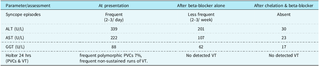
The patient was started on chelation therapy with penicillamine (250 mg daily), along with zinc, vitamin D (1000 IU/day), and vitamin B12 supplementation. Beta-blocker therapy was continued for 3 months, during which the syncopal attacks ceased. The response to combined therapy was dramatic and is summarised in Table 1. Repeat Holter monitoring showed no arrhythmia, and gradual withdrawal of the beta-blocker was successful without recurrence of arrhythmias. Liver function normalised with therapy (alanine aminotransferase 30 U/L, aspartate aminotransferase 23 U/L, gamma-glutamyl transferase 17 U/L, and total bilirubin 0.4 mg/dL) (Table 1). A follow-up fibroscan demonstrated only stage F1 fibrosis and no evidence of steatosis.
Sequential changes in clinical presentation, Holter 24-hour monitoring, and liver function tests

ALT = alanine aminotransferase, AST = aspartate aminotransferase, GGT = gamma-glutamyl transferase, PVCs = premature ventricular contractions, VT = ventricular tachycardia.
Family screening
Following the diagnosis, Wilson disease screening was carried out in the family. The patient’s twin sister also tested positive, while his older brother was negative. A cousin was also diagnosed with Wilson’s disease. Early detection allowed the timely initiation of therapy and prevention of complications in the affected relatives. A sequence of investigations and treatment was presented as Figure 2.
A flow chart summarises the sequence of investigations and the treatment given.

Discussion
Wilson disease is an autosomal recessive disorder caused by mutations in the ATP7B gene, resulting in impaired copper metabolism and the accumulation of copper in the liver, brain, and other organs. The clinical presentation is highly variable and often depends on the primary site of copper deposition. Reference Roberts and Schilsky3 In children, liver involvement is the most common early sign, while neurological and psychiatric features usually appear later. Cardiac issues, especially ventricular arrhythmias, are very rare. Reference Ertaş, Gul and Demirbas2,Reference Litwin, Gromadzka and Członkowska4 In this case, the child first showed recurrent syncope due to ventricular arrhythmias. Interestingly, no structural heart disease was detected by echocardiography or cardiac MRI, denoting that those electrocardiographic changes can occur in Wilson disease in a normal heart.
In one of the previous studies, Kuan et al. Reference Kuan5 examined 53 adult patients with Wilson disease and documented four types of cardiac manifestations: arrhythmias, cardiomyopathy, sudden cardiac death, and autonomic dysfunction. Electrocardiography abnormalities were present in approximately one-third of patients. Importantly, two patients died of cardiac causes-ventricular fibrillation and cardiomyopathy-underscoring the clinical importance of such complications. Furthermore, Hlubocka et al. Reference Hlubocka, Marecek and Linhart6 evaluated 42 patients with Wilson disease using echocardiography and 24-hour Holter monitoring, and they found increased left ventricular wall thickness and a spectrum of electrocardiography abnormalities, most frequently supraventricular tachycardia and ectopic beats.
The pathophysiology of cardiac arrhythmias in Wilson disease is multifactorial. One mechanism involves direct copper toxicity to the cardiac conduction system and myocardium, which leads to enhanced automaticity or triggered activity. Additionally, copper deposition increases oxidative stress, leading to damage in the cardiac muscle. Wilson disease may also cause dysautonomia, a dysfunction of the autonomic nervous system that controls heart rate, which in turn can cause arrhythmias. Reference Kuan5,Reference Hlubocka, Marecek and Linhart6 The observation that ventricular tachycardia resolved only after copper chelation, while beta-blocker therapy provided only partial control, supports a direct causal link between copper overload and arrhythmias. This case also illustrates that proper chelation therapy can result in a good prognosis and resolution of arrhythmias.
Our case report is important for two reasons: the patient’s young age and the life-threatening nature of the presenting symptom. Wilson disease is typically presented in older children with liver disease or neurological symptoms. Our patient had neither, and his primary complaint was cardiac syncope.
Moreover, persistently high liver enzymes prompted a hepatology referral, and subsequent genetic testing confirmed Wilson’s disease. The initial liver biopsy, while abnormal, was non-specific and directed the initial workup towards glycogen storage disease. Liver biopsy is not pathognomonic for Wilson disease, and definitive genetic and biochemical testing are required.
This case report emphasises the importance of considering a broad range of diagnoses when evaluating unexplained arrhythmias in children, especially if liver function is abnormal. Chelation therapy with penicillamine, along with zinc supplements, was effective in restoring liver function and eliminating arrhythmias. Reference Hedera7 Early diagnosis and treatment were vital, not only for the patient but also for family members identified through cascade screening. Finding Wilson disease in the child’s twin sister and cousin highlights the importance of genetic and biochemical screening in relatives at risk. Reference Litwin, Gromadzka and Członkowska4
Conclusion
This case highlights a rare presentation of Wilson disease, where ventricular arrhythmia-induced syncope was the initial and only manifestation in a young child before the development of classic hepatic or neurologic signs. The case emphasises the need for clinicians to consider metabolic and genetic causes in paediatric patients with unexplained cardiac arrhythmias, particularly when associated with elevated liver enzymes. Early recognition and initiation of chelation therapy can prevent disease progression and enable timely screening and management of affected family members.
Financial support
None.
Competing interests
None to declare.
Ethical standard
Ethical approval was waived in accordance with the institutional policy, and a written informed consent was obtained from the parents.