Permanent cardiac pacing is rarely uneventful throughout childhood and complications usually involve epicardial leads. Reference Fortescue, Berul, Cecchin, Walsh, Triedman and Alexander1,Reference Kubus, Materna and Gebauer2 Coronary artery compression is a life-threatening event but is rarely reported in the literature. Reference Brenner, Gaines, Cordier, Reiner, Haney and Gundry3-Reference Carreras, Duncan, Djurdjev and Campbell6 The real incidence might be higher because of atypical clinical presentations, diagnostic imaging limitations, and lack of awareness. Reference Carreras, Duncan, Djurdjev and Campbell6,Reference Mah, Prakash, Porras, Fynn-Thompson, DeWitt and Banka7
Case presentation
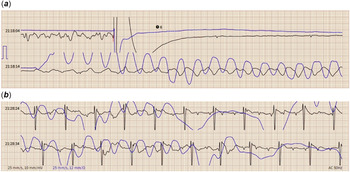
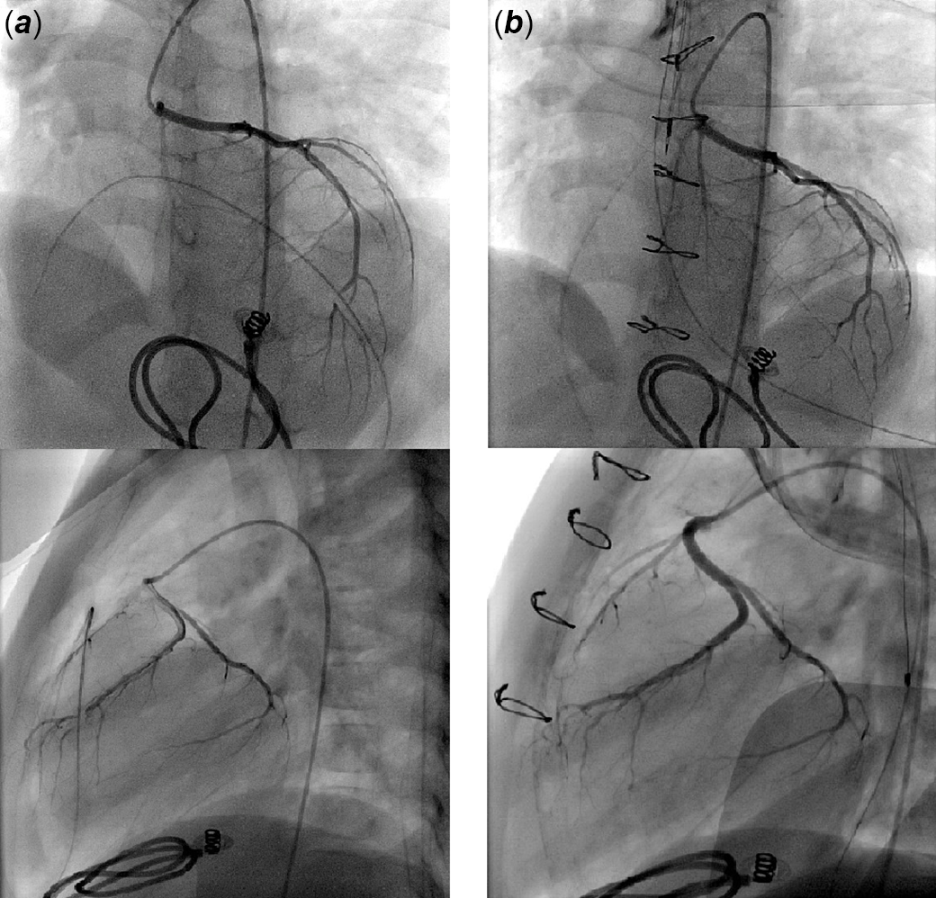
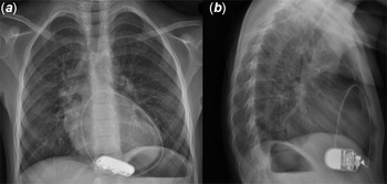
A patient with transposition of the great arteries, ventricular septal defect, and aortic coarctation had complete surgical repair at 3 days of life. Surgery was complicated with a complete atrioventricular block. Single-chamber pacemaker (MicronyTM, St. Jude Medical, Inc., Saint Paul, Minnesota, United States of America) connected to a monopolar, sutured-on left ventricular lead was implanted after one week. Routine coronary angioscan at 4 years of age showed no coronary ostial stenosis. The pacemaker was programmed in VVI-R mode with a minimal rate of 65 bpm, and routine interrogations demonstrated strict pace-dependent rhythm. At the age of 5, the previously asymptomatic patient was transferred to our centre after a resuscitated cardiac arrest. The rescue squad arrived 10 minutes after parental alert, started chest compressions and one electric shock was delivered by an automated external defibrillator. Upon ICU admission, patient was on respiratory and inotropic support while no stimulation spikes were visible on electrocardiogram. Device interrogation confirmed pacing failure secondary to lead fracture (lead impedance > 2000 Ohms). Given the paced-dependent status, a new bipolar lead was urgently implanted, and the generator was exchanged with an AzureTM XR SR MRI SureScanTM (Medtronic, Inc. Minneapolis, Minnesota, United States of America). The proximal part of the fractured lead was extracted but the distal portion was abandoned to avoid extensive pericardium dissection. The ICU stay was uneventful and the neurological assessment showed no sequelae. The automated external defibrillator report was recovered. Tracings revealed that the patient was initially in ventricular fibrillation that was converted into a strictly regular heart rhythm at 65bpm after the external shock was delivered (Fig 1). These findings revealed good functioning of the pacemaker lead at the time of ventricular fibrillation, and motivated a diagnostic coronary angiography. Dynamic compression of the distal part of the left descending artery by the non-functioning abandoned lead was demonstrated (Fig 2a). Patient underwent redo sternotomy for lead removal. Control coronarography was normal (Fig 2b). Patient had an uneventful recovery and is asymptomatic on 6-month follow-up. Retrospective review of the last chest X-ray performed during regular follow-up, 2 months before the cardiac arrest, revealed the alarming pattern of epicardial leads encircling the cardiac silhouette (Fig 3).
Electrocardiogram tracing upon automated external defibrillator application. Note the electric shock delivered over ventricular fibrillation (red mark) ( a ), and followed by a stable 65 bpm regular rhythm after 10 minutes of resuscitation ( b ).

Selective angiographies showing anterior coursing of the abandoned epicardial lead above the distal part of the left anterior descending coronary artery leading to dynamic compression with loss of contrast ( a ). Complete resolution following lead removal with no residual defects ( b ).

Last posteroanterior ( a ) and lateral ( b ) routine chest radiographs before the cardiac arrest. Note the pacing wire posterior looping around the heart.

Discussion
Permanent pacing therapy has been performed in paediatric patients since the late 1960s. Although transvenous lead implantation has been recently advocated for children, epicardial route remains the most popular approach in small babies due to vascular access limitations, or complicated intracardiac anatomies. Stimulation is rarely uneventful and complications are usually lead-related. This case report illustrates how the commonly occurring lead fracture concealed the rarely reported and potentially fatal coronary compression, misleading initial clinical management. Retrospectively, the patient presented ventricular fibrillation secondary to the compression of the left anterior descending coronary artery and was resuscitated by the external electric shock. Chest compressions fractured the lead afterward. Upon admission, lead failure was logically incriminated in the mechanism of the cardiac arrest leading to extreme bradycardia followed by shockable ventricular arrhythmia. Surgery was precipitated. Diagnostic coronarography was performed based on three arguments: coronary reimplantation history, automated external defibrillator tracings confirming lead good functioning at the time of ventricular fibrillation, and the rapidly favourable neurologic evolution incompatible with prolonged cardiac arrest as seen in ventricular fibrillation after lead dysfunction.
Coronary compression is a recognised mechanical complication of epicardial pacing but it is underdiagnosed. Reference Mah, Prakash, Porras, Fynn-Thompson, DeWitt and Banka7 The incidence is not well defined with only few reported cases. Reference Brenner, Gaines, Cordier, Reiner, Haney and Gundry3–Reference Carreras, Duncan, Djurdjev and Campbell6 An incidence of 5.5% was recently reported by Mah et al. Reference Mah, Prakash, Porras, Fynn-Thompson, DeWitt and Banka7 Clinical presentations vary from coronary compression in asymptomatic subjects to post-mortem diagnosis. None of the recognised diagnostic tools is perfect for assessment. Reference Carreras, Duncan, Djurdjev and Campbell6,Reference Mah, Prakash, Porras, Fynn-Thompson, DeWitt and Banka7 Chest radiography is a good surveillance tool as it can detect early suspicious patterns of leads implanted within the pericardium (Fig 3). Cine CT-scan can identify dynamic compression but must be confirmed by catheter angiography and myocardial scintigraphy, especially when surgery is being considered. Reference Mah, Prakash, Porras, Fynn-Thompson, DeWitt and Banka7 Lead placement is challenging as a mismatch between limited lead length and somatic growth must be avoided. The risk factors are not well established but placement of the excess lead in the anterior pericardium or around the ventricles has been associated with an increased risk. Reference Takeuchi and Tomizawa8,Reference Alhuzaimi, Roy and Duncan9 Janík et al recently published a forensic report on how an epicardial pacing wire retained in place for almost three decades led to the sudden cardiac death of a young adult. Reference Janík, Hejna, Straka, Krajčovič and Novomeský10 Clinicians must be aware that active surveillance needs to be continued even for abandoned non-functioning leads and prophylactic pacing systems placed within the pericardium. Lead removal should be considered whenever suspicious patterns are diagnosed or suspected.
Conclusion
Lead-related complications of epicardial pacing may be clinically trapping. Life-threatening coronary compression can be concealed by more obvious mechanical complications and can occur even with abandoned leads.
Acknowledgments
None.
Author contributions
RH collected clinical data and took the lead in writing the manuscript. All the authors have read and approved the final version of the manuscript.
Financial support
This research received no specific grant from any funding agency, commercial or not-for-profit sectors.
Conflicts of interest
None.
Ethical standards
The authors assert that all procedures contributing to this work comply with the ethical standards of the relevant national guidelines on human experimentation, and with the Helsinki Declaration of 1975, as revised in 2008. The patient’s legal guardians signed informed consent was obtained for the reported procedures.