Introduction
There are many types of aortic valve disease in children, which can be divided into aortic valvestenosis and aortic valveinsufficiency according to the transvalvular blood flow status. The incidence of congenital aortic valve stenosis accounts for 3%–6% of the total incidence of CHD, Reference Yasuhara, Schultz, Bigelow and Garg1 and bicuspid valve malformation is the main type. Congenital aortic valve insufficiency accounts for 2.3% of the total incidence of CHD. Reference Zhang, Yang, Chen, Dong and Li2
Complex aortic valve disease in children is a major challenge in the field of cardiac surgery. The main purpose of surgical repair is to improve the symptoms of children and avoid or delay prosthetic valve replacement and reoperation as much as possible. At the same time, surgical repair should take into account the continuity of surgical effect and the growth ability of aortic valve after surgery, and there is still a lack of consistent surgical repair concept and universal surgical repair strategy. Reference Xie, Wu, Liu and Dian3 Aortic valve replacement is not a good choice for children. After mechanical valve replacement, children need to face the risks of long-term anticoagulation, valve dysfunction, prosthesis-patient mismatch and reoperation, while after biological valve replacement, children need to face the problems of valve failure, prosthesis-patient mismatch, and multiple reoperation. Reference Myers, Mokashi and Horgan4,Reference Etnel, Elmont and Ertekin5 At present, homografic aortic valve replacement is rarely used in the world, but the existing small sample clinical evidence suggests that 40%–50% of patients still need aortic valve reoperation in the future Reference Sharabiani, Dorobantu and Mahani6,Reference Fukushima, Tesar and Pearse7 Ross procedure is considered the gold standard for the treatment of complex aortic valve disease. However, the Ross procedure is limited because it places both the aortic and pulmonary valves at risk of degeneration and reoperation. The reoperation rates of aortic valve and right ventricular outflow tract were 1.60% and 1.91% / year, respectively. Reference Etnel, Elmont and Ertekin5 In addition, some children with special conditions are not suitable for Ross procedure. Absolute contraindications include Marfan syndrome, pulmonary valve disease, immune disorders such as lupus and rheumatoid arthritis, and severe mitral valve disease. Relative contraindications (due to a higher risk of autograft dysfunction) include rheumatic valve disease and dysplastic aortic root dilation. Reference Mazine, El-Hamamsy and Verma8,Reference Wang, Zhang and Ma9 Given the large number of children with CHD in developing countries, suitable conduits for right ventricular outflow tract reconstruction, especially homograft, are scarce. For patients with a small annulus, there is a lack of suitable bioprosthetic or mechanical valves. Balloon dilatation was performed primarily in an interventional manner. Especially for young children with aortic valve stenosis, balloon dilatation can well relieve the stenosis state, prolong the use of autologous aortic valve, and avoid thoracotomy in a short time. However, there are also some problems such as valve tear and postoperative aortic valve regurgitation, especially in young children.
The effect of aortic valvuloplasty in children has been steadily improved recently, making its application in children with aortic valve disease more active and widespread Reference Zhu, Konstantinov, Wu, Wallace, Brizard and Buratto10,Reference Bouhout, Ba, El-Hamamsy and Poirier11 In infants and young children, compared with balloon dilatation, aortic valvuloplasty has the same safety and higher long-term freedom from aortic valve reoperation Reference Vergnat, Asfour and Arenz12,Reference Siddiqui, Brizard and Galati13 The concept and surgical strategies of aortic valvuloplasty are complex and variable. The main surgical strategies include leaflet thinning, junction incision, junction suspension/folding, leaflet extension, and leaflet reconstruction (Ozaki technique). Ozaki procedure is an innovative aortic valvuloplasty in which the diseased valve leaflets are completely removed and reconstructed with autologous pericardial, which provides an alternative treatment method for aortic valve disease. Reference Ozaki, Kawase, Yamashita, Uchida, Takatoh and Kiyohara14–Reference Unai, Ozaki and Johnston16 As a new aortic valve repair method, Ozaki procedure has expanded the surgical indications of complex aortic valve disease and has been applied as a promising alternative in the paediatric population. Reference Baird, Marathe and Del17 Compared with conventional mechanical or biological valves, it preserves the natural motion of the aortic annulus and preserves the coordination of the left ventricle, sinus of Valsalva, and aorta, so that the aortic root naturally expands during systole with the maximum effective orifice area. Reference Amabile, Krane and Dufendach18 Compared with the Ross procedure, the Ozaki procedure restores the physiological laminar flow pattern and shows similar haemodynamic results in the short term. Reference Secinaro, Milano and Ciancarella19 Because the aortic annulus cannot be fixed by the Ozaki technique, the nascent aortic annulus grows over time. A large coaptation height should allow the retention of a satisfactory coaptation surface, which decreases as the ring grows. Reference Sivalingam, Haranal and See20 In 2011, Professor Shigeyuki Ozaki initially described the aortic valve neo-cuspidization procedure (AVNeo) in a series of adult patients Reference Ozaki, Kawase and Yamashita15 They reported their initial experience of 88 patients with a mean age of 70.6 years with 65 patients having aortic stenosis. At 3 years of follow-up, freedom from reoperation was 100%. Subsequently, in 2014, they reported the long-term outcomes of 404 patients at their institution with a mean age of 69 years undergoing Ozaki procedure. The mean follow-up time was 23.7 ± 13.1 months. Freedom from reoperation was 96.2% at 53 months follow-up Reference Ozaki, Kawase and Yamashita21 In 2018, they described their experience on 850 patients with a median age of 71 years. The mean follow-up period was 53.7 ± 28.2 months. Actuarial freedom from death, cumulative incidence of reoperation, and that of recurrent moderate aortic regurgitation or greater were 85.9%, 4.2%, and 7.3%, respectively, with the longest follow-up of 118 months. Reference Ozaki, Kawase, Yamashita, Uchida, Takatoh and Kiyohara14 In addition to Asia, similar findings have been achieved in some heart centres in the United States that perform Ozaki procedure. Therefore, it is necessary to describe the application of Ozaki technique in the real perspective of paediatric population.
Ozaki technique received Food and Drug administration approval in the United States in 2014 and is commercially available through the Japanese Organization for Medical Device Development (JOMDD, Tokyo, Japan) company. The AVNeo system is based on single or multi-use sizing templates, and measurements are based on the aortic valve coaptation points and commissures being in the same plane with the ratio of distance between two commissures and free edge length being similar. The original sizers and template were manufactured in sizes 17 to 31 mm and the smaller paediatric sizes (13, 15 mm) were introduced in 2015. In the past decade, paediatric cardiac surgeons from different countries have applied Ozaki technique to the surgical treatment of congenital and acquired aortic valve disease in children and have achieved some encouraging results. This article reviews the past and present, surgical indications, surgical procedures, advantages, prognosis, and prospects of Ozaki technique in treating aortic valve disease in children.
Past and current status of Ozaki technique in the treatment of paediatric aortic valve disease
To date, there are few reports on paediatric patients with aortic valve disease undergoing Ozaki procedure. The Boston Children’s Hospital experience with Ozaki procedure began in 2014. Christopher W. Baird et al first reported their acceptable early outcomes of the Ozaki procedure for congenital aortic and truncal valve disease in 2020. Retrospective analysis was performed on all 57 patients with congenital aortic and truncal valve disease that had a three-leaflet Ozaki procedure. There were no hospital mortalities or early conversions to valve replacement. At discharge, 98% of patients had mild or less regurgitation and peak aortic gradient was 16.9 ± 9.5 mmHg. Two patients underwent aortic valve replacement. At median follow-up of 8.1 months, 96 and 91% of patients had less than moderate regurgitation and stenosis, respectively. Reference Baird, Cooney, Chavez, Sleeper, Marx and Del22 Marathe et al began to undergo the single aortic valve leaflet reconstruction attempting to retain native leaflet tissue, with several advantages including maintaining single valve disease, somatic growth potential, preserved dynamic annular motion, and limited need for long-term anticoagulation. Reference Marathe, Chavez and Sleeper23 Freedom from moderate or greater aortic valve regurgitation and aortic valve stenosis was 76 and 86% at 2 years in patients who underwent single leaflet reconstruction. At the 59th annual meeting of the Society of Thoracic Surgeons, Supreet P. Marathe et al reported early outcomes in young patients with small native aortic valve annuli who underwent a modified Ozaki procedure including annular enlargement. Reference Marathe, Chavez, Sleeper, Marx, Del and Baird24 Median native annulus diameter was 17 mm; sinus and annular enlargements were required in 22 and 9 patients, respectively. At mean follow-up of 11.9 months, 80 and 82% of patients had <moderate aortic valve regurgitation and aortic valve stenosis. Three patients required surgical re-intervention. In annular enlargement patients, mean annulus Z-score remained >0 at follow-up. Wiggins et al from the Great Ormond Street Hospital reported 58 patients with a median age of 14.8 years that underwent three leaflets and single leaflet reconstruction. Freedom from reoperation or moderate and greater aortic valve regurgitation was 79% at 3 years. Reference Wiggins, Mimic and Issitt25 Bambino Gesu` Children’s Hospital reported their medium-term results with Ozaki procedure performed in a paediatric patient population with both congenital and acquired isolated aortic valve disease. Reference Polito, Albanese, Cetrano, Cicenia, Rinelli and Carotti26 Among the 22 patients, the median age was 13.9 years. Effective treatment of aortic valve stenosis or aortic valve regurgitation was achieved and remained stable over a median follow-up of 11.3 months. Three patients required aortic valve replacement at 4.9, 3.5, and 33 months. At follow-up, an upward trend of both median indexed vena contracta jet widths and aortic peak and mean gradients were recorded, the latter associated with a failure to grow the aortic annulus. Predictor of such outcome turned out to be the use of bovine pericardium. A significant inverse linear correlation between aortic valve peak gradient at follow-up and preoperative aortic annular size (P = 0.008) was also demonstrated. Then, for the first time, their team directly compared the mid-term efficacy of Ozaki with that of the Ross procedure for isolated aortic valve disease in children. Thirty-eight consecutive patients with congenital or acquired aortic valve disease underwent either Ozaki (n = 22) or Ross (n = 16) operation. There was no significant difference in freedom from reoperation or death between the two groups. The medium-term outcome of Ozaki and Ross in paediatric patients is similar, despite an increased tendency of the former to develop aortic transvalvular gradient in the follow-up. Reference Polito, Albanese and Cetrano27 Murat Cicek et al. Reference Cicek and Ozdemir28 presented their early results with the Ozaki procedure in the treatment of congenital aortic vale disease. A total of 14 patients (median age: 13.9 years; range, 8.5 to 15 years) who underwent neoaortic valve reconstruction of three leaflets using Ozaki procedure were retrospectively analysed. There was no conversion to valve replacement, myocardial infarction, or mortality in the early postoperative period. The median follow-up time was 8.5 (range, 6 to 19) months. One patient who performed the Ozaki procedure with bovine pericardium underwent valve replacement eight months later. Jiaxi Huang et al from Children’s Hospital of Fudan University reported outcomes and midterm follow-up data of their confluent neo-cuspidization technique with pericardium (a modified Ozaki technique ) for aortic valve replacement in children. Reference Huang, Zhang and Jia29 Among the 20 patients, the median age was 7.5 years. The median postoperative follow-up time was 19 months. The peak pressure gradient across the aortic valve decreased from 81.0 ± 37.0 mmHg in aortic valve stenosis group and aortic valve stenosis and aortic valve regurgitation group before surgery to 25.9 ± 15.8 mmHg within 24 hours after surgery (P < 0.001) and was mostly around 25 mmHg during follow-up. All patients presented mild or less than mild regurgitation within 24 hours after surgery. There were no hospital mortalities. Three patients needed reintervention during follow-up. There was one late death related to mitral valve stenosis. Zawadi Edward Kalezi et al. Reference Kalezi, Simbila and Mongella30 reported clinical characteristics and early outcomes of children who underwent Ozaki procedure at Jakaya Kikwete Cardiac Institute. A total of 10 children underwent Ozaki procedure during the study period. Eight children had severe aortic valve regurgitation while 2 had severe aortic valve stenosis preoperatively. All children had either none or trivial aortic valve regurgitation immediately after surgery. None of them had redone operations throughout the follow-up period. There was no in-hospital mortality; however, one child died one year after surgery. The mean follow-up period was 1.6 years with the longest follow-up time of 4 years. Guan-Xi Wang et al evaluated the mid-term results of Ozaki technique compared with Ross operation for complex aortic valve disease in children at Fuwai Hospital. Reference Zhang, Jiang and Liu31 One hundred and seventeen children underwent either Ozaki (n = 64) or Ross (n = 53) operation from January 2017 to December 2023. During follow-up (20.4 ± 17.3 versus 22.7 ± 22.8 months, P = 0.526), five patients (7.8%) in Ozaki group but no patients in Ross group required reoperations. The incidence of moderate or severe aortic valve regurgitation (28.1% versus 3.1%) and aortic valve stenosis (31.3% versus 5.7%) were significantly higher than Ross group. Multivariate analysis identified lower age and extracorporeal membrane oxygenation installation to be risk factors for moderate or severe aortic valve regurgitation, and higher aortic transvalvular gradient before discharge was confirmed as the only risk factor for moderate or severe aortic valve stenosis (≥36 mmHg) at follow-up in Ozaki group. Sivakumar Sivalingam et al from National Heart Institute, Kuala Lumpur, Malaysia, analysed the factors influencing outcomes of Ozaki procedure in paediatric patients. Reference Ogami, Dufendach and Imran32 Ozaki procedure was performed in 53 patients with a mean age of 10.7 ± 3.91 years, of which 29 had congenital aortic valve disease. There was one early inhospital mortality. At discharge, the mean coaptation height was 9.9 ± 1.0 mm, the peak gradient was 12.0 mmHg, and less than mild regurgitation was observed. The median follow up period was 27.4 months. Forty-four patients (83%) remained event-free. A significant aortic annular growth was noted, with rheumatic aetiology patients showing the most stable coaptation height over time. Truncus arteriosus patients demonstrated rapid growth of the annulus leading to accelerated truncal valve regurgitation progression. Wen Zhang et al from Shanghai Children’s Medical Center evaluated the midterm outcomes of the Ross and Ozaki procedures as alternatives to mechanical valve replacement in children with aortic valve disease. Reference Yamamoto, Iino and Shintani33 The cohort comprised 54 patients, with 35 patients (65%) undergoing the Ross procedure and 19 undergoing (35%) the Ozaki procedure. The mean age at surgery was 8.5 years (standard deviation: 4.0). Patients undergoing the Ozaki procedure had more cases of predominant aortic valve regurgitation and large aortic valve annuli compared to the Ross group. The median follow-up time was 3.8 years. Freedom from moderate or greater aortic valve stenosis /aortic valve regurgitation was 92% at 2 years and 88% at 4 years in the Ross group, versus 59% at 2 years and 30% at 4 years in the Ozaki group (P < 0.001). Freedom from neoaortic valve reoperation was 100% at 2 years and 94% at 4 years in the Ross group, compared to 92% at 2 years and 71% at 4 years in the Ozaki group (P = 0.002). Overall freedom from any reoperation was similar between groups (P = 0.16). Table 1 summarises the results of the application of Ozaki technique in the treatment of paediatric aortic valve disease in heart centres around the world over the past decade.
Previous clinical studies of Ozaki technique in Childrenwith AVD

AVD = aortic valve disease; AS = aortic valve stenosis; AR = aortic valve regurgitation; yr = year; FF = freedom from.
Timing and indication of Ozaki technique in the treatment of aortic valve disease in children
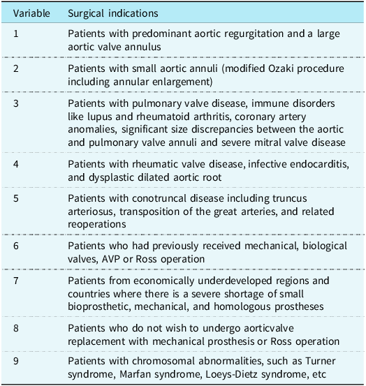
Theoretically, patients of all ages (adults and children) and those with acquired and congenital aortic valve disease, including those with prior mechanical or biologic valves, should be considered for Ozaki procedure. Reference Baird, Marathe and Del17 The Ozaki procedure was preferred for patients presenting with predominant aortic valve regurgitation and a large aortic valve annulus. Reference Wang, Zhang and Ma9,Reference Zhang, Jiang and Liu31 Children with hypoplastic aortic annulus are suitable candidates for Konno, Nicks, and/or Manougian aortic root enlargement procedures at the same time as Ozaki procedures. Consideration for performing annular enlargement was given to patients with annular Z-scores less than 2 and whose native annulus measured less than 15 mm. When enlarging the annulus, the patients’ age and weight should be taken into account and is often upsized to a Z-score of + 2. Reference Marathe, Chavez, Sleeper, Marx, Del and Baird24 In addition, children with certain conditions were not good candidates for Ross surgery. Absolute contraindications included Marfan syndrome, pulmonary valve disease, immune disorders like lupus and rheumatoid arthritis, coronary artery anomalies, significant size discrepancies between the aortic and pulmonary valve annuli and significant mitral valve disease. Relative contraindications (due to a higher risk of autograft dysfunction) included rheumatic valve disease, infective endocarditis and dysplastic dilated aortic root. The technique can also be used in the paediatric congenital population with conotruncal disease including truncus arteriosus, transposition of the great arteries and related reoperations. Reference Wang, Zhang and Ma9 In addition, Ozaki technique can also be considered in developing countries, given the large number of children with CHD, the lack of suitable conduit for right ventricular outflow tract reconstruction, especially homograft, and the lack of suitable biologic or mechanical valve for children with small valve annulus. Management of anticoagulants in young children can be challenging, increasing the risk of complications. Most parents were hesitant to choose mechanical valve replacement and wanted to wait until their children were older and able to self-administer anticoagulants. In this context, the Ozaki technique provides a new option to address these complex situations. For some older children with chromosomal abnormalities and obvious aortic root dilatation, root replacement should be performed at the same time as valve replacement, or aortic valve-sparing root replacement should be performed. Reference Zhang, Yang, Chen, Dong and Li2 It is also possible to do an AVNeo reconstruction followed by a valve-sparing root type replacement procedure (aka: David-Ozaki or Yacoub-Ozaki procedure) or a bio-Bentall with the AVNeo valve reconstructed within a tube graft prior to implant Reference Baird, Marathe and Del17 (Figure 1). Table 2 summarizes the indications for the Ozaki technique in the treatment of aortic valve disease in children.
Bio-bentall with the AVNeo valve reconstructed on the back table before cardiopulmonary bypass. ( a ) Inside view. ( b ) Outside view.

Surgical indications for Ozaki technique in the treatment of AVD in children

AVP = aortic valvuloplasty.
Surgical procedures
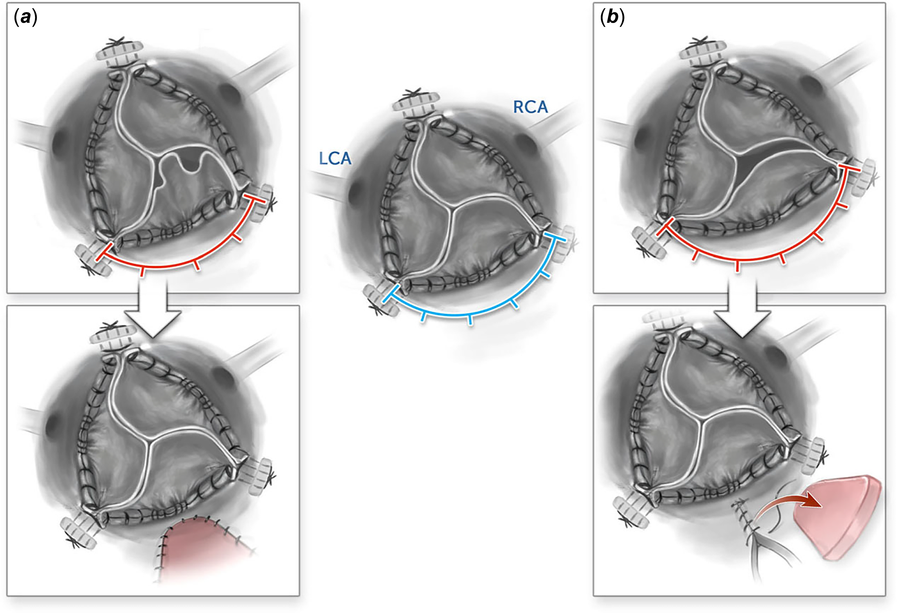
All procedures were performed through full sternotomy with cardiopulmonary bypass. Autologous pericardium was carefully harvested and then meticulously attached to a stainless-steel board. The outer surface of the pericardium was thoroughly cleansed of fat and other redundant tissue. Subsequently, the excised pericardium underwent treatment with a 0.6% glutaraldehyde solution for 5–10 minutes, followed by three rinses with physiological saline solution, each lasting 6 minutes. Following heparinization, cardiopulmonary bypass was established routinely through sequential cannulations of the ascending aorta, inferior vena cava, and superior vena cava. Finally, a transverse incision through the aortic root was made to expose the aortic valve. Inspected the pathological changes of leaflets carefully, and then determined whether to perform valve repair or leaflet reconstruction. The leaflets that were amenable to repair were meticulously excised. Different from three-leaflet or single-leaflet reconstruction technique, the number of leaflets to be reconstructed depended on the number of leaflets could not be repaired, maybe one, two, or three leaflets at our centre. Then, the distance between the commissures of the damaged leaflet was measured using the sizing apparatus originally invented by Ozaki and his colleagues (JOMDD, Tokyo, Japan), providing appropriate tension to reproduce the annulus during diastole, upsizing rather than downsizing if in between sizes. A new leaflet of the corresponding size was trimmed from the glutaraldehyde-treated autologous pericardium using an original template. If there was no sufficient autologous pericardium, bovine pericardium patch would be another choice. Finally, the annular margin of the pericardial leaflet was sutured with a running 4–0 or 5–0 prolene sutures to the annulus. The top of the commissural coaptation was secured with additional 5–0 prolene suture, and the new junction should be slightly higher than the original junction. As a result, a new tricuspid coaptation was created. When the patient presented with a bicuspid valve deformity and symmetric development, if only one leaflet was damaged, a bicuspid valve mould may be used to create a new leaflet to replace the impaired one. Consideration for performing annular enlargement was given to patients with annular Z-scores less than 2 and whose native annulus measured less than 15 mm. For patients requiring annular enlargement, Konno, Nicks and/or Manouguian incisions were made and patched with pericardium. The sizers were then used to mark the new annulus and the leaflets were sized as above. To avoid redundant leaflets, the leaflet size must be precisely determined and the sinus and sinus tube junction dimensions cannot be undersized. It is most frequently the non-coronary sinus that is undersized and can be enlarged with pericardium. Sometimes it is corrected by resecting a segment of the sinus to avoid a longer distance between the right/non and non/left commissures making the non-coronary sinus too large Reference Baird, Marathe and Del17 (Figure 2). Individualised surgical techniques were also used to treat the combined lesions. After eliminating the air in the heart, remove the crossclamp and withdraw cardiopulmonary bypass gradually. Transesophageal echocardiography was routinely arranged to confirm the haemodynamical results in the operation room. If there was significant residual regurgitation or stenosis, reoperation would be carried out.
( a ) A small aortic sinus can have the opposite effect whereby the leaflet “folds or buckles.”; enlarging the sinus and sinotubular junction allows improved coaptation by stretching the leaflet. ( b) Schematic of an enlarged aortic sinus can cause stretching of the leaflet, resulting in poor leaflet mobility; a triangular resection of the aortic wall from the non-coronary sinus with primary closure will decrease the aortic diameter, allowing more leaflet mobility and improved co-aptation.

Advantages of Ozaki technique in the treatment of aortic valve disease in children
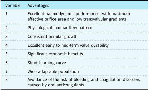
As a new aortic valve repair method, Ozaki technique expands the surgical indications of complex aortic valve disease and can be used as a promising alternative in children. It has several advantages over other surgical approaches (Table 3).
Advantages of Ozaki technique in the treatment of AVD in children

AVD = aortic valve disease.
1. Compared with conventional mechanical or biological valves, it preserves the natural motion of the aortic annulus and preserves the coordination of the left ventricle, sinus of Valsalva, and aorta, so that the aortic root naturally dilates during systole. Reference Ogami, Dufendach and Imran32 That leads to excellent haemodynamic performance, with maximum effective orifice area and low transvalvular gradients. Yoshikawa et al have reported the usefulness of measurements of the aortic annulus diameter using speckle-tracking transesophageal echocardiography. Compared with the measurement in an aortic valve replacement case, systolic expansion of the aortic annulus is well maintained in a case with AVNeo. Reference Ogami, Dufendach and Imran32
2. Compared with the Ross procedure, the Ozaki procedure restored the physiological laminar flow pattern and showed similar haemodynamic performance in the short term. Aurelio Secinaro et al. Reference Secinaro, Milano and Ciancarella19 analysed a patient population undergoing Ross or Ozaki procedure at their institution to compare the haemodynamic characteristics after the two procedures. All patients underwent haemodynamic assessment using 4D-flow Cardiovascular magnetic resonance, including aortic root and ascending aorta analysis. Cardiovascular magnetic resonance studies were performed at 1.5 T using a 4D flow-sensitive sequence acquired with retrospective electrocardiogram-gating and respiratory navigator. Post-processing of 4D-flow analysis was performed to calculate flow eccentricity and wall shear stress. No significant differences were observed in wall shear stress values measured at the level of the proximal ascending aorta between the two groups. The analysis of flow patterns showed no clear association between eccentric flow and the procedure performed. The Ozaki group showed just a slightly increased transvalvular maximum velocity.
3. Ozaki technique retains a certain degree of annular growth potential, which is helpful for delaying the age of valve replacement and reoperation, reducing the risk of bleeding and coagulation disorders caused by oral anticoagulants, and improving the quality of life after surgery. Since the Ozaki technique does not fix the aortic annulus, the neo-aortic annulus grows with time. A generous coaptation height should allow to retain a satisfactory coaptation surface as the annulus grows and the coaptation height reduces. Reference Kalfa, LaPar, Chai and Bacha34 In our experience, we have been able to demonstrate consistent annular growth. Reference Wang, Zhang and Ma9 This has also been confirmed by reports published by other paediatric heart centres. Reference Baird, Cooney, Chavez, Sleeper, Marx and Del22,Reference Marathe, Chavez, Sleeper, Marx, Del and Baird24,Reference Polito, Albanese, Cetrano, Cicenia, Rinelli and Carotti26
4. The Ozaki technique has good valve durability in the early and middle term. A recent consensus paper defined structural valve degeneration as deterioration of the leaflets or supporting structures of the bioprosthesis resulting in thickening, calcification, tearing, or disruption of the prosthetic valve materials, with subsequent associated haemodynamic dysfunction manifesting as progressive onset of stenosis or regurgitation. Reference Dvir, Bourguignon and Otto35 When comparing different prostheses, freedom from reoperation has been used as a proxy for durability, and there is a growing body of evidence supporting AVNeo in terms of early to mid-term durability (Table 1). Previously published studies have had small sample sizes and short follow-up periods, and further studies with larger samples and longer follow-up periods are needed to clarify late valve function and durability.
5. The promotion of Ozaki technique has significant economic benefits, especially in developing countries where homografts, small-sized biological valves or mechanical valves are seriously lacking but many children with CHD are economically very poor. Reference Wang, Ma and Li36 Because it takes the appropriate size of autologous pericardial tissue as the raw material for the preparation of leaflets, the material is convenient. The AVNeo system is reusable, simple to manufacture, and has a short learning curve, which is conducive to the proficiency of young surgeons.
6. It is suitable for a wide range of people. Theoretically, patients of all ages (adults and children) and those with acquired and congenital aortic valve disease (including those with prior mechanical or biologic valves) can be considered for Ozaki technique. Reference Baird, Marathe and Del17
Outcomes and factors of Ozaki technique in the treatment of aortic valve disease in children
Most of the experience with Ozaki technique in the treatment of aortic valve disease in children has been concentrated in a few volume paediatric heart centres, with small patient sample sizes and short follow-up periods, and most of the reports are single-center retrospective studies (Table 1). In paediatric populations, the Ozaki procedure has demonstrated promising short-term outcomes in several pioneering centres. Studie from Boston group reported that 96 and 91% of patients had less than moderate aortic valve regurgitationand aortic valve stenosis, respectively, at the last follow-up, with a median follow-up of 8.1 months. Reference Baird, Cooney, Chavez, Sleeper, Marx and Del22 Another study from Great Ormand Street Hospital in London confirmed its acceptable short-term haemodynamic results, with freedom from reoperation or moderate or greater aortic valve regurgitation at 1, 2 and 3 years being 94, 85 and 79%, respectively, with a median follow-up of 14.1 months. Reference Wiggins, Mimic and Issitt25 The Boston Children’s Hospital team also reported encouraging results with 80 and 82% of patients had <moderate aortic valve regurgitation and AS, at the last follow-up, with a median follow-up of 11.9 months, with the Modified Ozaki procedure including annular enlargement for small aortic annuli in 51 patients. Reference Marathe, Chavez, Sleeper, Marx, Del and Baird24 In 2017, we introduced Ozaki technique to treat aortic valve disease in children and achieved promising short-term results. Sixty four children underwent Ozaki operation from January 2017 to December 2023. During follow-up (20.4 ± 17.3 months), five patients (7.8%) in Ozaki group required reoperations. The incidence of moderate or severe AR and AS was 28.1% and 31.3%, respectively. Reference Wang, Zhang and Ma9 Shanghai Children’s Medical Center Reference Zhang, Jiang and Liu31 reported similar short-term results, with freedom from neoaortic valve reoperation was 92% at 2 years and 71% at 4 years in the Ozaki group. Ozaki technique is less effective in paediatric aortic valve disease than in adult aortic valve disease. Considering the vigorous basal metabolism in children, the content of calcium and phosphorus in blood is higher than that in adults, so the occurrence rate of valvular degeneration in children is faster than that in adults. However, the early and mid-term results in children are acceptable, and the long-term results need to be confirmed by longer follow-up. Given these findings, we recommend Ozaki technique as an alternative to delay valve replacement, Ross procedure and reoperation in paediatric aortic valve disease.
The incidence of recurrent aortic valve regurgitation after Ozaki technique is usually higher than that of aortic valve stenosis. Previous studies Reference Wang, Zhang and Ma9,Reference Sivalingam, Haranal and See20,Reference Polito, Albanese, Cetrano, Cicenia, Rinelli and Carotti26,Reference Bazylev, Tungusov and Mikulyak37,Reference Myers, Tissot, Christenson, Cikirikcioglu, Aggoun and Kalangos38 that lower age, extracorporeal membrane oxygenation installation, truncus arteriosus pathology, and asymmetric neocusps are risk factors for aortic regurgitation aggravation after Ozaki technique. However, higher aortic transvalvular gradient before discharge, smaller aortic annulus dimension, and use bovine pericardium were risk factors for aortic stenosis aggravation after Ozaki technique. Therefore, when performing Ozaki technique for aortic valve disease in children, attention should be paid to avoiding these related risk factors, which is beneficial to improve the long-term prognosis of patients, reduce the incidence of aortic valve disease recurrence, and delay the time of reoperation.
Application prospect of Ozaki technique in the treatment of aortic valve disease in children
Aortic valve neo-cuspidization using the Ozaki technique is an evolving procedure. Though the world experience is limited about the paediatric population, AVNeo sizers and templates will hopefully lead to a versatile solution with reproducible outcomes for young patients with congenital and acquired aortic valve disease. In recent years, with more and more paediatric heart centres carrying out Ozaki technique, the surgeons have become more and more skilled in the surgical techniques, which greatly shortens the cardiopulmonary bypass time and aortic cross-clamp time, and further improves the prognosis of the operation. Ranucci and associates Reference Ranucci, Frigiola, Menicanti, Castelvecchio, de Vincentiis and Pistuddi39 have demonstrated cross-clamp time being an independent predictor of severe cardiovascular-related morbidity, with an increased risk of 1.4% per 1-minute increase. In addition, with the improvement of surgical techniques, the spectrum of paediatric heart disease treated by Ozaki technique has been further expanded. For some older children with CHD, chromosomal abnormalities and obvious aortic root dilatation, root replacement should be performed at the same time of valve replacement, or aortic valve-sparing root replacement should be performed. It is also possible to do an AVNeo reconstruction followed by a valve-sparing root type replacement procedure (aka: David-Ozaki or Yacoub-Ozaki procedure) or a bio-Bentall with the AVNeo valve reconstructed within a tube graft prior to implant. Ozaki technique can be used during pulmonary valve replacement for tetralogy of Fallot and other right ventricular outflow tract obstruction diseases. In recent years, several studies have reported that the use of AVNeo system to create a new pulmonary valve directly in the autologous pulmonary valve annulus or artificial conduit has achieved some encouraging results Reference Baird, Marathe and Del17 (Figure 3). In our centre, we found that most of the children who underwent Ozaki technique were admitted to the hospital for reoperation after discharge, and the main reason was degenerative valve structure, which was attributed to the problem of manufacturing valve leaflet material. At present, it is generally believed that autologous pericardium tissue is superior to other biological tissue. Based on their experience using live right atrial appendage tissue for pulmonary valve reconstruction, Paighton C. Miller et al. Reference Miller, Miller, Mannie, Nath and Eghtesady40 described preliminary results using this tissue for aortic valve reconstruction. Right atrial appendage tissue was used to recreate the right coronary cusp in 6 patients. At the last follow-up (5–34 months), trivial to mild central aortic insufficiency was noted in 5 patients and mild to moderate insufficiency at the leaflet base in 1. These preliminary results suggest live atrial tissue can function in the aortic position, offering a possible new option for those requiring aortic valve interventions. Long-term results remain to be seen. In the future, the emergence of new anti-calcification leaflet materials may improve the long-term outcomes of Ozaki technique in children.
The right ventricular outflow tract after removal of the native leaflets and implantation of both leaflets. ( a ) Size 15 was used to measure the right ventricular outflow tract patch. ( b ) The autologous pericardium was transannular right ventricular outflow tract patch, and the annular width was confirmed using a sizer. ( c ) Creation of a neo-pulmonary valve using AVNeo technique.

Limitations
Of course, some limitations of this paper must be acknowledged. The sample size of previous studies was small, and the follow-up time was short. This view needs to be further clarified in multi-centre prospective studies with larger sample size and longer follow-up time.
Conclusions
In summary, with the steady improvement in the efficacy of Ozaki technique in children over the past decade, it is more active and widely used in children with aortic valve disease. Ozaki technique has obvious advantages over traditional prosthetic valve replacement. It preserves the natural motion of the aortic annulus, preserves the coordination of the left ventricle, the sinus of Valsalva and the aorta, naturally dilates the aortic root during systole, restores the physiological laminar flow pattern, and retains the continuous growth potential of the aortic annulus. It has good early and mid-term valve durability, no need for anticoagulation, short learning curve time, easy to promote, suitable for a wide range of people, and significant economic benefits, especially in developing countries with serious shortage of medical resources but a large number of CHD patients. Theoretically, Ozaki techniques can be considered in patients of all ages (adults and children) and those with acquired and congenital aortic valve disease, including those with previous mechanical, bioprosthetic, or Ross procedures. Modified Ozaki procedure for aortic root and sinus enlargement, David-Ozaki, Yacoub-Ozaki, or Bentall-Ozaki procedure, and creating a neo-pulmonary valve using the AVNeo technique further expand the spectrum of disease. We currently support the concept of using Ozaki technique as a surgical repair method for children with severe aortic valve disease, but the specific strategy should be made individually according to the patient’s condition. Further multicentre prospective studies with larger sample size and longer follow-up time are needed to confirm the findings.
Data availability statement
All data are incorporated into the article, and the data underlying this article are available in the article.
Acknowledgements
We acknowledge the roles of our colleagues, perfusionists, nurses, and others involved in the care of the study participants.
Author contribution
Zhangwei Wang: Conceptualisation; data curation; formal analysis; investigation; methodology; software; validation; visualisation; and writing – original draft. Honghao Fu: Writing – review & editing. Shoujun Li: Writing – review & editing.
Financial support
No funding was received for this work.
Competing interests
None declared.