Introduction
The gold standard for the treatment of locally advanced cervical cancer is definitive chemoradiation.Reference Marth, Landoni, Mahner, McCormack, Gonzalez-Martin and Colombo1, Reference Gupta, Parab and Kerkar2 In patients with large tumour volumes at presentation, there is a considerably higher tumour burden in the distal parametria and near the pelvic side walls, as opposed to earlier stages. This additional burden of peripheral tumour demands that care be taken during target volume definition and treatment delivery to ensure adequate doses to the pelvic side wall. Recurrence rates are higher among patients with locally advanced tumours compared with early stages. The incidence of locoregional relapse exceeds that of distant metastases in patients with larger tumour volumes at initiation of radiotherapy.Reference Perez, Grigsby and Camel3
Cervical cancer mostly presents at locally advanced stages in low-resource settings and is a major health problem for women in Southeast Asia. India, in particular, continues to be a major contributor to prevalence and mortality from the disease in this region.Reference Ferlay, Soerjomataram and Ervik4, Reference Bruni, Barrionuevo-Rosas and Albero5 Recommended doses to the high-risk clinical target volume (HRCTV) and intermediate-risk clinical target volume (IRCTV) in locally advanced cervical cancer are higher than that of early stages.Reference Pötter, Haie-Meder and Van Limbergen6 Owing to financial and infrastructure constraints, many centres in low-resource settings continue to provide intracavitary brachytherapy (ICRT) services, which contribute to lower pelvic side wall doses compared to interstitial applications. However, to improve local control in larger tumours, adequate coverage of the peripheral IRCTV cannot be overemphasised. In the absence of widespread facilities for interstitial brachytherapy (ISBT), the external beam parametrial boost (PMB) is a valuable tool to compensate for the under-dosing of the peripheral IRCTV.
However, reports on the PMB in literature indicate that organ-at-risk doses are higher and target coverage is suboptimal with midline blocked boost fields. Additionally, unacceptably high rates of radiation enteritis and proctitis have been observed in patients who received external beam PMB.Reference Huang, Wang, Hsu, Lin, Chen and Sun7, Reference Fenkell, Assenholt and Nielsen8 This often deterred physicians from administering external beam PMB to provide adequate doses to the distal parametria, and increased the chances of pelvic side wall recurrence. Mohamed et al. have demonstrated ISBT boost to be more conformal, resulting in lower normal-tissue volume exposure to intermediate doses of 60 Gy (V60).Reference Mohamed, Kallehauge, Fokdal, Lindegaard and Tanderup9 However, the impact of higher V60 in the clinic, in terms of organ-at-risk toxicity and pelvic side wall control is largely unexplored.
The primary objective of this study was to assess patient outcomes when midline shields of the 2D planned external beam PMB were individually customised based on the width of the 3D image-guided brachytherapy prescription isodose. The secondary objective was to evaluate patient outcomes with dose-escalated combined PMB using 3D image-guided ISBT and external beam radiotherapy.
Materials and Methods
This retrospective study included 242 women with histologically proven locally advanced carcinoma of the cervix (FIGO stage IIB–IVA).Reference Pecorelli10 The study population included patients from two different cancer centres; consecutive patients were included. Those from one centre received PMB, while those from the other did not. The two groups were compared for pelvic side wall recurrence and toxicity during a median duration of 24 months. The study extended from April 2007 to February 2014, to provide an adequate sample size for statistical analysis.
All patients received telecobalt external beam pelvic radiotherapy with weekly concomitant Cisplatin at doses of 40 mg/m2, followed by high-dose-rate (HDR) brachytherapy with Ir192 sources. A total of 2-Gy equivalent doses (EQD2) were calculated in all patients. Total pelvic side wall doses differed between the two groups. Patients in one group received PMB, while the other received no boost as per institutional protocols.
Patients receiving PMB (n = 112)
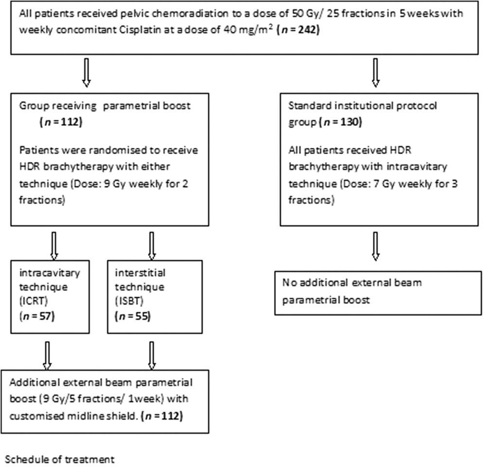
These women received pelvic chemoradiation with telecobalt to a dose of 50 Gy per 25 fractions in 5 weeks with weekly concomitant Cisplatin at a dose of 40 mg/m2. Subsequently, all patients received image-guided HDR brachytherapy with 3D image-based brachytherapy planning. Patients were randomly assigned following simple randomisation using computerised random numbers to receive HDR brachytherapy by either technique, that is, ISBT or ICRT. Randomisation eliminated physician bias and ensured allocation to either brachytherapy group irrespective of the initial extent of parametrial involvement. All patients received brachytherapy to a dose of 9 Gy per fraction, weekly, for 2 weeks. This HDR brachytherapy schedule was chosen as it reduced overall treatment time, and has been found to be safe and effective.Reference Patel, Rai, Mallick and Sharma11–Reference Sood, Gorla and Gupta13
Following brachytherapy, all patients in this group received an additional external beam PMB using a midline shield customised according to the width of the brachytherapy prescription isodose. The dose for PMB was 9 Gy per 5 fractions in 1 week. Since overall treatment time has a bearing on treatment compliance in our low-resource setting, a shorter brachytherapy and PMB fractionation schedule was chosen. Difficulties in transportation and socioeconomic disadvantages make longer schedules and more frequent hospital visits an inconvenience for most patients. In addition, restricted access to radiotherapy services for a large proportion of the population necessitates higher patient turnover in our overburdened practice.
Patients receiving no PMB (n = 130)
All patients received pelvic chemoradiation as per institutional protocol with telecobalt to a dose of 50 Gy per 25 fractions in 5 weeks with weekly concomitant Cisplatin at a dose of 40 mg/m2. Subsequent HDR brachytherapy was delivered by the ICRT technique. In all patients, the prescribed dose was 7 Gy per fraction, weekly, for 3 weeks. No additional PMB was delivered after brachytherapy.
The mean pelvic side wall dose, toxicities of Radiation Therapy Oncology Group (RTOG)Reference Cox, Stetz and Pajak14 grade ≥2 and pelvic side wall recurrence during the median duration of 24 months were compared between the boost versus no-boost groups. In patients receiving PMB, the pelvic side wall recurrence rates and toxicities between the two different brachytherapy subgroups were also compared. One subgroup received a dose-escalated combined ISBT and external beam PMB (ISBT/PMB) to the distal parametria and pelvic side wall (n = 55). The other subgroup (n = 57) received ICRT with PMB (ICRT/PMB) (Figure 1).
Schedule of treatment in either patient group.

External beam planning
As our institute had not implemented 3D external beam radiotherapy planning at the time of initiation of the study, 2D planning was utilised for all patients based on clinical gynaecologic examination findings and contrast-enhanced CT scan of the pelvis. Anteroposterior and posteroanterior parallel opposed beams were employed in most patients. The four-field box technique was reserved for patients in whom inter-field distance was ≥18 cm.
Brachytherapy procedure
A pre-planning CT scan was performed after external beam radiotherapy. This allowed assessment of the extent of residual parametrial disease and helped in planning ISBT needle placement. Following chemoradiation, residual parametrial disease was observed in 24 patients who received ISBT and 27 patients who received ICRT.
Intracavitary technique
For all intracavitary treatments, either Manchester three-channel or Fletcher-Suit applicators were used depending on the degree of vaginal stenosis and available space. The tandem was inserted under direct vision in all cases.
Interstitial technique
Interstitial applications were template-guided. The MUPIT (Martinez Universal Perineal Interstitial Template) or Syed Neblett template were employed in conjunction with rigid stainless steel needles or flexible needles (only for the Syed Neblett template).
Applicator geometry was pre-planned, accounting for the extent of disease as evaluated by speculum and bi-manual examination after completion of chemoradiation, in conjunction with findings on the pre-planning CT scan.
Post-applicator placement CT scans assisted in 3D volume-based planning. Geometrical optimisation without reference points, on a case-to-case basis, ensured optimal dose distributions. The plans were considered suitable for treatment after analysis of dose distribution in each image section, width of the reference isodose, bilateral pelvic side wall doses at the level of the external os, dose to HRCTV, and dose volume histograms of bladder and rectum. For intracavitary applications, the International Commission on Radiation Units and Measurements (ICRU) bladder and rectal point doses were also recorded.15
PMB planning
PMB was planned after both brachytherapy fractions were complete and the width of reference isodose was documented. 2D radiotherapy planning was employed for PMB fields. Treatment was delivered by anteroposterior–posteroanterior parallel opposed fields. The individually customised width of the midline shield equalled the width of brachytherapy prescription isodose at the level of the external os. Only the parametria extending from the brachytherapy reference isodose up to the lateral pelvic wall were included in the boost fields; pelvic nodes were excluded.
Follow-up
At each visit, patients were assessed with history, local examination, and appropriate radiological investigations. Locoregional and distant recurrence was confirmed using contrast-enhanced CT scans. To save costs, contrast-enhanced MRI was employed only in cases where CT scan findings were equivocal.
Statistical analysis
Statistical analysis was performed using MedCalc Statistical Software version 18.6 (MedCalc Software bvba, Ostend, Belgium; http://www.medcalc.org; 2018). Differences in recurrence rates and toxicities between the groups were analysed using the Fisher’s two-tailed t-test.
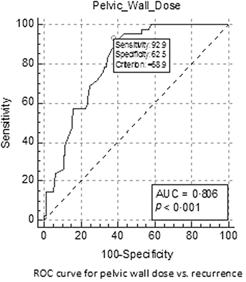
Receiver operating characteristic (ROC) curve analysis was performed to evaluate pelvic side wall doses and recurrence at 24 months, and to determine a cut-off dose that had significant impact on pelvic side wall recurrence in this cohort.
Results
This cohort included 242 women with locally advanced cervical cancer. A majority (97·5%) had non-keratinising squamous cell carcinoma; the remainder had other histologies; 82·2% had stage IIIB disease at presentation. The median age of this cohort was 39 years (range 25–69). A total of 213 women (88%) had completed at least 4 weeks of concomitant chemotherapy. The median duration of follow-up was 24 months (range 20–29) (Table 1).
Patient characteristics

The mean pelvic side wall doses (EQD2) between the two subgroups of women receiving PMB were 72·9 Gy (68·7–78·2 Gy) for those receiving escalated combined ISBT/PMB, and 68·1 Gy (62·3–70·7 Gy) for those receiving external beam PMB only (ICRT/PMB). In patients who received no PMB, the mean pelvic side wall dose was 57·4 Gy (55·2–58·6 Gy) (Table 2).
Mean pelvic side wall dose in each treatment group

At 24 months, 2/112 and 40/130 patients who received any PMB and no boost, respectively, had pelvic side wall recurrence (p < 0·000001); 0/55 and 2/57 patients in the ISBT/PMB (dose-escalated PMB) and ICRT/PMB (external beam PMB only) groups, respectively, had recurrences in the pelvic side wall (p = 0·49). Distant recurrence was observed in 5/112 and 7/130 patients in the boost and no-boost groups, respectively (p = 0·78).
Toxicities of RTOG grade ≥2 were found in 28/112 and 37/130 patients in the boost and no-boost groups, respectively (p = 0·56), and in 17/55 and 14/57 patients in the two subgroups of the PMB cohort, that is, escalated combined ISBT/PMB and ICRT/PMB, respectively (p = 0·47).
ROC curve analysis demonstrated a cut-off dose of 58·9 Gy to the pelvic side wall for significant clinical benefit. The area under the ROC curve was 0·81 (p < 0·0001). The Youden Index J for the analysis was 0·65 (Figure 2).
ROC curve showing optimal pelvic side wall dose that significantly reduced recurrence.

Discussion
The practice of using an external beam PMB had been much debated in the era of 2D planning. While the boost dose to the parametrium in locally advanced cervical cancer ensures adequate coverage of the IRCTV, higher normal-tissue toxicities and inadequate target coverage have been reported with external beam PMB. The advent of hybrid intracavitary and interstitial applicators has made dosimetry more predictable. However, in low-resource areas, a majority of centres continue to deal with a large burden of locally advanced cervix cancer without adequate infrastructure and funding for ISBT or hybrid applicators. Many centres are compelled to deliver ICRT, which under-doses peripheral IRCTV. An external beam PMB in these situations ensures adequate target coverage. However, in view of reports of unacceptable dosimetry and toxicities, these were largely avoided. Fenkell et al. reported that midline block fields led to unpredictable dosimetry for the target and organs-at-risk.Reference Fenkell, Assenholt and Nielsen8 Huang et al. found that higher cumulative rectal doses with external beam PMB caused a higher incidence of rectal toxicities. These toxicities arose from an unpredictable dosimetry of the standardised midline block. However, data on disease recurrence were not reported.Reference Huang, Wang, Hsu, Lin, Chen and Sun7 Wolfson et al. suggested that potential inhomogeneities in target doses were related to the relative positioning of the brachytherapy applicator with respect to standardised midline blocks, and that customising the midline block should be considered.Reference Wolfson, Abdel-Wahab and Markoe16 Popple et al. investigated various approaches of the midline block and observed that custom step wedge blocking using dynamic multileaf collimation provided a more homogeneous dose distribution compared to other techniques.Reference Popple, Kim, Pareek, Duan, Shen and Brezovich17 Yin et al. reported that an intensity-modulated radiation therapy (IMRT) PMB, in addition to ICRT, offered low organ-at-risk doses, and recommended its use in cases where ISBT was not feasible.Reference Yin, Wang and Lang18 Cheng et al. reported on the use of a simultaneous integrated boost to the parametrium using IMRT or VMAT. Either technique offered better dosimetry than the traditional midline block. However, the organ-at-risk doses were higher.Reference Cheng, Huang, Hsu and Wang19 None of these studies reported on the incidence of pelvic side wall recurrence and toxicities. In the present study, we intended to explore patient outcomes with an external beam PMB that used individually customised midline blocks, tailored according to the width of the 3D image-based brachytherapy reference isodose. All women received brachytherapy in addition to pelvic chemoradiation. The use of brachytherapy ensured that the central high-dose region, which is vital to the successful radiocurability of cervical cancer, was achieved. In the present study, different patient groups received different total pelvic side wall doses (EQD2). Significant benefit in local control was noted irrespective of PMB technique, that is, external beam radiotherapy or combined dose-escalated ISBT (p < 0·000001). Clearly, PMB using any technique was better than no boost. A retrospective study by Rajasooriyar and coworkers, which included nearly 200 patients staged with PET-CT scans, had concluded that doses up to 50 Gy are adequate to avoid isolated pelvic nodal failure, and that clinically involved parametria may be adequately treated without a PMB.Reference Rajasooriyar, Van Dyk, Lindawati, Bernshaw, Kondalsamy-Chennakesavan and Narayan20 Our findings suggest, however, that PMB achieved higher pelvic side wall doses, which led to significantly lower pelvic side wall recurrence. ROC analysis revealed the optimal pelvic side wall dose of 58·9 Gy for significant improvement in pelvic side wall control, which was achievable with customised PMB planning.
Although PMB was associated with significantly better outcomes compared to no boost, further dose escalation using combined ISBT and external beam radiotherapy offered no significant benefit compared to PMB alone (p = 0·49). Clearly, pelvic side wall dose escalation beyond recommended doses is not superior. Mohamed et al. reported higher normal-tissue doses and V60 using a non-customised midline block. However, patient outcomes in that cohort were not reported.Reference Mohamed, Kallehauge, Fokdal, Lindegaard and Tanderup9 Our findings show that toxicities of RTOG grade ≥2 were not significantly different between the boost versus no-boost groups (p = 0·56) and also between the subgroups that received dose-escalated combined PMB versus external beam only PMB (p = 0·47). Individually customising the width of the PMB midline shield probably minimised the chances of high dose to organs-at-risk. One of the major limitations of the present study was the unavailability of 3D external beam planning. Consequently, volumetric summation of brachytherapy and external beam plans to assess the association of dose volume parameters with clinical outcomes was not possible.
Our study also included a smaller number of patients, and the median duration of follow-up was short. This was mostly due to high attrition rates among follow-up cases. Socioeconomic disadvantages, financial constraints and inadequate resources often lead to higher rates of non-compliance during treatment and follow-up.
Despite these limitations, the results demonstrate that the individually customised external beam PMB offered adequate pelvic side wall control without additional toxicity, even with 2D external beam planning. Tailoring the width of the midline block to the width of the reference isodose obtained from 3D image-based brachytherapy planning probably improved safety. In the absence of resources for 3D planning, the individually customised external beam PMB provided a safe and practical solution in this low-resource setting.
Conclusion
Individually customised PMB fields with 2D planned external beam radiotherapy offered better outcomes than no boost. Therefore, higher V60 doses do not necessarily translate to significantly higher normal-tissue toxicities in the clinic. However, escalation of pelvic wall doses beyond the recommended limit is unnecessary. Further studies on customised PMBs with 3D planned external beam radiotherapy are warranted.
Acknowledgements
The authors are grateful to Mr Surojit Maity, Department of Medical Records, Chittaranjan National Cancer Institute, Kolkata, for his invaluable assistance during data collection.
Financial support
This research received no specific grant from any funding agency, commercial or not-for-profit sectors.
Conflicts of interest
None.
Ethical standards
The authors assert that all procedures contributing to this work comply with the National Ethical Guidelines for Biomedical and Health Research Involving Human Participants of the Indian Council of Medical Research and with the Helsinki Declaration of 1975, as revised in 2008, and has been approved by the institutional committee of the Medical College and Hospitals, Kolkata.
Sources of support
None.